This article contains AI generated content.
Posterolateral Fat Pad Impingement: 4 Genes and 7 Biomarkers to Track
If you're dealing with a nagging, deep ache at the back-outer corner of your ankle that flares with pointed-toe positions, downhill running, or pushing off hard, you've probably already heard the standard advice: rest, ice, maybe a cortisone injection, maybe surgery if it doesn't settle. That advice isn't wrong, but it's generic. It treats every posterolateral fat pad impingement as the same injury, when in practice the reason one person's soft tissue keeps getting pinched between the talus and fibula while another person's doesn't often comes down to things a standard exam doesn't measure — tissue quality, inflammatory load, joint laxity, and how well the body remodels collagen after repeated microtrauma.
That's the gap this article tries to close. Instead of repeating "strengthen your ankle and see a physio," it looks at the measurable biology underneath the diagnosis: which lab values and functional tests correlate with fat pad irritation and slow recovery, and which inherited traits quietly raise the odds of getting here in the first place. None of this replaces a proper orthopedic or podiatric evaluation, and no test in this article diagnoses posterolateral impingement on its own — imaging and a clinical exam still do that.
What better information does offer is a way to stop guessing. If you know your vitamin D is low, your joints are unusually lax, or you carry a collagen gene variant linked to soft-tissue injury, you can build a recovery and prevention plan around your actual biology rather than a generic protocol built for an average ankle. That's a meaningfully different starting point, and it's one you can act on this month.
This article covers two complementary angles. The main section walks through seven biomarkers worth tracking — inflammatory, structural, and functional — with realistic cost ranges and what to do if a result comes back unfavorable. A shorter section afterward looks at the genetic side: a handful of collagen and remodeling genes that influence how resilient your ligaments and fat pads are, and how to work around a less favorable variant. A book-based section and a rundown of complementary approaches with real supporting evidence round things out.
Summary
Posterolateral fat pad impingement happens when soft tissue behind the ankle — often a hypertrophied or fibrotic fat pad, sometimes alongside scarred synovium or an os trigonum — gets repeatedly pinched between the talus and fibula during forced plantarflexion. It's common in dancers, kickers, and anyone with a history of lateral ankle sprains. What most people don't realize is that two biological factors quietly decide who develops this and who recovers quickly: systemic inflammatory and connective-tissue biomarkers, and inherited collagen-remodeling genetics. Below, you'll find the seven blood, imaging, and functional markers worth checking — from an inexpensive vitamin D test to a specialty leptin panel that reflects how metabolically active your fat pad has become — plus the four genes (COL5A1, MMP3, VEGFA, COL1A1) most consistently linked to ligament and soft-tissue injury risk, and exactly what to do about each one, with or without supplements. A book recommendation on joint mechanics and a short list of complementary therapies with real trial support close out the piece.
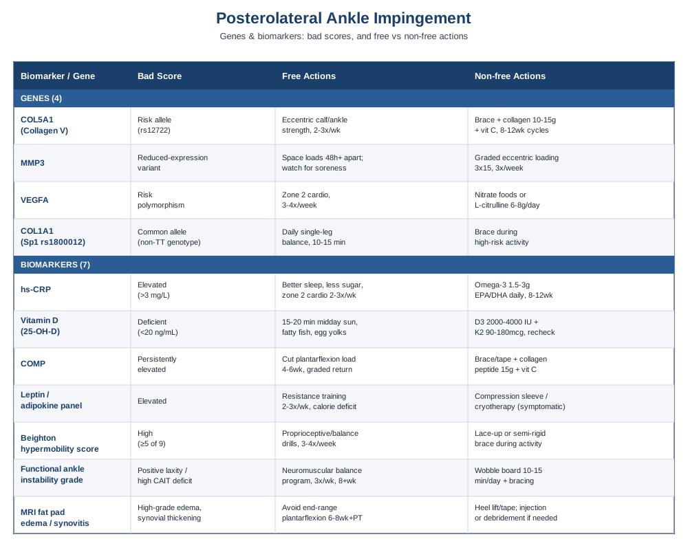
7 Biomarkers That Reveal What's Really Happening in Your Ankle
Posterolateral ankle impingement is fundamentally mechanical — a soft-tissue structure getting caught in a bony space during plantarflexion — but how much it hurts, how fast it resolves, and how likely it is to recur depend heavily on biology that a standard X-ray won't show you. The clinical literature describes this impingement as arising from fibrosis, synovial thickening, and fat pad hypertrophy in the space behind the posterior malleolus and posterolateral talus (Posterior Ankle Impingement: Clarification and Confirmation of the Pathoanatomy, Posterior ankle impingement syndrome: a systematic four-stage approach). The seven markers below help you see the biological terrain behind that mechanical problem — how inflamed you run, how well your tissue is built to remodel, and how lax your joints are to begin with.
High-sensitivity CRP (hs-CRP)
Why it matters: hs-CRP is the most accessible marker of systemic low-grade inflammation, and research on joint disease consistently shows a correlation between elevated hs-CRP and local synovial inflammation, pain, and slower functional recovery (Elevated C-reactive protein levels in osteoarthritis are associated with local joint inflammation, Severity and extent of osteoarthritis and low-grade systemic inflammation as assessed by hs-CRP). A fat pad that's chronically impinged behaves like a small, low-grade inflamed structure, so tracking your systemic baseline gives you a sense of how much your whole-body inflammatory load might be adding fuel to local irritation.
How to measure it: a standard blood draw, often bundled into a basic metabolic or cardiovascular panel. Cost ranges from about $15 to $40 through direct-to-consumer labs (Quest, LabCorp, or similar), and it's frequently covered by insurance when ordered by a physician. Erythrocyte sedimentation rate (ESR) is commonly ordered alongside it as a slower-moving companion marker.
If the score is bad, the plan without supplements: prioritize sleep regularity, reduce ultra-processed food and added sugar intake, and add two to three sessions per week of zone 2 cardio (30-45 minutes at a conversational pace), which reliably lowers hs-CRP over 8-12 weeks in most people. Reassess after 10-12 weeks rather than weekly — CRP is noisy day to day and reacts to unrelated illness or poor sleep the night before the draw.
If the score is bad, the plan with supplements or equipment: omega-3 fatty acids (1.5-3g EPA/DHA combined daily, with food) have the best-supported anti-inflammatory signal among accessible supplements; give it 8-12 weeks before rechecking. Side effects are mild and dose-dependent — fishy aftertaste, occasional loose stools, and a theoretical bleeding risk at high doses, so check with a physician if you're on blood thinners. Cycling isn't necessary for omega-3s; they're reasonable to take continuously.
Vitamin D (25-hydroxyvitamin D)
Why it matters: vitamin D receptors are present on tendon and ligament tissue, and deficiency has been linked to weaker tendon-ligament structures and slower healing after soft-tissue injury. One study found that patients with ligamentous or cartilaginous complaints had the highest rates of low vitamin D among lower-extremity injury categories (Prevalence of Vitamin D Insufficiency and Deficiency in Young Female Patients With Lower Extremity Musculoskeletal Complaints), and a systematic review found associations between vitamin D status and ACL injury rates and post-reconstruction function (Effect of vitamin D on anterior cruciate ligament injury rates and post-reconstruction function).
How to measure it: a simple blood test, $40-80 out of pocket, often included free in an annual physical. Target range is generally considered 30-50 ng/mL, with levels below 20 ng/mL considered deficient.
If the score is bad, the plan without supplements: 15-20 minutes of midday sun exposure on bare arms and legs several times a week (skin tone and latitude dependent), plus dietary sources like fatty fish, egg yolks, and fortified dairy. Recheck in 3 months, since sunlight-driven changes are slow and seasonal.
If the score is bad, the plan with supplements or equipment: vitamin D3, typically 2,000-4,000 IU daily for someone moderately deficient, taken with a fat-containing meal for absorption, paired with vitamin K2 (90-180 mcg) to support proper calcium utilization. Recheck levels after 8-12 weeks and adjust the dose rather than guessing. Side effects are rare at these doses but include hypercalcemia at very high, prolonged intakes — an actual reason to retest rather than simply continuing indefinitely.
Serum COMP (cartilage oligomeric matrix protein)
Why it matters: COMP is a structural glycoprotein released into circulation as cartilage and connective tissue undergo remodeling and degradation. It rises before radiographic changes appear, making it a useful early window into joint tissue turnover (Cartilage oligomeric matrix protein in monitoring and prognostication of osteoarthritis, Cartilage Oligomeric Matrix Protein: A Biomarker of Arthritis). For an impinged posterolateral joint under repetitive mechanical stress, a persistently elevated COMP can be an early signal that the joint is remodeling faster than it's recovering.
How to measure it: this isn't a routine test at a standard lab — it requires a specialty or research-oriented lab (such as those used in sports medicine or rheumatology research settings), typically $100-250, and usually needs to be specifically requested through a sports medicine physician or orthopedist.
If the score is bad, the plan without supplements: reduce the volume of high-plantarflexion loading (repeated jumps, sprinting, pointed-toe positions) for 4-6 weeks while maintaining pain-free range-of-motion and strength work, then reintroduce load gradually using a graded return-to-sport protocol.
If the score is bad, the plan with supplements or equipment: a supportive ankle brace or kinesiology taping during high-load activity to offload the posterolateral compartment while tissue settles, alongside collagen peptide (15g) with 50mg vitamin C taken about an hour before loading sessions, which has shown increased collagen synthesis in engineered ligament tissue (Efficacy of Vitamin C Supplementation on Collagen Synthesis and Oxidative Stress After Musculoskeletal Injuries). Use for 8-12 weeks, then reassess — there's no established reason to cycle it, and side effects are minimal (mild GI upset at high vitamin C doses).
Leptin and adipokine panel
Why it matters: fat pads aren't inert padding — they're metabolically active tissue that secretes inflammatory adipokines, especially leptin, directly into the joint. Research on the knee's infrapatellar (Hoffa's) fat pad — the best-studied analog to other joint fat pads — shows leptin is actively secreted into synovial fluid and drives cartilage-degrading enzyme production (The Corpus Adiposum Infrapatellare (Hoffa's Fat Pad) — the Role of the Infrapatellar Fat Pad in Osteoarthritis Pathogenesis, Infrapatellar fat pad as a source of biomarkers and therapeutic target for knee osteoarthritis). A hypertrophied, impinged fat pad in the ankle likely behaves similarly, so serum leptin gives an indirect read on how "activated" that fat pad tissue has become.
How to measure it: a specialty panel, typically $150-300, ordered through a lab that runs adipokine/metabolic research panels; not part of routine primary care blood work.
If the score is bad, the plan without supplements: body composition matters more here than body weight — reducing visceral and local adiposity through resistance training (2-3 sessions weekly) and a modest caloric deficit if overweight tends to lower circulating leptin over 3-6 months.
If the score is bad, the plan with supplements or equipment: there's no well-supported supplement that directly lowers leptin in a targeted way; the honest answer is that body composition change, not a pill, is what moves this number. A compression sleeve or targeted cryotherapy (10-15 minutes post-activity) may reduce local swelling and discomfort, but won't change the underlying adipokine profile.
Beighton hypermobility score
Why it matters: generalized joint hypermobility is a documented risk factor across a long list of soft-tissue injuries, including recurrent ankle sprains and instability, which are common precursors to posterolateral impingement (Signs and symptoms of foot and ankle dysfunction in children with joint hypermobility). If your ligaments are naturally more elastic, the talus and fibula have more room to move into positions that pinch the posterolateral soft tissue.
How to measure it: a free, 5-minute physical exam any physiotherapist, sports physician, or orthopedist can perform, scoring 9 possible points across thumb, pinky, elbow, knee, and trunk flexion tests. It has good inter-rater reliability when performed correctly (Interrater and Intrarater Reliability of the Beighton Score: A Systematic Review).
If the score is bad, the plan without supplements: this isn't a trait you reverse — it's one you train around. Prioritize proprioceptive and neuromuscular control work (single-leg balance progressions, wobble board drills) 3-4 times weekly rather than passive stretching, since hypermobile joints need stability training, not more range of motion.
If the score is bad, the plan with supplements or equipment: a lace-up or semi-rigid ankle brace during high-risk activities (court sports, uneven terrain) provides mechanical restraint that hypermobile ligaments don't supply on their own. There's no strong evidence that any supplement changes ligament laxity itself; collagen/vitamin C protocols may support tissue quality but won't meaningfully tighten already-lax ligaments.
Functional ankle instability grade
Why it matters: this combines a clinical laxity test (anterior drawer, talar tilt) with a validated patient-reported measure like the Cumberland Ankle Instability Tool. Chronic ankle instability is one of the most consistent precursors to posterolateral impingement, since repeated micro-instability drives the synovial and fat pad thickening that eventually gets pinched (Effect of Joint Mobilization in Individuals with Chronic Ankle Instability: A Systematic Review and Meta-Analysis).
How to measure it: a clinical exam plus a short questionnaire, generally $75-150 through a sports medicine physician or physical therapist, sometimes bundled into an initial evaluation at no extra cost.
If the score is bad, the plan without supplements: a structured neuromuscular and balance program, 3 sessions weekly for at least 8 weeks, is the best-evidenced non-pharmacological intervention for chronic ankle instability and, by extension, reducing the mechanical drivers of impingement.
If the score is bad, the plan with supplements or equipment: a wobble board or balance pad (10-15 minutes daily) accelerates proprioceptive retraining, and taping or bracing during sport-specific activity reduces re-injury risk while that retraining takes hold. No supplement directly addresses mechanical instability.
MRI-graded fat pad edema and synovitis
Why it matters: this is the closest thing to a direct structural biomarker for this specific condition — MRI can grade the degree of fat pad edema, synovial thickening, and any associated os trigonum or bony contribution, which correlates with symptom severity and helps guide whether conservative care or surgical debridement is more appropriate (Posterior ankle impingement syndrome: A systematic four-stage approach, Ankle impingement: a review of multimodality imaging approach).
How to measure it: an MRI of the ankle, generally $400-1,800 depending on region and insurance status; ultrasound is a cheaper ($150-400) but operator-dependent alternative that can still show fat pad thickening and dynamic impingement during plantarflexion.
If the score is bad, the plan without supplements: a structured period of activity modification avoiding end-range plantarflexion (pointe work, deep calf raises off a step, sprinting starts) for 6-8 weeks, combined with physical therapy focused on ankle mechanics, is first-line before considering injection or surgery.
If the score is bad, the plan with supplements or equipment: a heel lift or slightly restricted dorsiflexion taping technique can reduce end-range plantarflexion during daily activity while tissue settles. If conservative measures fail after 3-6 months, a corticosteroid injection or ultrasound-guided debridement becomes a reasonable next conversation with an orthopedic specialist — beyond the scope of self-directed care.
Blood work and imaging tell you what's happening now; genetics tell you why some ankles are more prone to this cycle in the first place, which is where the next section picks up.
What Your Genes May Be Telling You About Soft-Tissue Resilience
Consumer genomics has made it easy to pull up your raw genetic data and start asking pointed questions about injury risk — a trend popularized by researchers like Ali Torkamani, whose work at Scripps Research has pushed genomic risk profiling into mainstream use, and by figures like Gary Brecka, who built a public following around interpreting consumer gene panels for everyday health decisions. Neither of them has published specific work on posterolateral ankle impingement, but the underlying idea they popularized — that a handful of common gene variants meaningfully shift your baseline connective-tissue resilience — is well supported by tendon-ligament injury genetics research specifically (Tendon and Ligament Genetics: How Do They Contribute to Disease and Injury? A Narrative Review).
None of the genes below are unique to ankle impingement specifically — the condition itself hasn't been the subject of dedicated genetic studies — but they've been repeatedly linked to the broader category of soft-tissue and ligament injury that predisposes someone to chronic ankle instability and, downstream, to fat pad impingement. Importantly, none of these are "bad genes" in an absolute sense — they're common variants that shift probability, not certainty, and every one of them can be compensated for.
COL5A1 — collagen type V, alpha 1
What it may affect: COL5A1 helps regulate the diameter of collagen fibrils in tendons and ligaments. Certain variants (notably around rs12722) are associated with altered tendon-ligament injury risk across multiple meta-analyses, with some alleles linked to reduced risk and others to increased risk of soft-tissue injury (Association of COL5A1 gene polymorphisms and risk of tendon-ligament injuries among Caucasians: a meta-analysis). Mutations in this gene in more extreme forms also cause classic Ehlers-Danlos syndrome, underscoring its role in ligament integrity.
If the gene is bad, the plan without supplements: since a less favorable COL5A1 profile is associated with looser, more injury-prone connective tissue, the practical response is the same as for hypermobility — build neuromuscular control and strength around the joint rather than chasing more flexibility. Emphasize eccentric calf and ankle strengthening 2-3 times weekly, which improves tendon stiffness independent of genotype.
If the gene is bad, the plan with supplements or equipment: consider a period of ankle bracing during high-risk activity, and a collagen peptide (10-15g daily) plus vitamin C protocol taken before training sessions, cycling 8-12 weeks on with periodic reassessment of symptoms rather than continuous indefinite use. Side effects are minimal; the main consideration is that collagen supplements are not a substitute for load management if pain is present.
MMP3 — matrix metalloproteinase 3
What it may affect: MMP3 is central to remodeling the extracellular matrix during tendon and ligament repair. Reduced-expression variants have been linked to slower or impaired remodeling and higher tendinopathy risk, while other variants shift ACL and Achilles injury risk in different directions depending on the specific polymorphism studied (Association between matrix metalloproteinase-3 gene polymorphisms and tendon-ligament injuries: evidence from a meta-analysis).
If the gene is bad, the plan without supplements: since MMP3 variants affect repair speed rather than structural integrity per se, the most useful lever is giving tissue more recovery time between loading sessions than a "standard" protocol would suggest — spacing high-plantarflexion training by at least 48 hours and monitoring for lingering soreness before repeating.
If the gene is bad, the plan with supplements or equipment: there's no supplement shown to directly correct MMP3 activity in humans; the more defensible approach is mechanical — a graded eccentric loading program, which has strong evidence for improving tendon and soft-tissue remodeling regardless of genotype (The Role and Implementation of Eccentric Training in Athletic Rehabilitation), performed 3 sets of 15 reps, 3 times weekly, ramping load gradually over the first 3 weeks to avoid overloading tissue that remodels more slowly.
VEGFA — vascular endothelial growth factor A
What it may affect: VEGFA drives angiogenesis — new blood vessel formation — which matters directly for how well-vascularized (and therefore how well-healing) tendon and ligament tissue is. A meta-analysis found VEGFA polymorphisms associated with altered tendon-ligament injury risk (Correlation between vascular endothelial growth factor A gene polymorphisms and tendon and ligament injury risk: a systematic review and meta-analysis). Poorly vascularized tissue tends to heal more slowly after the repeated microtrauma that drives fat pad hypertrophy.
If the gene is bad, the plan without supplements: aerobic conditioning (zone 2 cardio, 3-4 sessions weekly) improves general tissue vascularization and healing capacity, and is a reasonable non-supplement lever for someone whose genetics suggest slower vascular adaptation.
If the gene is bad, the plan with supplements or equipment: some practitioners use nitrate-rich foods (beetroot, leafy greens) or L-citrulline (6-8g daily) to support nitric oxide-mediated blood flow, though direct evidence in tendon-ligament healing specifically is preliminary rather than established — worth trying for 8 weeks with no expectation of a dramatic effect. Side effects are minimal (occasional GI discomfort with citrulline); no cycling is required.
COL1A1 — collagen type I, alpha 1
What it may affect: the well-studied Sp1 polymorphism (rs1800012) in COL1A1 affects a transcription factor binding site and shifts the ratio of collagen chains produced. Interestingly, the research here shows the rarer TT genotype is associated with reduced ACL and ligament injury risk, while the more common allele carries a slightly higher risk — with no clear association found for tendinopathy specifically (Association of polymorphisms rs1800012 in COL1A1 with sports-related tendon and ligament injuries: a meta-analysis).
If the gene is bad, the plan without supplements: because this variant affects ligament rather than tendon tissue specifically, the most useful non-supplement lever is proprioceptive and balance training aimed squarely at reducing the ankle-rolling episodes that precede chronic instability and impingement — 10-15 minutes of single-leg balance work daily.
If the gene is bad, the plan with supplements or equipment: bracing during high-risk activity remains the most defensible equipment-based compensation, since it mechanically substitutes for ligament restraint the genotype may not fully provide. Collagen/vitamin C supplementation follows the same 8-12 week protocol described above, with no specific COL1A1-targeted supplement currently supported by human trial evidence.
Genetics set your baseline risk, but the day-to-day habits and mental models you bring to joint health still do most of the work — which is exactly the territory the next section covers.
The Book That Reframes How You Think About Joint Mechanics
Kelly Starrett's Becoming a Supple Leopard isn't written specifically about ankle impingement, but its central argument — that most "structural" joint pain is actually a positional and mechanical problem that can be retrained — is unusually well-matched to a condition like this one, where a soft-tissue structure gets repeatedly pinched because of how a joint moves under load, not because the tissue itself is inherently damaged. It challenges the passive-treatment default (rest, injections, imaging) by insisting that most people can restore normal joint mechanics themselves, with consistent, deliberate practice. Below are ten of its most useful ideas, reframed for someone dealing with posterolateral ankle impingement specifically.
1. Pain is often a downstream signal, not the root problem
Starrett's framework treats pain at a joint as frequently originating from a mechanical fault elsewhere in the kinetic chain — restricted hip extension or poor ankle dorsiflexion, for instance — rather than assuming the painful structure itself is the sole issue. For posterolateral impingement, this means checking hip and knee mechanics during running or jumping, not just treating the ankle in isolation.
2. "Tissue creep" explains why static stretching alone often fails
Connective tissue temporarily elongates under sustained low load (creep) but returns to its original length within hours. This is why a single stretching session before activity does little for chronic impingement — lasting change requires repeated loading over weeks, not one-off flexibility sessions.
3. Full range of motion should be trained, not just tested
Starrett argues that if you can't actively control a range of motion, you don't really "own" it — passive flexibility without control is a liability, not an asset. For an ankle with impingement, this means training controlled, loaded dorsiflexion and plantarflexion rather than simply checking whether the joint can passively move that far.
4. Joint capsule restriction is often the missing piece
Much of his system is built around restoring capsular mobility — the sliding and gliding of joint surfaces — using banded distraction techniques, arguing that muscle-only stretching ignores this layer entirely. Ankle-specific banded mobilizations that distract the joint before dorsiflexion work directly target this idea.
5. "Systems" thinking beats isolated exercises
Rather than prescribing one exercise per problem, Starrett frames the body as an interconnected system where a squat pattern, breathing mechanics, and ankle position are all linked — a useful mental model for understanding why generic ankle-only rehab sometimes plateaus.
6. Ten minutes a day beats one long session a week
His mobility protocols are built around short, frequent daily sessions rather than occasional long stretching blocks, on the premise that tissue adapts to consistent, repeated stimulus far better than infrequent large doses.
7. Position matters as much as the exercise itself
A recurring theme is that the same exercise performed in a subtly different joint position produces a completely different training effect — relevant for ankle work, where small changes in foot and knee alignment during calf raises change which tissues get loaded.
8. Self-assessment should precede self-treatment
Starrett pushes readers to test their own baseline range of motion and movement quality before choosing an intervention, rather than applying a generic protocol — echoing the biomarker-tracking philosophy of this article.
9. Chronic, low-grade restriction is often invisible until it isn't
Small mobility deficits accumulate silently for years before manifesting as an acute-feeling injury — a useful reminder that posterolateral impingement rarely appears out of nowhere; it's usually the visible endpoint of a slow accumulation of restricted mechanics.
10. Consistency outperforms intensity in tissue remodeling
The book's overarching message is that daily, unglamorous mobility work compounds over months in a way that occasional intense intervention (a single deep-tissue session, one aggressive stretch) never will — a principle directly reflected in the eccentric loading and neuromuscular protocols described earlier in this article.
Beyond training your own mechanics, a handful of complementary therapies also have real, condition-relevant trial evidence worth knowing about.
Complementary Approaches Worth Considering
The therapies below were chosen specifically because they have human clinical evidence tied to chronic ankle instability or posterior/posterolateral ankle impingement — not general joint pain — since that's the population closest to what this article addresses. Evidence quality varies, and this section says so plainly where it applies.
Tai chi
Tai chi is a slow, weight-shifting movement practice that emphasizes balance, controlled joint loading, and proprioceptive awareness — qualities directly relevant to posterolateral impingement, since much of the condition's risk stems from repeated micro-instability rather than a single traumatic event.
A randomized controlled trial gave patients with chronic ankle instability 12 weeks of Tai Chi training (one hour, twice weekly) and found significant improvements in dynamic balance (Star Excursion Balance Test) and self-reported instability compared to a no-intervention control group (Effects of 12 Weeks of Tai Chi Intervention in Patients With Chronic Ankle Instability: A Randomized Controlled Trial).
To apply this realistically, two sessions weekly of a beginner-friendly Tai Chi class or guided video routine for at least 12 weeks is a reasonable trial period, done alongside — not instead of — standard ankle strengthening. It's low-risk and low-cost, though it should be paused if any specific movement sharply reproduces impingement pain.
Massage therapy
Plantar and calf soft-tissue massage targets restricted ankle mechanics and altered sensorimotor input, both plausible contributors to the instability that precedes posterolateral impingement.
A comparative trial found that plantar-massage techniques produced meaningful improvements in single-limb postural control in people with chronic ankle instability, while joint mobilization produced the larger effect on weight-bearing dorsiflexion range — suggesting the two techniques address slightly different mechanical deficits (Comparative Effectiveness of Plantar-Massage Techniques on Postural Control in Those With Chronic Ankle Instability).
A realistic approach is a 10-15 minute plantar and calf massage 2-3 times weekly, either self-administered with a massage ball or via a licensed therapist, combined with the balance and strengthening work described earlier — massage alone is unlikely to resolve structural impingement but can meaningfully support the mechanics around it.
Biofeedback
Biofeedback uses real-time sensory input (visual, auditory, or pressure-based) to help retrain movement patterns that contribute to chronic ankle instability, addressing the neuromuscular deficits that often underlie repeated impingement episodes.
A systematic review and meta-analysis found that biofeedback interventions meaningfully altered biomechanical factors — including plantar pressure distribution and ground reaction forces — associated with chronic ankle instability, with auditory feedback showing particularly consistent effects on pressure control (Effects of biofeedback on biomechanical factors associated with chronic ankle instability: a systematic review with meta-analysis).
In practice, this usually means working with a physical therapist who has pressure-sensing insoles or motion-tracking equipment, typically over a 4-8 week program of twice-weekly sessions; the evidence is encouraging but still evolving, so treat it as an adjunct to — not a replacement for — standard strengthening and mobility work.
Joint mobilization (manual therapy)
Manual joint mobilization, a technique used within physical therapy and chiropractic-style manipulative care, applies graded manual pressure to restore normal glide and range of motion at a restricted joint — directly relevant given how consistently restricted ankle dorsiflexion shows up alongside posterior impingement.
A systematic review and meta-analysis of randomized trials found joint mobilization effectively improved ankle dorsiflexion range of motion and postural control in people with chronic ankle instability, though it found no significant effect on pain specifically (Effect of Joint Mobilization in Individuals with Chronic Ankle Instability: A Systematic Review and Meta-Analysis), a nuance worth knowing since it means mobilization is better suited to restoring mechanics than to directly relieving impingement pain.
A reasonable approach is a short course of 4-6 sessions with a physical therapist trained in ankle mobilization techniques, evaluated afterward by whether dorsiflexion range and balance measures actually improved — since the evidence supports function gains more clearly than pain relief.
Conclusion
Posterolateral fat pad impingement rarely happens for one isolated reason. It's usually the visible result of a slower-moving combination — inherited connective tissue tendencies, an inflammatory or nutritional status that affects how well tissue repairs itself, and mechanical instability that built up over months or years of small, repeated stress. Tracking the seven biomarkers above gives you a concrete read on the biology behind your symptoms, and understanding the genetic tendencies covered here helps explain why a generic rehab protocol might work well for one ankle and only partially for another.
None of this replaces a hands-on evaluation from an orthopedist, podiatrist, or sports medicine physician, especially if pain is worsening, catching, or not responding to a reasonable trial of conservative care. But walking into that appointment with actual numbers — your vitamin D, your hs-CRP, your Beighton score, your family or personal history of soft-tissue injury — turns a generic conversation into a specific one. Start with the two or three markers most relevant to your situation, retest on a sensible timeline, and let the data, not guesswork, guide what you try next.
Musculoskeletal: Bone Conditions Joint Conditions Tendon & Ligament Conditions