This article contains AI generated content.
Posteromedial Corner Injury: 5 Genes And 7 Biomarkers To Track
Introduction
If you're dealing with a posteromedial corner (PMC) injury, you've probably already noticed that most advice online treats it like a generic "knee sprain." Rest, brace, physical therapy, maybe surgery if instability persists. That advice isn't wrong, but it's built for an average case, and your knee isn't an average case. The posteromedial corner is a small, structurally complex area, made up of the posterior oblique ligament, the semimembranosus tendon, the oblique popliteal ligament, and the posterior horn of the medial meniscus, and it heals — or fails to heal — based on biology that varies a lot from one person to the next anatomy and management strategies for the posteromedial corner.
Generic advice can't account for the fact that two people with an identical MRI finding can have very different healing trajectories. One person's collagen turnover, inflammatory response, and micronutrient status support fast, sturdy repair. Another person's biology quietly works against them — low vitamin D, sluggish collagen cross-linking, a genetic variant that makes connective tissue more pliable or more injury-prone to begin with. None of that shows up on an X-ray, and none of it gets addressed by "wear the brace for six weeks."
This article takes a more specific approach. Instead of repeating generic rehab timelines, it looks at the measurable biology behind ligament and connective tissue resilience: the biomarkers that reflect how well your body is building and maintaining collagen-based tissue right now, and the genetic variants that may explain why your ligaments behave the way they do under stress. Neither is a diagnosis tool for the PMC injury itself — that's still an orthopedic exam and imaging job — but both can meaningfully shape how you rehab, what you supplement, and what you monitor over time.
None of this promises a faster tear repair or a guaranteed return to sport. What it offers is better information — the kind that turns "just rest it" into a plan you can actually track and adjust. The sections below start with the biomarker panel worth asking your doctor about, then cover the genetic variants with the most human evidence, a podcast episode that reframes how ligament and tendon healing actually works, and a short set of complementary approaches with real supporting data.
Summary
Before diving in: your ligament and connective tissue biology is more measurable than most PMC injury advice suggests. Below, you'll find seven blood and lab markers — from vitamin D to a lesser-known cartilage protein called COMP — that reflect how well your body is currently equipped to synthesize, cross-link, and repair collagen-based tissue like the posterior oblique ligament. Each comes with what a low or high score may mean, how it's tested, roughly what it costs, and two levels of a response plan: one built entirely around habits and diet, and one that adds supplements or equipment for people who want to move faster.
You'll also find the five genetic variants most consistently linked to ligament and tendon injury risk in published research, what each gene actually does, and how its effect can often be partially offset through training and nutrition even if you can't change the gene itself. After that, a research-backed podcast episode breaks down ten specific, sometimes counterintuitive findings about how tendons and ligaments really heal — several of which contradict standard "rest and ice" advice. Finally, a short section covers complementary approaches — like biofeedback, photobiomodulation, and tai chi — that have real, if sometimes limited, human evidence in ligament and knee rehabilitation. Together, these sections aim to answer the two questions generic advice skips: what does your biology actually look like right now, and what, specifically, can you do about it?
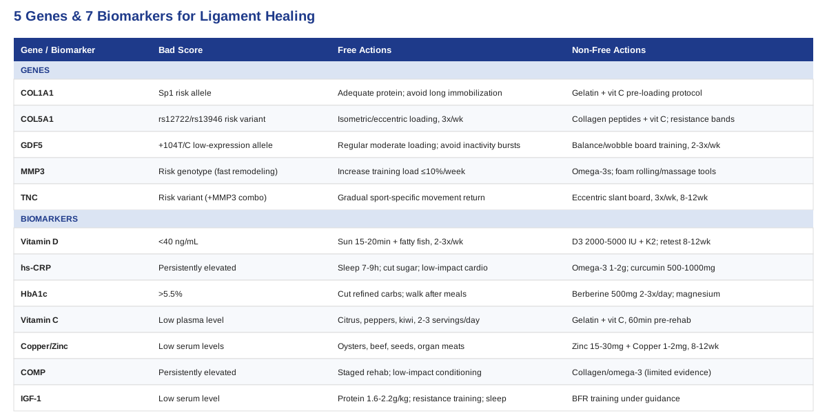
The Seven Biomarkers Worth Tracking After a Posteromedial Corner Injury
The posteromedial corner is built almost entirely from collagen-based tissue — ligament, tendon sheath, and fibrocartilage. That means its ability to heal after a sprain, partial tear, or post-surgical repair depends heavily on your body's collagen-building machinery: raw materials (protein, vitamin C, copper, zinc), signaling molecules (IGF-1), the inflammatory environment (CRP), and the metabolic conditions that either support or degrade collagen quality (blood sugar, vitamin D). The seven markers below aren't a substitute for an orthopedic evaluation, but together they give a realistic picture of whether your internal environment is set up to help or hinder the repair process.
Vitamin D (25-hydroxyvitamin D)
Vitamin D does more than manage calcium. It also modulates the inflammatory response involved in tendon and ligament healing, and low levels have been associated with poorer outcomes after connective tissue injury and surgery, including slower recovery and weaker post-surgical strength gains vitamin D and the immunomodulation of tendon injury. If you've had a PMC injury with surgical repair on the table, this is one of the cheapest and most actionable numbers to know.
How to measure it
A standard 25-hydroxyvitamin D blood test, available through most primary care providers or direct-to-consumer lab services. Cost typically runs from $15 to $50 out of pocket if not covered by insurance. Optimal range for tissue healing purposes is generally considered 40 to 60 ng/mL, not just "in range."If the score is low, the plan without supplements
Aim for 15 to 20 minutes of midday sun exposure on bare skin several times a week, weather permitting, and increase intake of fatty fish (salmon, sardines, mackerel) two to three times weekly. This is a slow lever — expect gradual improvement over two to three months, not weeks.If the score is low, the plan with supplements
Vitamin D3, typically 2,000 to 5,000 IU daily, paired with vitamin K2 (around 100 mcg) to support proper calcium utilization. Retest at 8 to 12 weeks rather than adjusting blindly. Side effects are rare at these doses but can include elevated blood calcium with very high, prolonged intake — a reason to retest rather than guess indefinitely.High-Sensitivity C-Reactive Protein (hs-CRP)
CRP rises sharply in the first days after any soft tissue injury as part of the normal healing cascade, but a persistently elevated CRP weeks or months out can indicate that inflammation isn't resolving the way it should, which can slow tissue remodeling. In shoulder injury research, circulating CRP alongside IL-6 and IL-10 has been linked to how much pain improves in the days following soft tissue injury, making it a useful window into whether your inflammatory response is on a normal trajectory circulating inflammatory biomarkers and pain recovery after soft tissue injury.
How to measure it
A high-sensitivity CRP blood test, distinct from the standard CRP used to detect acute infection. Cost is usually $10 to $30. Best interpreted a few weeks after the acute injury phase has passed, not immediately post-injury when it's expected to be elevated.If the score is high, the plan without supplements
Prioritize sleep consistency (7 to 9 hours), reduce ultra-processed food and added sugar intake, and keep low-grade aerobic activity (walking, cycling within pain limits) as a regular habit — deconditioning itself is pro-inflammatory. Reassess after 6 weeks of consistent changes.If the score is high, the plan with supplements
Omega-3 fatty acids (1 to 2 g combined EPA/DHA daily) have reasonable evidence for modestly lowering CRP over 8 to 12 weeks. Curcumin (500 to 1,000 mg daily with piperine or a phospholipid formulation for absorption) is a second option some people cycle for 8 weeks on, 2 to 4 weeks off. Both can mildly increase bleeding risk, which matters if surgery is being considered — flag supplement use to your surgeon beforehand.HbA1c and Fasting Glucose
Chronically elevated blood sugar accelerates the formation of advanced glycation end-products (AGEs) — irreversible cross-links that stiffen collagen fibers abnormally and impair the normal sliding and remodeling behavior of ligament and tendon tissue. This effect has been shown even in people without a diabetes diagnosis, with elevated HbA1c linked to altered tendon properties and reduced tissue quality effect of glucose dysregulation on tendon structure and function.
How to measure it
HbA1c blood test, roughly $15 to $40, or included in most annual metabolic panels. A result under 5.5% is generally favorable for tissue quality purposes, even though the clinical "normal" cutoff is higher.If the score is high, the plan without supplements
Reduce refined carbohydrate and added sugar intake, walk for 10 minutes after larger meals to blunt glucose spikes, and prioritize protein and fiber at the start of meals. These changes tend to move HbA1c meaningfully within 3 months.If the score is high, the plan with supplements
Berberine (500 mg, two to three times daily with meals) has clinical evidence for improving glucose control comparable to some first-line medications, typically cycled for 8 to 12 weeks with a break, and can cause GI upset initially. Magnesium glycinate (200 to 400 mg nightly) supports insulin sensitivity. Anyone on glucose-lowering medication should coordinate with their physician before adding berberine, since the combined effect can push blood sugar too low.Vitamin C (Ascorbic Acid)
Vitamin C is a required cofactor for the enzymes that hydroxylate proline and lysine during collagen synthesis — without adequate vitamin C, the body simply cannot assemble stable collagen fibers, regardless of how much protein is available. A randomized crossover trial found that consuming vitamin C-enriched gelatin an hour before exercise roughly doubled a marker of collagen synthesis compared to placebo, directly supporting a practical timing strategy for connective tissue repair vitamin C-enriched gelatin supplementation augments collagen synthesis.
How to measure it
Plasma vitamin C level, a less common but available test, roughly $30 to $60 through specialty or functional medicine labs. Not part of routine panels, so it needs to be specifically requested.If the score is low, the plan without supplements
Citrus fruit, bell peppers, kiwi, and strawberries are all vitamin C-dense; two to three servings daily is usually enough to correct mild insufficiency within a few weeks.If the score is low, the plan with supplements
15 to 20 g of hydrolyzed collagen or gelatin combined with 50 to 100 mg of vitamin C, taken about 60 minutes before rehab exercises or physical therapy sessions, on training days only. This is a targeted, activity-linked protocol rather than a daily supplement — no meaningful side effects at these doses beyond mild GI upset in some people if taken on an empty stomach.Copper and Zinc
Both minerals are essential cofactors for lysyl oxidase, the enzyme responsible for cross-linking collagen fibers into strong, load-bearing structures. Without adequate lysyl oxidase activity, newly synthesized collagen stays weak and disorganized, since cross-linking is what gives ligament tissue its tensile strength lysyl oxidase activity is required for ordered collagen fibrillogenesis by tendon cells.
How to measure it
Serum copper and plasma zinc, often $20 to $40 each. Ceruloplasmin can be added as a secondary marker of copper status. These aren't routine panel items, so they typically require a specific request.If the score is low, the plan without supplements
Oysters, beef, pumpkin seeds, and legumes for zinc; organ meats, shellfish, nuts, and seeds for copper. Because zinc and copper compete for absorption, a diet naturally balanced across food sources is usually safer than fixing one in isolation.If the score is low, the plan with supplements
Zinc, 15 to 30 mg daily, paired with 1 to 2 mg of copper to maintain the ratio (high-dose zinc alone can induce a copper deficiency over months). Take with food to avoid nausea. This is typically a 8 to 12 week correction protocol followed by retesting, not an indefinite regimen, since excess copper and zinc both carry their own toxicity risks.Cartilage Oligomeric Matrix Protein (COMP)
COMP is a structural protein released into joint fluid and blood when cartilage and ligament tissue is mechanically stressed or damaged. After a knee injury involving the cruciate or collateral ligaments, COMP levels rise and can stay elevated, and persistently high levels have been associated with ongoing joint tissue turnover and elevated risk of early cartilage degeneration — relevant for PMC injuries given the meniscal and capsular tissue involved release of COMP into joint fluid after knee injury.
How to measure it
Serum COMP is not a routine test; it's typically ordered through sports medicine research labs or specialty orthopedic clinics, and cost varies widely ($50 to $150) since it isn't standardized in most commercial labs. This is the most advanced marker on this list and worth discussing directly with an orthopedic specialist rather than ordering independently.If the score is high, the plan without supplements
Graduated, criteria-based rehabilitation rather than early aggressive loading — following a physical therapist's staged return-to-load protocol reduces repeated mechanical stress on already-turnover-heavy tissue. Low-impact conditioning (swimming, cycling) while formal rehab progresses.If the score is high, the plan with supplements
Evidence here is thinner than for the other markers. Some clinicians use collagen peptides and omega-3s as general joint-support adjuncts, but there's no strong trial data showing supplementation directly lowers COMP. This is a marker to track trends on over months rather than something to try to "fix" acutely with a supplement stack.Insulin-like Growth Factor 1 (IGF-1)
IGF-1 is produced locally within tendon and ligament tissue and is one of the more potent stimulators of fibroblast proliferation and collagen type I synthesis during repair. Both dietary and mechanical factors (loading) influence local IGF-1 activity, and delivery of IGF-1 has been shown across studies to increase collagen content, tissue stiffness, and healing quality in injured connective tissue effects of IGF-1 delivery strategies on tendon healing.
How to measure it
Serum IGF-1, roughly $40 to $90, sometimes bundled with growth hormone axis panels. Levels naturally vary with age, sex, and sleep, so a single low reading is less informative than a trend.If the score is low, the plan without supplements
Adequate total protein intake (around 1.6 to 2.2 g per kg bodyweight daily during rehab), consistent resistance training within pain-free range once cleared, and protecting sleep — IGF-1 production is closely tied to deep sleep and overall caloric sufficiency. Chronic under-eating during rehab is a common, overlooked cause of low IGF-1.If the score is low, the plan with supplements/equipment
No supplement reliably and safely raises IGF-1 in healthy people, and hormonal interventions in this space carry real risk and require medical supervision — this isn't a self-directed supplement category. Where equipment helps: blood flow restriction (BFR) training, used under professional guidance during early-stage rehab, allows low-load exercise to produce a disproportionately large local growth factor and metabolic stimulus, which is one reason it's increasingly used in post-ligament-injury rehab protocols.Taken together, these seven markers describe a fairly complete picture: raw materials (vitamin C, copper, zinc), regulatory environment (vitamin D, HbA1c, CRP), and direct tissue signals (COMP, IGF-1). None of them diagnose a PMC injury, but retested every 8 to 12 weeks during rehab, they can reveal whether your biology is quietly helping or working against the healing timeline your surgeon or physical therapist has laid out.
What Your Genes May Reveal About Ligament Resilience
Biomarkers describe your current internal state; genetics describes your baseline tendencies. Genomics researchers such as Ali Torkamani have spent years cataloguing how common gene variants shift disease and injury risk at a population level, while health educators like Gary Brecka have popularized the idea that a genetic predisposition isn't a sentence — it's a heads-up that usually points toward a specific, actionable nutrient or training adjustment. That framing applies well to connective tissue injury genetics, where most of the research is on collagen and matrix-remodeling genes rather than exotic markers.
It's worth being direct about the evidence level here: gene-injury association studies in sports medicine are mostly candidate-gene studies and meta-analyses with moderate sample sizes, and results sometimes conflict between populations (Caucasian, Asian, and African cohorts don't always show the same direction of effect). This is real, published human research, but it's nowhere near as mature as, say, cardiovascular genetics. Treat the findings below as directional risk information, not deterministic fate.
COL5A1: Ligament Elasticity and Stiffness
COL5A1 encodes a component of type V collagen, a minor but structurally important collagen that regulates the diameter and organization of the much more abundant type I collagen fibers in ligaments and tendons. Certain COL5A1 variants (notably around the rs12722 and rs13946 sites) have been repeatedly linked to altered ligament and tendon injury risk, generally by influencing tissue stiffness — some variants appear protective against ACL injury, others increase risk, and the effect seems population-dependent COL5A1 rs13946 polymorphism and anterior cruciate ligament injury: systematic review and meta-analysis.
If the variant is unfavorable, the plan without supplements
Since the risk variants are linked to tissue that's mechanically more compliant (less stiff) under load, the practical countermeasure is progressive tendon and ligament loading — isometric holds and slow eccentric work performed consistently, 3 times weekly for 8 to 12 weeks, which has been shown to increase tissue stiffness independent of genotype.If the variant is unfavorable, the plan with supplements or equipment
Collagen peptides plus vitamin C timed before loading sessions (see the vitamin C section above) support the raw-material side of adaptation. Resistance bands or a cable system for controlled eccentric loading make consistent, gradual overload easier to standardize than bodyweight alone.COL1A1: Base Collagen Structure
COL1A1 encodes the primary chain of type I collagen, the dominant structural protein in ligaments, tendons, and bone. A regulatory (Sp1 binding site) polymorphism in COL1A1 has been associated with tendon injury risk, thought to work by altering how much type I collagen is transcribed relative to more elastic collagen types, shifting the stiffness-to-elasticity balance of the tissue Sp1-binding site polymorphism within the COL1A1 gene and Achilles tendon injuries.
If the variant is unfavorable, the plan without supplements
Adequate total daily protein (prioritizing complete protein sources across meals) supports baseline type I collagen availability. Avoiding prolonged immobilization beyond what's medically necessary matters too, since collagen synthesis drops quickly with disuse.If the variant is unfavorable, the plan with supplements or equipment
The same gelatin-plus-vitamin-C, pre-exercise timing protocol described earlier applies directly here, since it's specifically targeting type I collagen synthesis. No specialized equipment is required beyond consistent, progressive rehab loading.GDF5: Growth Signaling for Ligament Homeostasis
GDF5 (growth differentiation factor 5) is part of the TGF-beta signaling family and plays a role in joint and ligament development and ongoing tissue maintenance. A specific promoter variant (+104T/C) has been associated with reduced GDF5 expression in ligament tissue and a higher rate of ACL rupture in some populations, theorized to work through weaker baseline ligament homeostasis the GDF5 gene and anterior cruciate ligament rupture.
If the variant is unfavorable, the plan without supplements
Since GDF5 supports baseline tissue turnover, regular, moderate mechanical loading (rather than long stretches of inactivity followed by high-intensity bursts) helps keep ligament tissue in an active remodeling state. This is more about training consistency than any specific exercise.If the variant is unfavorable, the plan with supplements or equipment
There's no supplement that directly restores GDF5 signaling. The more relevant equipment-based approach is neuromuscular training — proprioceptive and balance work (wobble boards, single-leg stability drills) 2 to 3 times weekly, which has separately been shown to reduce ligament injury rates by improving how joints absorb load, compensating behaviorally for weaker underlying tissue signaling.MMP3: Tissue Remodeling Speed
MMP3 encodes an enzyme that breaks down and remodels extracellular matrix components, including collagen, as part of normal tissue turnover and repair. Certain MMP3 variants are associated with altered remodeling rates, and specific genotype combinations with COL5A1 have shown the strongest and lowest risk profiles for tendon-ligament injury, suggesting these genes interact rather than act independently association between MMP3 gene polymorphisms and tendon-ligament injuries: a meta-analysis.
If the variant is unfavorable, the plan without supplements
Avoid rapid spikes in training load (a common trigger for remodeling to fall behind breakdown) by following the standard sports medicine guidance of increasing weekly training load by no more than about 10% at a time.If the variant is unfavorable, the plan with supplements or equipment
Omega-3 fatty acids have some evidence for moderating excessive matrix metalloproteinase activity in inflamed tissue; 1 to 2 g daily is a reasonable, low-risk addition. Foam rolling or massage tools to manage tissue quality between sessions may help support consistent, moderate mechanical stimulus rather than replacing it.TNC: Matrix Organization Under Mechanical Stress
Tenascin-C is a matrix glycoprotein that gets strongly upregulated in tendon and ligament tissue in response to mechanical loading and injury, helping organize the extracellular matrix during repair. Variants in the TNC gene, particularly in combination with MMP3 genotype, have been linked to elevated tendinopathy risk in high-performance athletes tenascin-C–MMP3 phenotype and the risk of tendinopathy in high-performance athletes. A broader review of tendon and ligament genetics covers this and the genes above in more depth tendon and ligament genetics: how do they contribute to disease and injury.
If the variant is unfavorable, the plan without supplements
Gradual exposure to the specific movement patterns relevant to your sport or activity, rather than jumping straight back to full intensity, gives tenascin-C-dependent matrix reorganization time to keep pace with mechanical demand.If the variant is unfavorable, the plan with supplements or equipment
No direct supplement targets tenascin-C expression. Eccentric-loading equipment (a slant board or decline setup for lower-body tendon work) is the most evidence-supported non-drug tool for improving matrix organization under controlled stress, typically used 3 times weekly for a minimum of 8 to 12 weeks before reassessing.Genetics and biomarkers answer different questions, but they connect directly: a COL5A1 or MMP3 variant might explain why your baseline risk is elevated, while your current vitamin D, CRP, and vitamin C status determine how well you're compensating for that risk right now. Reviewing both gives a fuller picture than either alone — and it's exactly the kind of thinking behind the research discussed next.
The Podcast Episode Reshaping How Ligament and Tendon Healing Actually Works
Dr. Keith Baar, a physiologist at UC Davis who has spent his career studying tendon and ligament adaptation, laid out some genuinely practice-changing findings in a lengthy conversation on The Tim Ferriss Show. Several of his points directly contradict standard "rest, ice, and wait" advice for connective tissue injuries, and they're backed by his own published research rather than speculation. Below are the ten most useful takeaways for anyone managing a posteromedial corner or similar ligament injury.
1. Rest slows tendon and ligament healing more than people realize
Complete immobilization reduces collagen synthesis in the injured tissue. Some controlled, pain-appropriate loading — not passive rest — is what actually stimulates the fibroblasts responsible for laying down new collagen.2. Isometric holds may matter more than eccentric exercises for early rehab
While eccentric loading protocols became popular in tendon rehab over the past two decades, Baar's research suggests isometric contractions (holding a position under tension without movement) can be just as effective, and are often more tolerable for painful, recently injured tissue.3. Timing collagen and vitamin C intake around exercise matters
This is the practical output of his own AJCN study: consuming vitamin C-enriched gelatin about an hour before a loading session roughly doubled a blood marker of collagen synthesis compared to placebo, suggesting the body responds to nutrient timing much like it does with protein and muscle protein synthesis.4. Inflammation is a necessary part of healing, not just a problem to suppress
Baar argues that reflexively icing and taking anti-inflammatories immediately after an injury can blunt the signaling that triggers tissue repair. This doesn't mean skipping pain management, but it reframes inflammation as a phase to work with rather than eliminate outright in the first days.5. Tendon and ligament tissue adapts far more slowly than muscle
Muscle can meaningfully strengthen in a matter of weeks; collagen-based tissue often needs many months of consistent loading to remodel. This is one reason rehab timelines that feel "too slow" against muscle recovery standards are actually normal for ligament tissue.6. Growth hormone and IGF-1 both matter, but through different pathways
Baar distinguishes between systemic growth hormone (which has broader, more indirect effects) and locally produced IGF-1 within the tissue itself, which appears to be the more directly relevant driver of tendon and ligament matrix repair — reinforcing why local mechanical loading (which stimulates local IGF-1) tends to outperform systemic interventions.7. Heat and blood flow before loading may prime tissue for adaptation
Warming the tissue and increasing local blood flow before a loading session appears to improve the collagen synthesis response, giving a physiological rationale for a proper warm-up beyond just injury prevention.8. Simple, boring exercises often beat complex rehab programs
According to Baar, the biggest predictor of tendon and ligament recovery isn't the sophistication of the exercise selection, it's consistency of appropriate mechanical loading over months — a message that runs counter to the appeal of complex rehab protocols.9. JAK inhibitors and certain anti-inflammatory drugs may impair tendon healing
Baar raises concerns that some anti-inflammatory medications, including certain classes used for other conditions, may interfere with the tendon and ligament healing process, an important conversation to have with a prescribing physician if you're on long-term anti-inflammatory therapy during rehab.10. "Listen to load, not just pain" as a rehab principle
Rather than using pain alone as the signal to stop or continue an exercise, Baar suggests monitoring how tissue responds to load over the following 24 hours — increased stiffness or swelling the next day is a more reliable overload signal than momentary discomfort during the exercise itself.Whether or not every claim here becomes standard clinical guidance, the core theme is worth sitting with: connective tissue healing responds to smart mechanical and nutritional inputs, not just time and avoidance. That segues naturally into a few complementary, lower-risk approaches with genuine supporting evidence for knee ligament rehab.
Complementary Approaches Worth Considering
None of the following should replace orthopedic care, physical therapy, or surgical management when indicated for a posteromedial corner injury. They're additions with real, if sometimes preliminary, human evidence — worth discussing with your treatment team rather than adding on your own without context.
Biofeedback
Electromyographic (EMG) biofeedback gives real-time visual or acoustic feedback on muscle activation, which is particularly useful for retraining muscles like the vastus medialis that often shut down (arthrogenic muscle inhibition) after knee ligament injury or surgery. For a PMC injury, where the semimembranosus and surrounding musculature play a stabilizing role, restoring normal activation patterns matters as much as ligament healing itself.
A randomized controlled trial in patients recovering from ACL reconstruction found that adding EMG biofeedback for the vastus medialis to standard rehab improved knee extension and quadriceps strength recovery compared to standard rehab alone influence of electromyographic biofeedback therapy on knee extension following ACL reconstruction. The protocol used acoustic and visual feedback sessions during the first week or two of rehab, layered onto normal physical therapy rather than replacing it.
Practically, this usually requires a physical therapy clinic with EMG biofeedback equipment, typically used for short, focused sessions two to three times weekly during the early rehab window (weeks 1 to 6). It's a reasonable add-on to ask your physical therapist about directly, especially if you notice your quadriceps or hamstring activation feels noticeably weaker or delayed on the injured side.
Low-Level Laser Therapy / Photobiomodulation
Photobiomodulation uses low-level laser or LED light believed to stimulate mitochondrial ATP production in cells, potentially supporting fibroblast activity, collagen synthesis, and reduced inflammation in healing tissue. The theoretical mechanism is well described, but clinical results in tendon and ligament tissue specifically have been mixed.
A review on photobiomodulation for acute tissue injury and sport recovery outlines the mechanism and summarizes generally favorable but inconsistent findings across tissue types photobiomodulation as medicine: low-level laser therapy for acute tissue injury or sport performance recovery. It's worth noting that at least one double-blind, placebo-controlled trial in tendon injuries of the hand found no significant benefit over placebo, so the evidence genuinely varies by tissue and protocol.
Given the mixed evidence, this is best treated as a low-risk adjunct rather than a core treatment — something to try through a physical therapy or sports medicine clinic that already has the equipment, generally in short sessions two to three times weekly for several weeks, rather than a reason to purchase home devices or delay other evidence-based rehab.
Tai Chi
Tai chi combines slow, controlled movement with weight shifting and balance challenges, and has accumulated the strongest evidence of any mind-body practice for knee joint conditions, primarily in osteoarthritis but with clear relevance to post-ligament-injury proprioception, which is often impaired after PMC and other knee ligament injuries.
A pilot randomized controlled trial specifically examined tai chi's effect on knee proprioception in patients with knee osteoarthritis and found measurable improvements in joint position sense after a structured program the influence of tai chi exercise on proprioception in patients with knee osteoarthritis. Since impaired proprioception is a known consequence of ligament injury (not just osteoarthritis), the mechanism is plausible even though this particular trial wasn't conducted in an acute ligament injury population specifically.
In practice, this means a beginner tai chi class or instructor-led program, two to three sessions weekly for at least 8 to 12 weeks, once cleared for weight-bearing balance work by a physical therapist. It pairs naturally with formal proprioceptive rehab (single-leg balance, wobble board work) rather than replacing it.
Massage Therapy
Manual therapy techniques, including cross-fiber and instrument-assisted massage, are commonly used in post-surgical knee rehab to manage scar tissue mobility, swelling, and secondary muscle tightness that develops around an injured or repaired ligament.
Human evidence specific to ligament injury is still limited and mostly case-based rather than large randomized trials; one published case report describes a massage therapy protocol used for patellofemoral pain following ACL reconstruction, showing symptomatic improvement in that individual case massage therapy protocol for post-ACL reconstruction patellofemoral pain syndrome. Earlier animal research has shown promising effects of instrument-assisted cross-fiber massage on perfusion and healing ligament tissue, but that evidence hasn't yet been confirmed in large human trials.
Given the limited but plausible evidence, massage is reasonable as a comfort- and mobility-focused adjunct — typically once or twice weekly during active rehab phases — rather than something expected to accelerate ligament tissue healing itself. It's worth coordinating timing with your physical therapist so it complements rather than interferes with progressive loading work.
Conclusion
A posteromedial corner injury responds to the same rehab fundamentals as any ligament injury — appropriate loading, patience, and good clinical care — but how well and how fast that happens depends on biology that's rarely discussed in generic recovery advice. The seven biomarkers covered here, particularly vitamin D, HbA1c, vitamin C status, and the copper-zinc pair behind collagen cross-linking, give you a concrete, retestable way to check whether your body's raw materials and inflammatory environment are actually supporting repair. The five gene variants add context on your baseline tendencies, and in most cases point toward the same practical answer: consistent, progressive mechanical loading paired with adequate collagen-building nutrients, regardless of which specific variant you carry.
None of this replaces an orthopedic evaluation, a structured physical therapy program, or surgical consultation when instability persists — it supplements that care with information most people never think to ask for. The calm, useful next step isn't to overhaul everything at once. It's to pick one or two markers from this list that you haven't checked before (vitamin D and HbA1c are the easiest starting points), get them tested, and bring the results to your physical therapist or physician as part of the conversation about your rehab plan.