This article contains AI generated content.
· UpdatedPrimary Hyperoxaluria Genes And Biomarkers: 3 Genes And 6 Biomarkers To Track
Introduction
If you or someone you care for has primary hyperoxaluria, you probably already know how isolating the diagnostic path can be. Recurrent kidney stones that start in childhood, imaging that shows dense calcium deposits where they should not exist, and a progression of kidney decline that standard kidney-stone advice cannot slow — and yet, for years, specialists may only suggest drinking more water and cutting back on spinach. That advice is not wrong, but it is nowhere near sufficient for what is actually happening metabolically.
Primary hyperoxaluria is a rare inherited disorder in which the liver produces far too much oxalate — a compound the kidneys must excrete but that builds up in tissues when production exceeds capacity. The root cause is genetic, and which gene is affected matters enormously. The disease behaves differently depending on the specific mutation, the residual enzyme activity, and the downstream metabolites that accumulate. Generic kidney-stone guidance was never designed with these distinctions in mind.
What actually moves the needle for most people with PH is understanding the genetic driver precisely and tracking the right biomarkers systematically. A confirmed genetic variant tells you whether pyridoxine will be useful, whether a newer RNA-silencing drug is appropriate, and what metabolite to watch in urine. A panel of six carefully chosen biomarkers gives a running picture of how well the liver, kidneys, and urine chemistry are holding up — and how well any intervention is working.
This article covers both layers: first, the three main genes responsible for primary hyperoxaluria and practical plans for when each is abnormal; second, the six biomarkers that give the clearest signal of disease activity and response to treatment. Beyond those two pillars, there is a look at recent science that is reshaping how clinicians and patients think about PH, and a brief review of evidence-backed complementary strategies. Better information does not cure the disease, but it does make every clinical conversation sharper and every decision better grounded.
Summary
This article examines primary hyperoxaluria from two complementary angles. On the genetics side: three genes — AGXT, GRHPR, and HOGA1 — account for essentially all known cases, and each carries a distinct prognosis, distinct metabolite signature, and distinct response to treatment. For AGXT mutations (the most severe and most common form), pyridoxine responsiveness can be tested, RNA-silencing drugs now reduce oxalate production dramatically, and a concrete plan exists whether or not supplements are an option. GRHPR and HOGA1 mutations follow their own logic, and knowing which one is in play changes what to monitor and what to expect.
On the biomarkers side: six measurements — 24-hour urinary oxalate, plasma oxalate, urinary glycolate, eGFR plus creatinine, urinary citrate, and urinary calcium — together provide an actionable picture of disease load, kidney function trajectory, and response to both dietary changes and medical treatment. For each one, this article explains what a bad score means, how to move it without supplements, and what targeted supplements or interventions have genuine evidence behind them.
Beyond genes and biomarkers, the article covers 10 key research insights from the RNA-interference science that is transforming PH treatment, plus evidence on gut-microbiome-directed therapy and mindfulness-based approaches that have specific relevance for people managing this condition long-term.
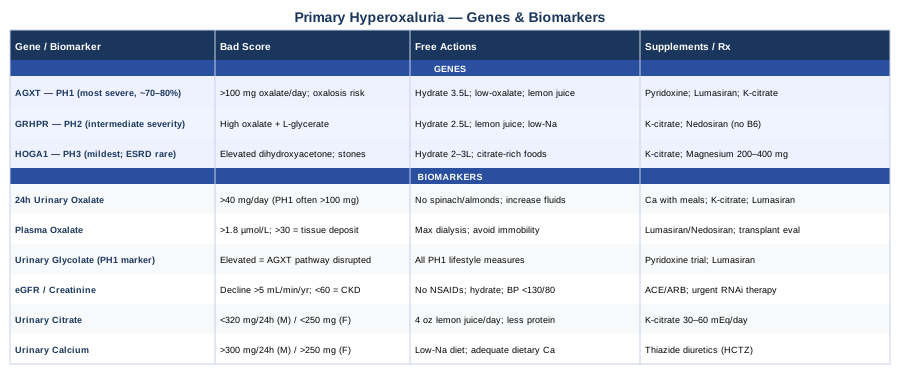
The Three Genes Behind Primary Hyperoxaluria — And What To Do About Each
Every case of primary hyperoxaluria traces back to one of three genetic defects in the liver's glyoxylate metabolism pathway. Glyoxylate is a normal metabolic intermediate, but in PH it is not converted correctly into glycine or glycolate — instead it is oxidized into oxalate, a compound the body cannot break down and that crystalizes in the kidneys and elsewhere. Which enzyme is defective determines the type of PH, the severity of oxalate overproduction, and which therapeutic options apply.
Understanding your specific genetic variant is not just academic. It determines whether pyridoxine will help, whether lumasiran or nedosiran is indicated, and which urine metabolites are most informative. This section goes through each gene in practical depth.
Gene 1: AGXT — The Most Common and Most Severe Driver
The AGXT gene encodes alanine-glyoxylate aminotransferase (AGT), an enzyme that lives inside liver peroxisomes and converts glyoxylate into the harmless amino acid glycine. When AGXT is mutated, AGT activity drops — sometimes to zero — and glyoxylate floods the oxalate production pathway. This is primary hyperoxaluria type 1 (PH1), which accounts for roughly 70–80% of all PH cases and is the most severe form.
More than 200 pathogenic AGXT mutations have been catalogued. The most common, p.Gly170Arg (also written G170R), accounts for approximately 30% of disease alleles and has an unusual feature: the mutant AGT protein is not only less active but is also mistargeted — it ends up in mitochondria instead of peroxisomes, where it cannot function properly even when its intrinsic activity is partially preserved. A second common mutation, c.33dupC, creates a truncated, completely nonfunctional protein. Knowing which specific mutation is present matters because the G170R variant, especially when combined with a co-segregating p.Pro11Leu polymorphism, shows partial responsiveness to pyridoxine (vitamin B6), while c.33dupC does not.
The clinical consequences of untreated PH1 are serious. Urinary oxalate can reach three to five times normal levels, and calcium oxalate crystals deposit not only in kidneys but also in bones, blood vessels, the heart, and the eyes — a systemic condition called oxalosis. Without treatment, many PH1 patients reach end-stage renal disease in their twenties or thirties, and sometimes in childhood.
If the AGXT gene is mutated: the plan without supplements
Even without pharmaceutical intervention, there are meaningful non-supplement strategies that reduce stone formation and slow kidney damage in PH1. The foundation is aggressive, sustained hydration. Adults with PH1 are typically advised to maintain urine output above 3 liters per day — meaning fluid intake of 3.5 to 4 liters daily or more, spread evenly throughout waking hours, including a glass before bed to reduce overnight concentration. Children are dosed by weight (at least 50 mL per kilogram per day of urine output).
A low-oxalate diet helps reduce the exogenous load. While endogenous hepatic overproduction is the primary driver in PH, dietary oxalate still adds to the burden. Keeping dietary oxalate below 50 mg per day — avoiding high-load foods like spinach, rhubarb, almonds, and wheat bran — is a reasonable starting point. Limiting added ascorbic acid (vitamin C supplements above 250 mg/day) matters because ascorbate is converted to oxalate in the body, which is particularly problematic in PH.
Reducing dietary sodium to below 2,300 mg per day lowers urinary calcium, which reduces the supersaturation driving calcium oxalate crystallization. Adequate dietary calcium intake (from food, not supplements between meals) actually helps by binding dietary oxalate in the gut before it is absorbed. Moderate animal protein restriction (below 0.8 g/kg/day) reduces the acid load that lowers urinary pH and urinary citrate.
Citrate is a natural inhibitor of calcium oxalate crystallization. Drinking 4 to 8 ounces of fresh lemon juice daily provides substantial dietary citrate and helps raise urinary pH without medication. This is not curative, but it reduces the risk of stone events and buys time between stone-forming episodes.
If the AGXT gene is mutated: the plan with supplements or equipment
Pyridoxine (vitamin B6): For patients with pyridoxine-responsive mutations (primarily G170R combined with P11L, and a few others), a supervised trial of high-dose pyridoxine is a first-line intervention before considering newer drugs. The standard starting dose is 5 mg/kg per day, rising to 10–20 mg/kg per day (capped at 500–1000 mg/day in adults) over a 3-month trial period. Response is defined as a ≥30% reduction in 24-hour urinary oxalate. Frequency: daily, continuous — this is not cycled. Critical side effect warning: peripheral sensory neuropathy occurs with sustained doses above 200 mg/day and is dose-dependent. Neurological monitoring is essential. Some specialists prefer maintaining doses under 100 mg/day unless clearly responsive, minimizing neuropathy risk. Patients who do not show measurable response at 3 months should stop — non-responders gain nothing and accrue the neuropathy risk.
Potassium citrate: This is indicated for essentially all PH1 patients regardless of pyridoxine responsiveness. The standard dose is 1 to 4 mEq/kg per day divided into three doses with meals. It raises urinary citrate (which inhibits crystallization), alkalinizes urine toward the target pH of 6.5 to 7.0, and reduces the ionic calcium available to form crystals. Side effects: gastrointestinal discomfort (take with food to minimize), bloating, and — at high doses — hyperkalemia, so potassium levels should be monitored. Frequency: three times daily, continuous.
Lumasiran (Oxlumo): FDA-approved in November 2020, lumasiran is an RNA interference (RNAi) drug delivered by subcutaneous injection that silences the LDHA gene (lactate dehydrogenase A) in hepatocytes, reducing the conversion of glyoxylate to oxalate. The ILLUMINATE-A and ILLUMINATE-B trials showed reductions in urinary oxalate of approximately 65% from baseline. The standard adult dosing is 3.5 mg/kg subcutaneously monthly for three loading doses, then 3.5 mg/kg every three months. Pediatric dosing is weight-adjusted. Side effects: injection-site reactions (redness, swelling, pain), flu-like symptoms shortly after injection. Lumasiran is indicated for PH1 specifically. It does not cure the genetic defect but dramatically reduces oxalate output in most patients.
Nedosiran (Rivfloza): FDA-approved in September 2023, this is a second RNAi drug also targeting LDHA, with monthly subcutaneous dosing. It is being studied across PH1, PH2, and PH3, making it potentially relevant for patients with GRHPR or HOGA1 mutations as well.
Liver transplantation: Because the defect is hepatic, a liver transplant corrects the enzyme deficiency and stops oxalate overproduction. Combined liver-kidney transplantation is considered when kidneys have already sustained significant damage. This is a major surgical intervention reserved for patients not responding adequately to medical therapy or with rapidly progressing renal failure.
Gene 2: GRHPR — The Underdiagnosed Middle Path
Primary hyperoxaluria type 2 (PH2) results from mutations in the GRHPR gene, which encodes glyoxylate reductase/hydroxypyruvate reductase. This enzyme has a dual role: it reduces glyoxylate to glycolate and reduces hydroxypyruvate to L-glycerate. When GRHPR is absent or dysfunctional, glyoxylate is shunted to oxalate, and hydroxypyruvate accumulates and is converted to L-glycerate — producing the disease's biochemical signature: elevated urinary oxalate combined with elevated urinary L-glycerate. This dual elevation, when present, is highly diagnostic.
GRHPR is located on chromosome 9q11 and around 90 pathogenic variants have been described. PH2 is generally considered less severe than PH1 — fewer patients progress to end-stage renal disease, and the age of onset tends to be later. However, it is not benign. Significant stone burden, recurrent stone events, and some degree of kidney function decline do occur, especially in patients who go undiagnosed for years. It is also underdiagnosed: because PH2 does not produce elevated urinary glycolate (unlike PH1), and because genetic testing is not universal, some PH2 patients are missed or labeled as idiopathic hyperoxaluria for years. Urinary L-glycerate testing, when it is included in the metabolite panel, makes diagnosis straightforward.
If the GRHPR gene is mutated: the plan without supplements
The non-supplement approach for PH2 closely parallels PH1 management. Hydration remains the cornerstone — urine output above 2.5–3 liters per day in adults, titrated to maintain urine oxalate concentration below 1.5 to 2 mmol per liter. Low-oxalate diet (below 50 mg/day), low-sodium diet, and avoiding high-dose ascorbic acid supplements all apply. Unlike PH1, there is no proven benefit from avoiding specific dietary glycerate precursors, but limiting fructose intake is sometimes suggested given fructose's role in glyoxylate generation.
Citrate-rich dietary choices — fresh lemon juice, fresh lime juice — should be integrated daily. The goal is urinary citrate above 300 mg/24 hours in men and 250 mg/24 hours in women. Monitoring urine chemistry every 3 to 6 months helps confirm that the dietary plan is actually shifting the key numbers.
If the GRHPR gene is mutated: the plan with supplements or equipment
Pyridoxine: High-dose B6 is not an established intervention for PH2. Unlike PH1 where AGT has B6 as a cofactor, GRHPR does not use pyridoxine, so there is no mechanistic basis and no consistent clinical evidence for supplementation. This is an important distinction — PH2 patients should not be given long-term high-dose B6 based on a PH1 protocol without confirmed mutation typing.
Potassium citrate: This is the most evidence-backed pharmacological supplement for PH2. Same dosing as PH1: 1–4 mEq/kg/day in three divided doses. It reduces stone risk by raising urinary pH and citrate. Side effects and monitoring are identical to PH1.
Nedosiran: This RNAi drug targeting LDHA is being studied for PH2 and PH3 (the PHYOX trials included broader PH populations). As of 2024, it is an evolving option worth discussing with a specialist. The LDHA pathway is active in PH2 oxalate generation, making this a logical target. Lumasiran, designed specifically for PH1, has less established benefit in PH2.
Gene 3: HOGA1 — The Mildest Form With a Mechanistic Twist
Primary hyperoxaluria type 3 (PH3) results from mutations in HOGA1, which encodes 4-hydroxy-2-oxoglutarate aldolase — an enzyme involved in the hydroxyproline degradation pathway inside mitochondria. When HOGA1 is deficient, its substrate accumulates and appears in urine as elevated dihydroxyacetone and L-2-hydroxyglutarate. There is also evidence that the accumulated substrate inhibits GRHPR activity, creating a secondary blockade in glyoxylate metabolism similar to — though less severe than — PH2.
The most commonly identified HOGA1 mutation is c.700+5G>T, an intronic variant that causes exon skipping and is found in a substantial portion of affected European and American patients. PH3 is the second most common form numerically but is the least severe clinically: end-stage renal disease is rare, spontaneous resolution of urinary oxalate elevation has been observed in some adolescents, and many patients have a relatively stable course. That said, stone burden and associated kidney damage are real concerns, and follow-up should not be dismissed based on severity alone.
If the HOGA1 gene is mutated: the plan without supplements
Given the milder course of PH3, lifestyle-first management is often the primary approach, especially in younger patients or those with moderately elevated oxalate. Hydration targets are the same in principle — urine output of 2–3 liters per day — but many PH3 patients do not require the ultra-aggressive volumes needed in PH1. Low-oxalate diet, low-sodium diet, and citrate-rich foods all reduce stone risk meaningfully.
Because dihydroxyacetone accumulates in PH3, some researchers have speculated about dietary influences on the hydroxyproline pathway (reducing gelatin and collagen-heavy foods may limit the precursor load), though clinical evidence for this specific dietary modification remains limited. It is worth discussing with a metabolic specialist familiar with PH3 specifically.
Regular urine monitoring every 6 months — 24-hour urinary oxalate and citrate at minimum — allows early detection of worsening. Some PH3 patients see spontaneous improvement through adolescence; tracking this objectively confirms whether the trajectory is favorable.
If the HOGA1 gene is mutated: the plan with supplements or equipment
Potassium citrate: Also the primary supplement for PH3. Dosing is typically at the lower end of the range — 1 to 2 mEq/kg/day — since the disease burden is usually less severe. Still given in divided doses with meals. Monitor potassium and urine citrate levels every 3 to 6 months.
Nedosiran: Given that PH3's elevated oxalate depends in part on GRHPR inhibition and LDHA activity, LDHA-targeting RNAi has theoretical relevance. The PHYOX trial data includes some PH3 patients. This remains a conversation-with-specialist territory rather than an established standard for PH3.
Magnesium supplementation: Magnesium binds oxalate in urine and reduces calcium oxalate supersaturation. Evidence for PH specifically is limited, but several randomized trials in recurrent calcium oxalate stone formers support its use. Dose: 200–400 mg/day of elemental magnesium (as glycinate or citrate to minimize GI side effects). Frequency: daily with food. Side effects: loose stool at high doses, especially magnesium oxide. Start low and titrate.
Moving from what the genes tell you to how you track the results — biomarkers give the ongoing signal that genetics alone cannot provide.
Six Biomarkers That Tell You How Primary Hyperoxaluria Is Behaving
Knowing your genetic variant is the foundation. But the six biomarkers below are what tell you week-to-week and month-to-month how well management is working, whether kidneys are holding their own, and whether it is time to escalate or adjust treatment. They are not interchangeable — each captures a different piece of the picture, and together they provide far more clarity than any single test.
Biomarker 1: 24-Hour Urinary Oxalate
This is the primary functional biomarker for primary hyperoxaluria. It measures how much oxalate the kidneys are excreting over a full day and reflects the combination of hepatic overproduction and dietary oxalate absorption. In healthy adults, the upper limit is approximately 40 mg (0.45 mmol) per 24 hours. In PH1 patients, values of 100–200 mg (1.1–2.2 mmol) or more per day are common without treatment. A treatment response to lumasiran, for example, is defined by bringing this number down toward the normal range.
How to measure it
A properly collected 24-hour urine sample preserved with hydrochloric acid (or a preservative container provided by the lab) is sent for oxalate analysis. Most hospital or commercial labs offer this. Cost: $30–$80 for the oxalate portion, or $100–$200 as part of a full 24-hour urine stone profile. Frequency for PH patients: every 3 months during active treatment adjustment, every 6 months when stable. Collection errors (incomplete collection, not using preservative, eating unusually high or low oxalate on collection day) are common — two separate collections give a more reliable picture.
If the score is high: the plan without supplements
A 24-hour urinary oxalate persistently above 40 mg/day despite reasonable hydration signals that dietary measures need to be tightened further. Systematically audit dietary oxalate with a food diary for one week. The biggest leverage points are eliminating high-oxalate foods (spinach, almonds, beets, rhubarb, sweet potatoes, peanuts, chocolate) and removing any ascorbic acid supplements over 250 mg/day. Increase daily fluid intake incrementally until urine output reaches 2.5–3 liters per day, confirmed by measuring urine output for one day. Increase dietary lemon juice. Monitor with a repeat collection 8–12 weeks after each change.
If the score is high: the plan with supplements or equipment
Calcium carbonate or calcium citrate taken with meals (not between meals) binds dietary oxalate in the gut, reducing absorption. Dose: 500 mg elemental calcium with each main meal. This approach has evidence in idiopathic hyperoxaluria and is often reasonable in PH as a dietary-oxalate-reducing measure. It is not a substitute for addressing hepatic overproduction. Potassium citrate addresses the crystal-forming environment. For confirmed PH1, discussing lumasiran or nedosiran with a specialist is the highest-yield next step when urinary oxalate remains significantly elevated.
Biomarker 2: Plasma Oxalate
When kidney function begins to decline, 24-hour urine oxalate becomes an unreliable proxy for hepatic oxalate production — a failing kidney excretes less oxalate, so the urine value looks better while plasma levels rise and systemic oxalosis worsens. Plasma oxalate fills this gap. It measures the oxalate concentration in blood and is the relevant biomarker once eGFR falls below 45 mL/min/1.73m² and becomes critical below 30.
Normal plasma oxalate is below 1.8–2 µmol/L. In untreated or poorly controlled PH1, plasma values can reach 50–100 µmol/L or higher. Above 30 µmol/L, deposition in soft tissues accelerates. Even in patients with preserved kidney function, plasma oxalate may be measured as a more sensitive reflection of treatment response on RNAi therapies.
How to measure it
Plasma oxalate measurement requires a specialized laboratory; it is not available in most standard labs. In the US, the Mayo Clinic Metabolic Kidney Disease Lab and a handful of specialized biochemical genetics labs offer this test. Cost: $100–$250 per test. Sample handling is critical — blood must be spun and frozen quickly. Frequency: every 3–6 months in patients with eGFR below 45 or those on RNAi therapy.
If the score is high: the plan without supplements
Plasma oxalate above 10–15 µmol/L with preserved or mildly reduced kidney function signals the need for urgent specialist review and likely indicates inadequate control. Non-supplement optimization: maximize dialysis clearance if already on dialysis (oxalate is dialyzable but requires frequent sessions), and aggressively increase urine output if kidney function allows. Avoid prolonged immobility (bone deposition worsens with immobility). Ophthalmology evaluation for retinal oxalate deposition.
If the score is high: the plan with supplements or equipment
At this level, medical escalation — lumasiran or nedosiran if not already initiated, and transplant evaluation if kidney function is rapidly declining — is the priority intervention. Supplements alone cannot meaningfully reduce plasma oxalate when it is severely elevated and hepatic production is unaddressed. Potassium citrate continues to reduce stone-forming risk in whatever residual kidney function remains.
Biomarker 3: Urinary Glycolate
Urinary glycolate is an upstream metabolite specific to the AGXT pathway. In PH1, when AGT is non-functional, glyoxylate cannot be converted to glycine and backs up into alternative pathways — including conversion to glycolate by lactate dehydrogenase. Glycolate is then excreted in urine at elevated levels. This makes high urinary glycolate a specific biomarker for PH1 that is not typically elevated in PH2 or PH3.
This marker is valuable in two ways: it helps confirm a PH1 diagnosis when genetic testing is inconclusive, and it can reflect the degree of AGXT pathway disruption over time. Some labs include it in organic acid urine panels.
How to measure it
Urinary glycolate is measured as part of a urine organic acids panel or a dedicated hyperoxaluria metabolite panel. Reference labs that specialize in metabolic disease testing (Mayo Clinic Genetics, ARUP Laboratories in the US) include this. Cost: $100–$300 as part of an organic acids panel. Frequency: at diagnosis to confirm type; then annually or whenever PH1 treatment is being evaluated or changed.
If the score is high: the plan without supplements
Elevated glycolate with elevated oxalate, combined with AGXT mutation confirmation, clarifies the diagnosis as PH1 and guides the management decision toward PH1-specific interventions. On the lifestyle side, all the general PH1 measures apply. Glycolate itself does not form dangerous crystals the way oxalate does, but its elevation confirms that the AGT enzyme is severely compromised and that aggressive oxalate-lowering intervention is warranted.
If the score is high: the plan with supplements or equipment
For pyridoxine-responsive mutations, a positive response to high-dose B6 will typically reduce both glycolate and oxalate in parallel. If a B6 trial is undertaken and glycolate does not fall alongside oxalate, that is additional evidence of limited responsiveness. For non-responders, this biomarker reinforces the case for lumasiran — which works specifically by blocking the LDHA-driven conversion of glyoxylate to oxalate (and secondarily, the glyoxylate-to-glycolate conversion).
Biomarker 4: eGFR and Serum Creatinine
The estimated glomerular filtration rate (eGFR) is the most direct measure of how much kidney function remains. It is calculated from serum creatinine, age, and sex. In primary hyperoxaluria, monitoring eGFR trajectory — not just a single value, but the direction and speed of change — is essential for gauging how urgently to escalate treatment and when transplant planning should begin.
PH1 patients who are not treated can lose kidney function at rates of 5–15 mL/min/1.73m² per year during periods of active disease. Any decline faster than 3–5 mL/min/1.73m² per year warrants investigation and, in most cases, treatment intensification.
How to measure it
Serum creatinine is one of the most widely available and lowest-cost laboratory tests — included in a basic metabolic panel (BMP) for $15–$40 or less. eGFR is calculated automatically. For PH patients, eGFR should be checked every 3 months during active disease progression or treatment changes, and every 6 months when stable. Cystatin C-based eGFR formulas are more accurate in patients with unusual muscle mass (very lean patients, children, athletes) and may be worth using as an alternative or complement.
If the score is declining: the plan without supplements
Avoid all nephrotoxic exposures: NSAIDs (ibuprofen, naproxen), contrast dye if imaging can be done without it, excessive protein intake, and dehydration. If an infection requires antibiotics, confirm with the prescriber that renal dosing adjustments are appropriate. Low-sodium diet and blood pressure control (target below 130/80 mmHg) reduce glomerular hyperfiltration stress. Maintain generous hydration to keep urine dilute, reducing crystal deposition in tubules.
If the score is declining: the plan with supplements or equipment
ACE inhibitors or ARBs are typically initiated when proteinuria develops, regardless of blood pressure — they reduce glomerular pressure and slow nephron loss. This is a prescription medication requiring physician management. Any patient with PH1 and declining eGFR who is not yet on lumasiran or nedosiran should be urgently referred for assessment of RNAi therapy eligibility. These drugs are the most powerful available tools to halt the underlying hepatic oxalate production and are specifically indicated when kidney function is at risk.
Biomarker 5: Urinary Citrate
Urinary citrate is often overlooked but mechanistically critical. Citrate is a powerful natural inhibitor of calcium oxalate crystallization — it binds calcium in urine, preventing it from forming complexes with oxalate. Low citrate is an independent risk factor for stone formation, and many PH patients are hypocitraturic (low in citrate) because of the acid load generated by oxalate overproduction and because chronic kidney disease itself reduces citrate excretion.
Normal 24-hour urinary citrate is approximately ≥320 mg/day in men and ≥250 mg/day in women. Values below 150 mg/day represent significant hypocitraturia and meaningfully increase stone-forming risk.
How to measure it
Included in the standard 24-hour urine stone profile alongside oxalate, calcium, uric acid, sodium, and creatinine. Cost: $100–$200 for the full panel. Given that citrate measurement requires no special handling beyond normal 24-hour urine collection, it should be a routine part of every PH patient's monitoring panel. Frequency: every 3–6 months, aligned with urinary oxalate measurements.
If the score is low: the plan without supplements
Fresh lemon juice is the most potent dietary citrate source. Studies in idiopathic hypocitraturia show that 120 mL (4 oz) of fresh-squeezed lemon juice daily can raise urinary citrate by 100–200 mg/24h. This is not trivial. Alternatively, 240 mL of low-sugar lemonade made from fresh lemons achieves a similar effect. Lime and grapefruit juice contain citrate as well, though grapefruit interacts with many medications and should be used cautiously. Reducing animal protein intake also raises urine citrate (animal protein generates acid load that promotes citrate reabsorption in the kidneys).
If the score is low: the plan with supplements or equipment
Potassium citrate is the direct pharmacological fix for hypocitraturia in PH. Starting at 10–20 mEq per dose three times daily (30–60 mEq/day total), adjusted by monitoring. It simultaneously raises citrate, alkalinizes urine to pH 6.5–7.0, and reduces ionic calcium. Side effects: GI discomfort (minimize by taking with food), and hyperkalemia risk at high doses — check potassium every 3 months. Potassium magnesium citrate is an alternative that adds the additional benefit of magnesium. Sodium citrate or sodium bicarbonate can be used when potassium-sparing is needed, but these add sodium load, which counterproductively raises urinary calcium.
Biomarker 6: Urinary Calcium
Calcium oxalate crystals require both calcium and oxalate. Addressing only oxalate without considering the calcium side of the equation misses an important lever. 24-hour urinary calcium above 250 mg/day in women or 300 mg/day in men (hypercalciuria) substantially multiplies the stone-formation risk from any given oxalate level. In PH patients, hypercalciuria and hyperoxaluria together create a very high supersaturation state.
Importantly, the relationship between dietary calcium and stone risk is counterintuitive: adequate calcium with meals reduces stone risk by binding dietary oxalate in the gut. Calcium restriction — a common but misguided advice — actually increases oxalate absorption and urinary oxalate.
How to measure it
Part of the 24-hour urine stone panel. No special collection requirements. Reviewed every 3–6 months alongside oxalate and citrate, or after any significant dietary or supplement change. Spot urine calcium-to-creatinine ratio can be used for rapid screening between 24-hour collections, especially in children.
If the score is high: the plan without supplements
Reduce dietary sodium to below 1500–2000 mg/day — sodium drives urinary calcium excretion, and low-sodium diet can reduce urinary calcium by 50–100 mg/day, a meaningful improvement. Adequate potassium intake from fruits and vegetables reduces renal calcium excretion as well. Maintain normal dietary calcium (1000–1200 mg/day from food, distributed across meals) — do not restrict it. Avoid calcium supplements taken between meals. Reduce animal protein. Maintain hydration.
If the score is high: the plan with supplements or equipment
Thiazide diuretics (hydrochlorothiazide 12.5–25 mg/day, or indapamide 1.25–2.5 mg/day) are the established pharmacological treatment for hypercalciuria in stone-forming patients. They reduce urinary calcium by 40–80 mg/day in most patients. Side effects: hypokalemia (often countered by combining with potassium citrate, which PH patients may already be taking), hyponatremia, glucose intolerance at high doses. Blood pressure and electrolytes require monitoring. This is a prescription medication; discuss with your nephrologist or urologist before initiating.
What the RNA Interference Revolution Is Teaching Us About Primary Hyperoxaluria
The past five years have seen a dramatic shift in the primary hyperoxaluria research landscape, driven largely by the development of RNA interference drugs targeting hepatic oxalate production. The findings from the ILLUMINATE and PHYOX clinical trial programs, together with advances in understanding PH's metabolic biology, contain ten insights that challenge the conventional picture of PH as a disease manageable only by hydration, diet, and, eventually, transplant.
1. The liver, not the kidneys, is the real target
Primary hyperoxaluria is a liver disease that damages the kidneys. Treating only the kidneys — with stone procedures, dialysis, or even kidney transplant alone — leaves the oxalate source untouched. Kidney-only transplants in PH1 almost universally fail because the new kidney is immediately exposed to the same toxic oxalate levels. This reframing, now firmly established in clinical guidelines, changes how specialists approach every stage of management.
2. Urinary oxalate can be normalized in most PH1 patients
The ILLUMINATE-A trial, published in The New England Journal of Medicine, showed that approximately 84% of adult and adolescent PH1 patients achieved 24-hour urinary oxalate levels at or below the upper limit of normal after 6 months of lumasiran. This was previously considered an unreachable target for a genetic disease.
3. RNAi works in infants and young children, too
ILLUMINATE-B extended lumasiran's evidence to children under six years, including infants, showing comparable reductions in oxalate even in very young patients with rapidly progressing disease. Early treatment may prevent the irreversible kidney damage that historically occurred before diagnosis.
4. Plasma oxalate saturation predicts soft-tissue disease
Research has established that plasma oxalate above approximately 30 µmol/L correlates with saturation of calcium oxalate in blood, meaning tissue deposition is likely occurring. Below this threshold, systemic oxalosis is far less likely. This gives the plasma oxalate measurement a concrete clinical action threshold.
5. Dialysis cannot keep up with hepatic overproduction
In advanced PH1 with kidney failure, dialysis can only partially clear plasma oxalate. The liver continues producing oxalate faster than dialysis can remove it, leading to progressive soft-tissue deposition even on aggressive dialysis. This is why RNAi drugs are being investigated even in dialysis-dependent patients — addressing the source, not just the clearance side, is the only complete solution short of liver transplant.
6. Nedosiran broadens the treatable population
While lumasiran is approved specifically for PH1, nedosiran's PHYOX trials enrolled patients with PH1, PH2, and rare/uncharacterized PH. This broadens the potential treatable population and offers hope for PH2 and PH3 patients who currently have no approved targeted therapy.
7. Spontaneous stone events can occur even with normalized oxalate
During the initial phase of treatment with lumasiran, as plasma oxalate falls from very high levels, calcium oxalate crystals already deposited in kidney tissue can mobilize and pass as stones. Patients initiating RNAi therapy need to be counseled about this paradoxical early stone-passing phase and to maintain high hydration through it.
8. Vitamin B6 responsiveness testing should precede drug therapy in PH1
In a cost-and-risk framework, a structured 3-month B6 trial is still recommended before initiating lumasiran in newly diagnosed PH1 patients where mutation pyridoxine-responsiveness cannot be confirmed from mutation data alone. The trial is cheap, low-risk, and — when positive — can meaningfully reduce urinary oxalate without the cost and logistical burden of quarterly injections.
9. The AGXT mistargeting mechanism has a partial dietary link
Research has shown that the G170R mistargeting of AGT to mitochondria is influenced by a concurrent p.Pro11Leu polymorphism, and that AGT enzymatic activity can be partially restored by pyridoxine because the peroxisomal import signal is partly preserved. This mechanistic insight explains why the same G170R mutation can have variable clinical severity depending on which alleles co-segregate — underscoring the value of knowing the complete genetic picture, not just one mutation.
10. Genetic diagnosis is achievable and changes management in over 90% of cases
Population studies from the Rare Kidney Stone Consortium and European registries show that sequencing of AGXT, GRHPR, and HOGA1 together identifies a causative mutation in over 90% of patients with confirmed biochemical PH. This means that for the vast majority of PH patients, a firm genetic diagnosis — with all its management implications — is within reach through commercial genetic testing.
Complementary Strategies Worth Knowing About
Primary hyperoxaluria is a genetic disease, and the core management is medical and biochemical. That said, two evidence-informed complementary approaches are worth understanding, one with direct mechanistic relevance to oxalate metabolism and one addressing the psychological and quality-of-life burden that a chronic rare disease imposes.
Microbiome-Directed Therapy: The Case for Oxalobacter Formigenes
Oxalobacter formigenes is an anaerobic gut bacterium whose sole energy source is oxalate. It degrades oxalate in the intestinal lumen, reducing the amount available for absorption into the bloodstream. In healthy individuals colonized with O. formigenes, intestinal oxalate secretion may even occur — the gut actively exports oxalate from blood into the intestinal lumen for bacterial degradation. Studies have found that PH patients are significantly less likely to be colonized with O. formigenes than healthy controls, likely because repeated antibiotic courses and the hostile oxalate-rich gut environment eliminate this bacterium.
A phase 2 clinical trial of O. formigenes as a probiotic (OXABACT) in PH patients showed reductions in urinary oxalate in a subset of patients, though results were mixed. The organism is difficult to cultivate and deliver reliably as a commercial product. More broadly, gut-focused strategies — avoiding unnecessary antibiotics, supporting a diverse microbiome through fiber-rich foods, considering probiotic strains that include Lactobacillus acidophilus and Bifidobacterium which have modest oxalate-degrading capacity — represent low-risk adjuncts that are biologically logical, even if the evidence for PH specifically remains limited.
Practically: minimize antibiotic use to confirmed bacterial infections; if antibiotics are necessary, follow with 4–8 weeks of a multi-strain probiotic (Lactobacillus acidophilus NCFM and Bifidobacterium lactis Bi-07 are among those studied for oxalate degradation). Increase dietary fiber from vegetables and legumes to support gut microbial diversity. These are not replacements for medical treatment but are sensible additions within an overall management strategy.
Mindfulness-Based Stress Reduction: Managing the Burden of a Rare Chronic Disease
Living with primary hyperoxaluria involves managing not only the physical complications but a sustained psychological burden: diagnostic uncertainty, fear of stone events, anxiety about kidney function trajectory, the complexity of treatment regimens, and — particularly for PH1 — the prospect of liver and kidney transplantation. This burden is real and has measurable health consequences, including elevated cortisol that affects fluid balance, blood pressure, and immune function.
Mindfulness-Based Stress Reduction (MBSR) has been studied in chronic kidney disease populations, including stone-forming patients, and shows consistent improvements in quality of life, depression, anxiety, and perceived pain. The standard 8-week MBSR program — 2.5-hour weekly group sessions plus 45 minutes of daily home practice — is the most studied format. Online versions have become widely available and show comparable outcomes to in-person delivery.
For PH patients specifically, the practical application is about building resilience for a condition that requires lifelong management. A formal MBSR course is the most evidence-backed entry point, but even 10–15 minutes of daily breath-focused or body-scan meditation has been shown to reduce physiological stress markers including blood pressure and cortisol in sustained practice over 8 weeks. This is not a disease-specific intervention, but for someone managing PH long-term, psychological stability translates directly into adherence to treatment protocols, medication timing, and dietary consistency — all of which have measurable clinical benefit.
Conclusion
Primary hyperoxaluria is not a condition where general advice and patience are sufficient. It is a genetically specific, metabolically precise disorder that requires equally precise monitoring and management. Three genes — AGXT, GRHPR, and HOGA1 — account for virtually all cases, and knowing which one is driving the disease changes everything about what to do next. Six biomarkers — urinary oxalate, plasma oxalate, urinary glycolate, eGFR, urinary citrate, and urinary calcium — together give a running picture of disease activity, kidney function, and treatment response that no single test can provide.
The emergence of RNA interference therapies has fundamentally changed the outlook for PH1 in particular, making normal urinary oxalate levels achievable for the majority of patients. For PH2 and PH3, targeted options are expanding. Alongside the medical interventions, attention to the gut microbiome and psychological resilience supports a more complete approach to living with this condition.
The most productive next step for most people reading this is to confirm genetic typing if it has not yet been done, request a full 24-hour urine stone profile and plasma oxalate measurement, and bring both to a specialist in metabolic kidney disease or rare kidney disorders. Precise information leads to precise decisions — and in primary hyperoxaluria, precision is everything.
Urological Endocrine & Metabolic
Digestive: Liver & Gallbladder Conditions
Urological: Kidney Conditions