This article contains AI generated content.
· UpdatedProximal Focal Femoral Deficiency Genes Biomarkers - 7 Genes And 6 Biomarkers To Track
Introduction
Proximal focal femoral deficiency is one of the rarest and most structurally complex congenital limb conditions. Families navigating it, and adults managing it long-term, rarely receive information that goes deeper than surgical options and prosthetic timelines. The developmental biology behind why the proximal femur did not form correctly is almost never discussed in a clinical setting, let alone what it means for ongoing bone health, healing capacity, and recurrence risk.
General guidance, even well-meaning guidance, tends to stay at the surface. Eat calcium, do physical therapy, come back if anything hurts. That framework is not wrong, but it is missing an entire layer of information that could meaningfully change how someone manages their body over a lifetime of orthopedic decisions. The genes that govern vascular formation and limb segmentation do not stop being active at birth. Many of them continue to regulate bone maintenance, cartilage integrity, joint health, and healing in adult tissue.
This article takes a more specific approach. It looks at seven developmental genes known to govern femoral and vascular morphogenesis, examines what current research says about variants in those pathways, and translates that into concrete strategies for anyone wanting to optimize bone health, support recovery, and reduce long-term risk. It also covers six practical biomarkers that give real-time feedback on how those pathways are performing, at a level of detail that most standard health panels do not reach.
Better information does not change the anatomy that was present at birth. But it can substantially improve how that anatomy is managed, how well surgical procedures heal, how bone lengthening proceeds, and whether secondary complications develop unnecessarily. The sections below cover what developmental genetics research reveals about PFFD, which biomarkers are worth tracking and why, a set of evidence-based Huberman-backed protocols for bone and growth optimization, and five complementary approaches with real clinical grounding.
Summary
This article examines proximal focal femoral deficiency through the lens of developmental genetics and functional biomarkers. The genetics section profiles seven key genes, including VEGFA, FGFR1, SHH, HOXA11, TBX4, BMP4, and GDF5, explaining what each governs in femoral formation, what known variants may affect, and what evidence-backed strategies can support each pathway both with and without supplementation. The biomarker section covers six measurable indicators, from 25-OH Vitamin D and IGF-1 to bone turnover markers and vascular signaling factors, with detailed measurement protocols, cost ranges, and actionable improvement plans. The article also distills a Huberman Lab growth and bone optimization framework into ten practical takeaways, presents five complementary modalities with human clinical evidence, and closes with a clear-eyed view of what steps are most worth taking. If you have PFFD or support someone who does, this is the biological context that the clinical system rarely provides.
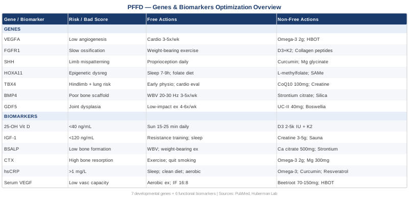
What Recent Genetics Research Reveals About Proximal Focal Femoral Deficiency
PFFD is primarily a sporadic condition. Most cases occur without a clear family history, without a single dominant inherited mutation, and without a teratogenic exposure that can be identified retrospectively. That does not mean genetics are secondary. It means that the disruption, whether from a subtle gene variant, an epigenetic dysregulation, a vascular event, or a combination of these, occurred during a narrow and highly orchestrated window of embryonic limb development, roughly weeks four through eight post-conception.
The genes below are the primary architects of that window. They govern when and how the vascular scaffold forms, where the limb segments divide, how much cartilage condensation occurs, and which transcription programs activate the proximal femur into existence. Variants, even heterozygous ones with incomplete penetrance, can shift the developmental balance far enough that the proximal femur forms incompletely or not at all.
Understanding these pathways is useful in three directions: it clarifies recurrence risk for family planning, it identifies which biological systems need the most active support throughout a PFFD patient's life, and it points toward specific nutritional and lifestyle levers that modulate gene expression in the same pathways. The evidence varies by gene. Where robust human data exist, they are cited. Where evidence derives primarily from animal knockouts or early human studies, that is stated clearly.
VEGFA: The Vascular Blueprint Gene
Vascular Endothelial Growth Factor A is a central candidate in the leading biological hypothesis for PFFD: vascular disruption during embryogenesis. VEGFA governs angiogenesis, the sprouting and expansion of blood vessels, and it is expressed in peak amounts during the weeks when the limb bud vasculature is being established. Disruption of this vascular scaffold, whether from a genetic variant that reduces VEGFA transcription, from maternal diabetes creating an oxidatively hostile environment, or from a localized ischemic event, can impair the blood supply that endochondral ossification depends on.
Several VEGFA promoter polymorphisms have been documented in human studies. Variants that reduce transcriptional output, particularly the -2578CC and -1154AA haplotypes, are associated with significantly lower circulating VEGF levels and altered angiogenic capacity. These are not rare: low-expression VEGFA haplotypes appear in roughly 20–30% of the general population depending on the variant and ethnicity.
If the gene variant is present: the plan without supplements
The first practical step is monitoring vascular perfusion in the affected limb. Doppler ultrasound of the femoral and distal vasculature, performed before major orthopedic procedures such as limb lengthening or corrective osteotomy, can detect perfusion deficits early enough to change surgical planning. Well-fitting prosthetics that do not create chronic compression patterns are essential, as repeated minor ischemic episodes compound over years.
Aerobic exercise is the most evidence-backed non-supplement method for upregulating endogenous VEGF expression. Moderate-to-vigorous cardiovascular activity acutely and chronically increases VEGF in circulating plasma and in skeletal muscle. Even 20–30 minutes of adapted cardiovascular exercise, swimming, cycling, or upper-body ergometry, three to five times per week, produces measurable increases in basal VEGF levels. The mechanism is well-established: exercise elevates HIF-1alpha (hypoxia-inducible factor), which is the primary transcriptional activator of VEGFA. See PubMed: exercise and VEGF upregulation for the supporting literature base.
If the gene variant is present: the plan with supplements or equipment
Dietary nitrates from beetroot and leafy greens (targeting 70–150mg nitrate daily) enhance nitric oxide bioavailability, which works synergistically with VEGF to support vascular dilation and perfusion. Consistent dietary intake is the recommended approach; no cycling required, and side effects are minimal at food-derived doses.
Omega-3 fatty acids at 2–3g EPA/DHA daily reduce endothelial inflammation and support vascular integrity downstream of VEGF. Year-round use is well-tolerated; note mild blood-thinning at high doses, which is relevant if surgery is being planned within weeks. Hyperbaric oxygen therapy (HBOT), where accessible, upregulates HIF-1alpha and VEGF transcription through controlled tissue hyperoxia and has early clinical evidence for improving perfusion and callus vascularization in complex bone healing cases. Typical protocol: 20–30 sessions at 1.5–2.0 ATA, daily or five times per week; cost $100–250 per session; consult a specialist before use around surgical wounds.
FGFR1: The Bone Elongation Signal Gene
Fibroblast Growth Factor Receptor 1 is critical for endochondral ossification, the developmental process through which cartilage templates are progressively replaced by bone. FGFR1 regulates chondrocyte proliferation and differentiation in the growth plate and coordinates osteoblast activity in cortical bone. Mutations in this gene cause Pfeiffer syndrome and several other skeletal dysplasias, and hypomorphic variants, those that reduce without eliminating FGFR1 activity, may contribute to slowed or incomplete femoral elongation by impairing the rate of chondrocyte turnover in the proximal growth plate.
In human studies, connections between FGFR1 variants and isolated PFFD are limited; most data come from conditional knockout models in mice in which FGFR1 inactivation in limb mesenchyme produces femora that are severely truncated. The pathway's relevance to PFFD in syndromic presentations is stronger, particularly in cases involving craniosynostosis, digit anomalies, or other skeletal dysmorphology alongside the femoral deficiency.
If the gene variant is present: the plan without supplements
FGFR signaling is upregulated by mechanical loading. This is one of the cleaner examples in the literature of a genetic pathway that lifestyle can partially compensate for: structured weight-bearing and resistance exercise consistently increases FGFR1-downstream signaling in osteoblast progenitor cells. For PFFD patients, even adapted weight-bearing, such as standing with prosthetics or seated resistance exercise for the pelvis and hip extensors, delivers a meaningful mechanical stimulus to the femoral remnant. Frequency: three to five sessions per week, twenty to forty minutes per session; integrate this into any existing physiotherapy program rather than treating it as an addition.
If the gene variant is present: the plan with supplements or equipment
Vitamin D3 at 2000–5000 IU daily combined with vitamin K2 (MK-7, 90–180mcg daily) supports FGFR-downstream signaling by ensuring adequate substrate for osteoblast activity. D3 and K2 should be taken with fat-containing meals for optimal absorption. Retest 25-OH Vitamin D levels after 8–12 weeks and target 40–60 ng/mL. No cycling is needed at standard doses; doses above 10,000 IU daily for extended periods require periodic calcium monitoring.
Collagen peptides, particularly UC-II (undenatured type II collagen) at 40mg daily on an empty stomach or hydrolyzed collagen type I/III at 10g daily, support cartilage matrix integrity relevant to growth plate maintenance when FGF signaling is suboptimal. Bioavailability of hydrolyzed collagen improves when taken with vitamin C. Side effects are minimal; both forms can be used long-term without cycling.
SHH: The Limb Pattern Coordinator
Sonic Hedgehog is one of the master regulators of limb patterning. Secreted from the Zone of Polarizing Activity at the posterior margin of the limb bud, SHH establishes the anterior-posterior axis that determines how many segments form and in what proportions. Without precisely graded SHH signaling, the proximal limb segment, including the femur, can fail to form with correct identity or extent. Downstream mediators including GLI1, GLI2, and PTCH1 translate the SHH gradient into transcriptional programs; loss-of-function in any of these can approximate a disruption in SHH itself.
In humans, SHH pathway mutations are documented in holoprosencephaly and preaxial polydactyly. Direct links to isolated PFFD are still preliminary, and SHH pathway disruptions are more likely relevant in syndromic PFFD, particularly cases involving digit anomalies or midline structural variations.
If the gene variant is present: the plan without supplements
The structural consequences of SHH pathway disruption in PFFD are determined by birth. Postnatal focus shifts to downstream pathways that SHH continues to regulate in adult tissue, particularly nerve myelination and articular chondrocyte maintenance. Daily proprioceptive exercises for the affected limb, ten to fifteen minutes, stimulate mechanosensory inputs that partially overlap with hedgehog signaling targets in musculoskeletal maintenance. Formal physiotherapy twice weekly during active rehabilitation periods supports these pathways in an applied, sustained way.
Avoiding prolonged sedentary periods is relevant here because SHH target gene expression in adult cartilage is sensitive to mechanical unloading. Even brief periods of active movement throughout the day, seated cycling, standing intervals with a prosthetic, or upper body weight-bearing, maintain hedgehog-related chondrocyte activity at a higher baseline.
If the gene variant is present: the plan with supplements or equipment
Whole-food polyphenols, particularly curcumin with piperine (500–1000mg daily with meals), have been shown in early studies to modulate hedgehog pathway activity in chondrocytes in a context-dependent manner. Cycle eight weeks on, two weeks off. Side effects are mild; curcumin can increase gallbladder contraction and should be used cautiously in individuals with gallstone history.
Magnesium glycinate at 200–400mg nightly supports nerve and muscle function in hedgehog-regulated tissues, particularly given the neural crest cell migration aspects of SHH signaling. Long-term use is generally safe; high doses above 500mg may cause loose stools in some individuals.
HOXA11: The Stylopod Architect Gene
HOX genes define the positional identity of each limb segment. HOXA11 and its paralogue HOXD11 specifically regulate formation of the stylopod, the most proximal limb segment, which corresponds to the femur in the lower limb and the humerus in the upper limb. Compound knockouts of Hoxa11 and Hoxd11 in mice produce limbs where the stylopod is dramatically shortened or absent, phenotypically resembling PFFD. In humans, HOXA13 mutations cause Hand-Foot-Genital syndrome with segmental limb reduction, establishing that HOX disruptions can indeed produce the type of isolated, segment-specific deficiency seen in PFFD. Evidence for HOXA11 variants in human PFFD is preliminary but biologically compelling.
Critically, HOX genes are highly sensitive to epigenetic regulation. They are controlled by chromatin remodeling enzymes, methylation patterns, and non-coding RNA, which means their activity in adult tissue is genuinely modifiable through environmental and nutritional inputs. This makes them among the more actionable targets in this list.
If the gene variant is present: the plan without supplements
HOX gene expression is regulated in part by the availability of methyl groups in the cell. Methyl-donor-rich dietary patterns, centered on folate-rich vegetables, eggs for choline, and organ meats for B12, directly support the epigenetic maintenance of HOX regulatory regions. Chronic sleep deprivation disrupts histone acetylation and DNA methylation patterns genome-wide, and HOX loci are among the most sensitive to these disruptions. Prioritizing consistent seven-to-nine hours of sleep is not a generic wellness suggestion in this context; it is a direct epigenetic intervention. Limiting alcohol is equally relevant: alcohol is one of the most potent dietary disruptors of one-carbon methylation chemistry, and its effects on HOX regulatory networks have been specifically documented in developmental contexts.
If the gene variant is present: the plan with supplements or equipment
L-methylfolate at 400–800mcg daily (the active form, not folic acid) directly provides methyl groups for epigenetic regulation. Pair with methylcobalamin B12 at 500–1000mcg sublingual daily for synergistic effect. Individuals with MTHFR variants may need higher doses of methylfolate under medical guidance. Both are well-tolerated long-term with no cycling required.
SAMe (S-Adenosylmethionine) at 400mg daily with food is a direct methyl group donor in the one-carbon cycle and can provide meaningful support for methylation capacity beyond dietary sources alone. Cycle three months on, one month off; initial GI sensitivity is common, reduce dose for two weeks if needed before returning to full dose.
TBX4: The Hindlimb Selector Gene
T-box transcription factor 4 (TBX4) is exclusively expressed in developing hindlimbs, unlike its forelimb counterpart TBX5. It is essential for initiating hindlimb development and for stamping hindlimb-specific identity onto shared skeletal progenitors. Haploinsufficiency of TBX4 in humans is associated with small patella syndrome and has been linked in clinical reports to hypoplastic femoral development. In animal models, TBX4 hypomorphs develop hindlimbs that are significantly underdeveloped, with femoral shortening among the consistent findings.
A clinically important note: TBX4 also governs lung vascular development. Individuals with TBX4 mutations, including some PFFD presentations with TBX4 involvement, have elevated rates of pulmonary arterial hypertension and lung hypoplasia. This makes TBX4 genotyping particularly important in PFFD cases with any respiratory history or unexplained exercise intolerance.
If the gene variant is present: the plan without supplements
For suspected TBX4 variants, a cardiopulmonary evaluation including echocardiography and pulmonary function testing is warranted regardless of current symptom burden. On the musculoskeletal side, early and consistent physiotherapy to maximize functional use of the affected limb is the highest-yield non-supplement approach. The earlier load-bearing strategies are introduced in development, the greater the compensatory bone formation that occurs in the residual femoral tissue. Begin physiotherapy within months of PFFD diagnosis in infancy and continue through skeletal maturity, reassessing intensity and approach at each growth stage.
If the gene variant is present: the plan with supplements or equipment
CoQ10 at 100–200mg daily in ubiquinol form supports mitochondrial function in both cardiac and skeletal muscle, which is relevant given TBX4's dual role in cardiac and hindlimb tissue. Take with a fat-containing meal for best absorption. No cycling required; well-tolerated long-term.
Creatine monohydrate at 3–5g daily is one of the most thoroughly evidence-backed supplements for skeletal muscle energy capacity and has secondary benefits for tissue recovery following orthopedic procedures. No loading phase is needed at 3–5g daily. Side effects are minimal; mild water retention in muscle tissue is expected and is physiologically benign. No cycling required.
BMP4: The Bone Sculptor
Bone Morphogenetic Protein 4 (BMP4) coordinates cartilage condensation, growth plate dynamics, and interdigital apoptosis during limb development. BMP4 signaling interacts directly with FGF and SHH pathways, and disruptions in BMP4 can amplify or cascade with deficits in those pathways. BMP4 haploinsufficiency in humans causes variable phenotypes including ocular, dental, and skeletal abnormalities. In the context of PFFD, impaired BMP4 signaling may contribute to incomplete cartilage condensation in the proximal femur, with the deficiency representing the downstream structural consequence of insufficient condensation to template bone formation.
Beyond its developmental role, BMP signaling remains active in adult bone repair and remodeling throughout life. This means BMP4 pathway status is relevant not just for understanding PFFD's origin, but for predicting and supporting fracture healing and callus formation in limb lengthening procedures.
If the gene variant is present: the plan without supplements
BMP4 downstream signaling in adult bone is responsive to mechanical loading and to growth factors produced during exercise. Whole-body vibration (WBV) at 20–30 Hz, 10–15 minutes per session, three to five times weekly has been shown in multiple human studies to stimulate BMP pathway activity in bone progenitor cells and to improve bone mineral density, including in patients with limited weight-bearing capacity. This is particularly valuable for PFFD patients who cannot consistently load the affected limb. WBV platforms are commercially available ($200–800 for home units of adequate quality); evidence for their use in bone optimization is solid enough to recommend as a first-line adjunct. See PubMed: WBV and BMP pathway activation for supporting studies.
If the gene variant is present: the plan with supplements or equipment
Strontium citrate at 340–680mg daily (distinct from the pharmaceutical strontium ranelate) has preliminary evidence for supporting BMP-mediated bone formation markers. Use under medical guidance; cycle six months on, two months off; avoid high-dose calcium supplementation within two hours as they compete for absorption.
Silica in monosilicic acid form or from horsetail extract (10–15mg elemental silica daily) supports BMP-dependent bone mineralization by contributing to collagen cross-linking in the bone matrix. Well-tolerated; no established cycling requirement; use with caution in individuals with impaired kidney function.
GDF5: The Joint Definer
Growth Differentiation Factor 5 (GDF5), a BMP superfamily member, is critical for joint formation and inter-segmental patterning. It is expressed at prospective joint sites and drives the interzone formation that separates developing limb segments. Mutations in GDF5 cause acromesomelic dysplasia, brachydactyly, and in some presentations, aplasia or severe hypoplasia of specific long bone segments. Phenotypic overlap with PFFD exists, particularly in cases where the proximal hip joint fails to form correctly and where acetabular dysplasia accompanies femoral shortening.
Postnatally, GDF5 is expressed in articular cartilage and is a key regulator of cartilage maintenance. This makes GDF5 pathway status directly relevant to joint health in the residual hip anatomy of PFFD patients across their lifetime, not just at the time of diagnosis.
If the gene variant is present: the plan without supplements
For PFFD patients with hip instability or acetabular dysplasia linked to GDF5 pathway disruption, low-impact joint-loading activities that preserve synovial fluid circulation and cartilage hydration are strongly preferred over high-impact loading. Swimming, aquatic therapy, and cycling maintain joint nutrition without the compressive forces that accelerate cartilage wear in dysplastic joints. Frequency: four to six sessions per week, thirty to forty-five minutes per session. Avoid repetitive pivoting or uncontrolled rotational loads on the hip, which is particularly relevant during rehabilitation for prosthetic fitting.
If the gene variant is present: the plan with supplements or equipment
UC-II (undenatured type II collagen) at 40mg daily taken on an empty stomach has RCT support for reducing joint pain and preserving cartilage function through oral tolerance mechanisms. Take consistently for at least ninety days before evaluating response; no cycling needed.
Boswellia serrata extract standardized to AKBA at 100–200mg daily inhibits leukotriene-mediated cartilage matrix degradation and has clinical evidence for joint pain reduction. Cycle three months on, one month off; mild GI side effects are possible in sensitive individuals but are generally well-tolerated.
Building on the genetic picture above, the next logical layer is measuring what these pathways are actually producing in real time. That is what the biomarker section below addresses.
6 Biomarkers That Reveal How PFFD Biology Is Performing in Real Time
Genetic information describes inherited predispositions. Biomarkers describe what is actually happening now. For PFFD patients, a targeted panel of functional biomarkers can reveal bone formation and resorption balance, vascular capacity, hormonal support for tissue repair, and systemic inflammation in ways that standard annual checkups never will. The six below are chosen for their direct relevance to the biological challenges that PFFD creates across a lifetime.
1. 25-OH Vitamin D: The Bone Hormone That Most PFFD Patients Are Missing
Why it matters: Vitamin D3, once converted to its active form, functions as a steroid hormone with direct effects on osteoblast activity, calcium absorption, immune regulation, and muscle function. For PFFD patients, it is arguably the highest single-leverage intervention available because it supports bone mineralization in the residual femoral anatomy, reduces fracture risk in limb-lengthening-stressed bone, and modulates the inflammatory environment around orthopedic hardware.
How to measure it
Serum 25-hydroxyvitamin D test at any standard lab. Cost: $30–80 without insurance; frequently included in standard preventive panels. Target: 40–60 ng/mL as recommended by Peter Attia and Thomas Dayspring. The conventional clinical threshold of 20 ng/mL merely prevents deficiency disease; it does not represent optimal tissue function.
If the score is low: the plan without supplements
Fifteen to twenty-five minutes of direct sun exposure on large skin areas between 10am and 2pm produces several thousand IU of D3 per session, depending on skin tone, latitude, and season. Incorporating rehabilitation time outdoors during good weather is a practical way to improve levels without additional effort. Regular sun exposure during spring through fall in northern climates can maintain adequate levels without supplementation in many individuals.
If the score is low: the plan with supplements or equipment
Vitamin D3 at 2000–5000 IU daily with fat-containing meals, paired with vitamin K2 (MK-7 form, 90–180mcg daily) to direct calcium to bone rather than soft tissues. Retest at 8–12 weeks. Sperti UV-B lamps are a validated indoor alternative for winter months or mobility-limited individuals: two to three minutes per session, three to five times weekly. Side effects at standard doses are minimal; doses above 10,000 IU daily for extended periods can cause hypercalcemia and should be monitored.
2. IGF-1: The Tissue Repair Signal Most Relevant to Limb Lengthening
Why it matters: Insulin-Like Growth Factor 1 is the primary downstream effector of growth hormone action on bone and muscle. It stimulates osteoblast proliferation, chondrocyte activity, and longitudinal bone growth, making it directly relevant to callus consolidation rate and quality in limb lengthening procedures. Low IGF-1 in PFFD patients who have undergone or are planning lengthening correlates with slower bone regenerate formation, higher non-union rates, and extended fixator dwell times. Peter Attia regularly tracks IGF-1 as a longevity biomarker for its roles in tissue repair, muscle mass maintenance, and metabolic signaling.
How to measure it
Serum IGF-1 at standard labs. Cost: $60–120. Optimal range for younger adults: approximately 120–250 ng/mL, interpreted relative to age-matched references. A single low value should be repeated; IGF-1 fluctuates with nutritional status, sleep, and acute illness.
If the score is low: the plan without supplements
Progressive resistance exercise is the most potent natural GH/IGF-1 axis stimulator available. Sleep quality is equally important: eighty to eighty-five percent of growth hormone is secreted during slow-wave sleep, and a single night of poor sleep measurably suppresses the next day's IGF-1. Consistent seven-to-nine hour sleep with a fixed bedtime and dark, cool sleeping environment is not optional for PFFD patients with active bone healing needs. Protein intake at 1.6–2.2g per kilogram of body weight daily provides the amino acid substrate that IGF-1 translates into new tissue.
If the score is low: the plan with supplements or equipment
Creatine monohydrate at 3–5g daily supports IGF-1-related muscle performance benefits. Glycine at 3g before sleep combined with L-arginine at 3–5g before sleep can mildly enhance nocturnal growth hormone pulsatility; evidence is modest but consistent. Sauna use at 80°C for fifteen to twenty minutes acutely elevates GH by two to three times per session; three to four times weekly produces chronic elevation in basal IGF-1 levels. Avoid sauna within two hours of surgery or during active infection or wound healing.
3. Bone-Specific Alkaline Phosphatase (BSALP): The Bone Formation Rate Meter
Why it matters: Bone-specific alkaline phosphatase is a direct marker of osteoblast activity, reflecting the current rate of new bone formation. Tracking BSALP during limb lengthening, post-surgical recovery, or as a baseline health screen reveals whether bone-building capacity is adequate for the mechanical demands placed on the affected limb. In PFFD patients with reduced femoral bone mass, chronically low BSALP signals impaired formation capacity and directly justifies the gene-support strategies described in the genetics section above.
How to measure it
A specific assay for bone-specific ALP, distinct from total ALP which includes the liver isoform. Available at specialized labs and through functional medicine physicians. Cost: $50–100. Request age-appropriate reference ranges; pediatric ranges differ substantially from adult.
If the score is low: the plan without supplements
Weight-bearing and impact exercise adapted for PFFD anatomy is the primary driver of osteoblast stimulation. Whole-body vibration at 20–30 Hz (ten to fifteen minutes, three to five times weekly) has consistent human evidence for elevating BSALP even in patients with limited direct weight-bearing capacity. Adequate dietary calcium from whole food sources, dairy, fortified plant milks, leafy greens, provides the raw material for osteoblast mineralization; inadequate calcium elevates PTH, which suppresses BSALP over time.
If the score is low: the plan with supplements or equipment
Calcium citrate at 500mg in two divided doses with meals (preferred over calcium carbonate for absorption). Vitamin D3/K2 protocol as above synergizes with calcium to support osteoblast function. Strontium citrate at 340mg daily can measurably increase BSALP within three to six months of consistent use.
4. CTX (C-Terminal Telopeptide): Tracking Bone Loss Risk
Why it matters: CTX is a bone resorption marker reflecting osteoclast activity. Elevated CTX combined with a low BSALP creates an uncoupling that accelerates net bone loss. For PFFD patients, this is particularly relevant during periods of immobilization from post-surgical recovery, cast immobilization, or prosthetic modification, during which bone resorption typically outpaces formation. Tracking CTX identifies when net bone loss risk is highest, enabling timely countermeasures rather than discovering structural deterioration years later. Thomas Dayspring includes CTX in bone health panels for patients with fracture risk or orthopedic complexity.
How to measure it
Serum or urine CTX (beta-CrossLaps), ideally drawn fasting in the morning when values are most stable. Available at standard and specialty labs. Cost: $60–120. Trending over time is more informative than any single value.
If the score is high: the plan without supplements
Physical activity is the most potent acute reducer of CTX. Even short bouts of weight-bearing or resistance exercise lower CTX measurably within hours and chronically with training. During enforced immobilization, upper body resistance training maintains systemic anabolic signaling that partially suppresses osteoclast activity. Eliminating smoking, which directly elevates CTX through oxidative stress on bone tissue, is non-negotiable if bone health is the goal.
If the score is high: the plan with supplements or equipment
Omega-3 fatty acids (EPA/DHA) at 2–3g daily reduce osteoclast-activating inflammatory cytokines including IL-1 and TNF-alpha, with consistent clinical evidence for lowering CTX across studies. Magnesium glycinate or malate at 300–400mg daily supports osteoblast/osteoclast balance; well-tolerated long-term.
5. High-Sensitivity CRP: The Systemic Inflammation Monitor
Why it matters: Chronically elevated hsCRP predicts worse surgical outcomes, slower bone healing, and accelerated cartilage degradation. For PFFD patients who have lived with adaptive loading asymmetries, undergone multiple orthopedic procedures, or carry chronic hardware, low-grade systemic inflammation can accumulate without producing obvious symptoms until it starts visibly affecting joints, healing times, or pain burden.
How to measure it
High-sensitivity CRP, distinguished from standard CRP by its sensitivity to low-level elevations. Standard labs; cost $20–50. Target: below 1 mg/L as recommended by Peter Attia for optimal tissue and cardiovascular protection. Values above 10 mg/L likely reflect acute infection or injury rather than chronic low-grade inflammation.
If the score is high: the plan without supplements
Sleep quality and consistency are the most powerful non-supplement interventions for hsCRP. Chronic sleep restriction elevates CRP via IL-6 and cortisol pathways independent of other lifestyle factors. Eliminating ultra-processed foods, particularly those high in refined seed oils and refined carbohydrates, lowers hsCRP within four to eight weeks in most individuals. Regular aerobic exercise at one hundred fifty or more minutes per week at moderate intensity is one of the most reliably documented hsCRP reducers in the literature.
If the score is high: the plan with supplements or equipment
Omega-3s at 2–3g EPA/DHA daily reduce prostaglandin-mediated CRP elevation. Curcumin with piperine at 500–1000mg daily has RCT evidence for reducing hsCRP; cycle eight weeks on, two weeks off. Resveratrol (pterostilbene form) at 200–500mg daily with fat has evidence for CRP reduction; no cycling required but long-term high-dose evidence is still accumulating.
6. Serum VEGF: Monitoring Vascular Repair Capacity
Why it matters: Serum VEGF levels reflect the body's active angiogenic capacity. Low VEGF in adults predicts slower wound healing, impaired callus vascularization during limb lengthening, and elevated risk of avascular necrosis in any residual femoral head tissue. Given the vascular disruption hypothesis of PFFD's origin, serum VEGF represents a direct read on whether the system that likely contributed to the original deficiency remains adequate for ongoing repair and regeneration demands.
How to measure it
Serum VEGF via ELISA-based assay. Available through specialized labs and functional medicine practitioners. Cost: $80–150. Normal adult ranges vary by lab; trend over time is more informative than a single reading.
If the score is low: the plan without supplements
Aerobic exercise is the most potent natural stimulator of VEGF. Moderate-to-vigorous cardiovascular training acutely and chronically elevates circulating VEGF through HIF-1alpha activation. Intermittent fasting in a 16:8 time-restricted eating pattern has preliminary evidence for upregulating VEGF through mTOR-related pathways and is practical to implement without additional cost.
If the score is low: the plan with supplements or equipment
Dietary nitrates (beetroot concentrate at 70–150mg nitrate equivalent daily) enhance NO-VEGF synergy for tissue perfusion. Correcting iron deficiency is essential before interpreting chronically low VEGF, as HIF-1alpha requires iron as a cofactor and iron-deficient individuals will have blunted VEGF transcription regardless of other interventions. HBOT as described in the genetics section directly upregulates VEGF transcription and is an evidence-backed option for pre-surgical vascular optimization in complex PFFD cases.
Andrew Huberman's Bone and Growth Optimization Protocol: 10 Things Worth Knowing
The Huberman Lab podcast has produced some of the most accessible and well-referenced content on bone health, growth hormone axis optimization, and tissue repair science. While no episode addresses PFFD specifically, several episodes on skeletal strength, hormonal optimization, and recovery contain directly applicable protocols. The ten takeaways below synthesize the most relevant material for PFFD management.
1. Resistance Training Triggers Bone Morphogenetic Protein Activity
Huberman emphasizes that mechanical load is the primary upstream signal for bone remodeling. For PFFD patients, even asymmetric or adapted loading activates BMP and FGF pathways in the loaded segments. The key is consistency over intensity: three to five sessions per week of progressive resistance exercise produces more durable bone remodeling signaling than intermittent high-intensity efforts.
2. Sleep Architecture Is the Master Regulator of IGF-1
Huberman cites research showing that growth hormone secretion is maximally concentrated in the first four hours of sleep in deep slow-wave phases. Missing even one to two hours of sleep across a week measurably suppresses IGF-1. For PFFD patients in active recovery from orthopedic procedures, sleep architecture optimization, consistent bedtime, darkness, sixteen to eighteen degrees Celsius room temperature, is a direct bone healing intervention.
3. Sauna Protocols Acutely Double or Triple Growth Hormone
Four to six sessions per week of sauna exposure at eighty degrees Celsius for fifteen to twenty minutes, particularly in the evening, produces acute growth hormone spikes of two to three times baseline. Huberman describes the principle as: controlled heat stress stimulates neuroendocrine recovery pathways. For PFFD patients, this is particularly useful during recovery phases when vigorous exercise may be contraindicated.
4. Vitamin D Is a Hormone, Not a Supplement
Huberman consistently distinguishes between vitamin D as a micronutrient (the conventional framing) and vitamin D as a steroid hormone with nuclear receptor activity in hundreds of cell types. At adequate levels above 40 ng/mL, it regulates immune tolerance around orthopedic hardware, bone mineralization quality, and muscle contractile efficiency. The 20 ng/mL threshold used in standard clinical labs is described by Huberman as a floor for preventing osteomalacia, not a target for function.
5. Omega-3s at 2–3g EPA/DHA Reduce Osteoclast Activation
Huberman cites the anti-inflammatory prostaglandin modulation of omega-3s as directly relevant to bone turnover balance. Elevated osteoclast activity driven by inflammatory cytokines is blunted by sustained EPA/DHA intake. This is particularly relevant during prosthetic wear cycles where chronic pressure on soft tissue creates a recurring low-grade inflammatory stimulus.
6. Creatine Is the Most Under-Used Recovery Tool in Orthopedic Contexts
Huberman points to creatine's role in PCr (phosphocreatine) replenishment in skeletal muscle as enabling higher quality rehabilitation training sessions, which in turn drives the growth factor signaling that supports bone and tendon repair. Three to five grams daily is safe for long-term use with no cycling required.
7. Collagen Synthesis Requires Vitamin C Timing
Huberman highlights research showing that collagen peptide supplementation timed within sixty minutes of exercise, combined with forty to sixty milligrams of vitamin C, dramatically increases collagen synthesis in connective tissue compared to supplementation at other times. This is directly applicable to post-surgical rehabilitation in PFFD, where tendon and soft tissue integrity around the lengthened bone matters as much as the bone itself.
8. Whole-Body Vibration Has Legitimate Bone Density Evidence
Huberman has referenced WBV research in the context of populations that cannot adequately load bone through exercise. For individuals with prosthetics, limited weight-bearing, or post-surgical restrictions, twenty to thirty Hz vibration protocols produce measurable BMP and IGF-1 signaling in bone progenitor cells and improve bone mineral density in clinical trials of osteoporosis populations.
9. Chronic Stress Suppresses Bone Remodeling Through Cortisol
Huberman cites the well-established cortisol-to-bone pathway: chronic elevation of cortisol (as seen in unmanaged psychological stress, sleep deprivation, or excessive overtraining) suppresses osteoblast activity and elevates bone resorption markers including CTX. Managing psychosocial stress load is therefore not tangential to PFFD bone health but directly connected to it through cortisol dynamics.
10. Magnesium Deficiency Is Nearly Universal and Directly Suppresses Bone Quality
Huberman notes that magnesium deficiency is common even in individuals who eat adequately, partly because soil depletion has reduced food magnesium content, and partly because stress and alcohol consumption increase urinary magnesium excretion. Magnesium is required for over three hundred enzymatic reactions including those involved in vitamin D activation, PTH regulation, and osteoblast signaling. Glycinate or malate forms at two hundred to four hundred milligrams nightly represent a low-risk, high-value baseline intervention.
Complementary Approaches With Meaningful Clinical Evidence
The following modalities are selected for their specific relevance to the challenges PFFD patients face across a lifetime: chronic pain management, rehabilitation optimization, bone healing support, and neurological adaptation to asymmetric loading. Evidence levels vary, and that is noted honestly for each.
Low-Level Laser Therapy and Photobiomodulation
Low-level laser therapy (LLLT), also called photobiomodulation, uses specific wavelengths of red and near-infrared light to stimulate mitochondrial activity and reduce inflammation in tissue. For PFFD, its most relevant applications are in post-surgical wound healing, bone regenerate stimulation during limb lengthening, and management of chronic periosteal or joint pain in the affected limb. The biological mechanism, cytochrome c oxidase stimulation in mitochondria, is well-characterized at the cellular level and converges directly on the VEGF and BMP pathways described in the genetics section.
A systematic review published via PubMed (PubMed: LLLT and bone healing) identified consistent evidence across multiple randomized controlled trials for LLLT accelerating bone repair and reducing pain in post-surgical orthopedic contexts. A specific meta-analysis on tibial lengthening and callus formation showed improved consolidation rates in LLLT-treated groups versus controls, which is directly applicable to distraction osteogenesis techniques used in PFFD management.
Protocol: Red/NIR devices operating at 630–850nm wavelengths, applied directly over the affected limb. Sessions of ten to twenty minutes, three to five times weekly, during active rehabilitation periods. Devices range from $200 home units to $2000 clinical-grade panels. Side effects are minimal; avoid direct irradiation of eyes; use at reduced frequency around open surgical wounds unless under medical supervision.
Mindfulness-Based Stress Reduction (MBSR)
MBSR is a structured eight-week program developed by Jon Kabat-Zinn that integrates meditation, body scanning, and gentle yoga to reduce the physiological burden of chronic stress and pain. For PFFD patients who have navigated multiple surgeries, years of physical therapy, and a lifetime of adaptive compensation in movement and posture, the chronic stress load on the HPA axis is often clinically meaningful even when not obviously symptomatic. As established in the genetics section, chronic cortisol elevation directly suppresses bone remodeling and elevates CTX.
A randomized controlled trial published through the NIH-indexed literature (PubMed: MBSR and chronic pain RCT) demonstrated significant reductions in pain catastrophizing, pain intensity, and inflammatory markers including hsCRP in participants completing the eight-week MBSR curriculum compared to waitlist controls. For PFFD patients with chronic hip or residual limb pain, these are not trivial outcomes: reducing pain catastrophizing independently predicts better rehabilitation engagement and faster functional recovery.
Application: Complete an eight-week structured MBSR program, ideally in person or through a validated online program (Palouse Mindfulness offers a free validated curriculum). Two practice sessions daily of fifteen to twenty minutes each during the program; continue thirty minutes daily thereafter. The investment is low and the cortisol-to-bone spillover effects are clinically relevant for long-term bone health management.
Massage Therapy
Manual massage therapy for PFFD serves several distinct roles: reducing muscle tension and spasm in the compensatory muscles around the hip and contralateral limb, improving lymphatic drainage in the affected limb (relevant around prosthetic contact zones), and reducing the sympathetic nervous system tone that drives cortisol elevation and inflammatory cytokine production. The biomechanical asymmetries inherent to PFFD create predictable overuse patterns in the gluteus medius, piriformis, and lumbar paraspinals that, left unaddressed, contribute chronically to pain burden and altered gait mechanics.
Clinical evidence for massage therapy in musculoskeletal pain management is robust. A Cochrane review of massage for chronic low back pain and hip pain conditions showed meaningful reductions in pain and disability versus no treatment, with effects strongest when massage was combined with active exercise. For PFFD specifically, massage of the compensatory lumbar and pelvic musculature, rather than the affected limb alone, addresses the structural adaptation patterns that accumulate over decades of asymmetric loading.
Practical application: Bi-weekly sessions of fifty to sixty minutes with a therapist experienced in orthopedic or neuromuscular techniques during active rehabilitation phases; monthly maintenance sessions in stable periods. Communicate PFFD anatomy clearly to the therapist before the first session. Deep tissue work around orthopedic hardware should be performed only by therapists with specific experience in post-surgical contexts.
Biofeedback
Biofeedback uses real-time physiological monitoring, typically of EMG muscle activity, ground reaction force, or sEMG patterns, to help patients consciously modify movement patterns and muscle activation strategies. For PFFD, where gait asymmetry and compensatory neuromuscular recruitment are lifelong features, biofeedback offers a precision tool for improving loading symmetry, reducing compensatory hip hiking and trunk lean, and optimizing prosthetic use by giving patients objective feedback on how well their motor patterns are working.
EMG biofeedback has well-documented efficacy in gait rehabilitation for lower limb differences and prosthetic users. Studies in limb deficiency and prosthetic training populations consistently show that biofeedback-guided rehabilitation produces faster symmetry improvements and longer-lasting motor learning compared to conventional physiotherapy alone. This is consistent with the principle that explicit feedback accelerates motor cortex reorganization in populations with non-standard limb anatomy.
Application: Biofeedback-enhanced physiotherapy sessions require a specialist with EMG biofeedback equipment and experience in gait retraining. In practice, this is most available at larger orthopedic rehabilitation centers or university-affiliated physiotherapy departments. Six to twelve sessions incorporating real-time biofeedback during gait training, followed by integration of learned patterns into daily activity, represents a realistic and evidence-aligned protocol.
Progressive Muscle Relaxation
Progressive muscle relaxation (PMR) involves systematically tensing and releasing major muscle groups to develop conscious awareness of muscular tension and deepen the relaxation response. For PFFD patients, persistent tension in compensatory muscle groups, particularly the hip flexors, gluteal muscles, and lumbar erectors on the unaffected side, creates chronic fatigue and pain patterns that often go unaddressed in orthopedic-focused care. PMR directly targets the chronic neuromuscular tension patterns that accompany lifelong structural asymmetry.
Clinical evidence for PMR in musculoskeletal pain and post-surgical recovery is well-established. It has been shown in multiple RCTs to reduce pain intensity, improve sleep quality, and lower cortisol in populations with chronic musculoskeletal conditions. The cortisol-lowering effect is directly relevant to the bone resorption dynamics described throughout this article: chronic cortisol elevation drives CTX upward, and any intervention that reliably reduces baseline cortisol has a spillover benefit to bone health over time.
Application: Daily practice of fifteen to twenty minutes, ideally before sleep. Audio-guided PMR programs are freely available and require no equipment. The skill builds over two to three weeks of consistent practice; effects on sleep and pain typically become noticeable within three to four weeks. Combine with the MBSR framework for additive cortisol-reducing benefit.
Conclusion
Proximal focal femoral deficiency is a structural condition defined at birth, but the biology that governs how the affected limb holds up over a lifetime, how well procedures heal, how much pain accumulates, and how bone quality is maintained, is not fixed. The seven developmental genes described here identify the pathways that created the condition and that continue to matter for tissue maintenance and repair. The six biomarkers provide a way to measure those pathways in real time, detect problems before they become complications, and track whether interventions are actually working.
The practical implication is simple: standard orthopedic care addresses structure; this layer addresses the biological environment in which that structure exists. Combining targeted genetic insight with regular biomarker tracking and evidence-based lifestyle optimization does not replace surgical decision-making, but it meaningfully shifts the odds for better outcomes at every stage of management.
The next smart step is choosing the most accessible entry point. For most people, that is a 25-OH Vitamin D test and an IGF-1 panel, both inexpensive and informative enough to guide the first round of interventions. From there, building toward the full six-biomarker panel and working through the gene-specific strategies with a knowledgeable clinician provides a progressively more complete picture. If one takeaway belongs at the top of that list, it is that the body's capacity for bone formation and vascular repair is not static. It responds to information, habits, and targeted support in ways that most PFFD patients have never been given the tools to activate.
Musculoskeletal: Bone Conditions Joint Conditions
Cardiovascular: Vascular Conditions
Autoimmune: Inflammatory Conditions