This article contains AI generated content.
· UpdatedProximal Tibiofibular Joint Cyst Genes Biomarkers - 4 Genes And 6 Biomarkers To Track
Introduction
A cyst forming at the proximal tibiofibular joint is not a common diagnosis, and that rarity is part of what makes it so frustrating to manage. You may have received a cortisone injection, been told to wait and see, or found almost nothing useful online beyond surgical case reports. If you have experienced the persistent ache at the outer knee, intermittent tingling down the lower leg, or a subtle sense of something quietly wrong near the fibular head, you already know how poorly the standard response lands.
What most people are not told is that a proximal tibiofibular joint cyst does not appear randomly. These synovial or ganglion cysts form when joint fluid escapes and accumulates in a localized sac, typically because of underlying joint capsule weakness, chronic low-grade inflammation, or altered synovial fluid dynamics — and all three of those factors are deeply individual. They are shaped by your genetics, your metabolic health, and biological patterns that a standard orthopedic evaluation is not designed to investigate.
Generic advice — anti-inflammatories, rest, watch for nerve involvement — addresses symptoms without touching what drives them. This article takes a different approach: by identifying specific measurable biomarkers and genetic variants that influence joint tissue integrity, inflammatory signaling, and synovial fluid behavior, it becomes possible to build a more precise picture of your individual biology and take more targeted action.
None of this replaces a structural evaluation or surgical consultation when warranted. What it can do is give you a clearer biological map, one that identifies actionable levers and helps you move from passive uncertainty to informed decision-making. The strategies below cover six trackable biomarkers, four genetic variants with practical plans, key insights from a landmark longevity medicine book, and four complementary approaches with real clinical evidence — each one chosen for its specific relevance to this condition.
Summary
This article covers six biomarkers — hs-CRP, uric acid, vitamin D, MMP-3, serum hyaluronic acid, and IL-6 — that are measurable, affordable at standard labs, and directly linked to the biology of joint cyst formation and persistence. For each one, you will find cost ranges, optimal targets used by leading clinicians, and specific plans both with and without supplements if results come back unfavorable. The article then explores four genes — COL1A1, MMP3, VEGF, and IL1B — whose variants influence connective tissue strength, matrix breakdown, fluid dynamics, and inflammatory amplification at the joint level, with actionable protocols for each genotype. A synthesis of ten key insights from Peter Attia's Outlive provides the broader metabolic and longevity framework behind why these markers matter over time. Finally, four complementary modalities — photobiomodulation, massage therapy, MBSR, and tai chi — are reviewed for their specific evidence in joint and nerve compression management. Whether you are dealing with an active cyst, recovering after aspiration, or trying to prevent recurrence, this article gives you a more productive starting point than most conventional consultations will offer.
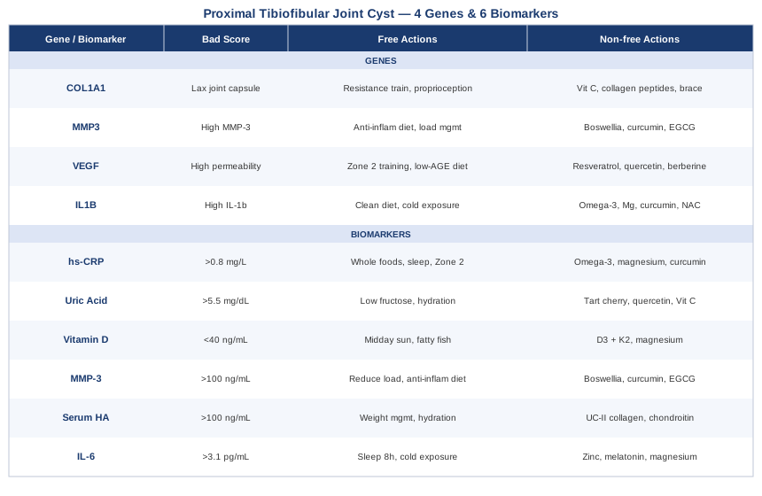
6 Biomarkers That May Reveal What Is Driving Your Proximal Tibiofibular Joint Cyst
The biology of a synovial or ganglion cyst at the proximal tibiofibular joint involves three overlapping processes: localized inflammation, structural changes in the extracellular matrix of the joint capsule, and dysregulation of synovial fluid production and resorption. Each of the biomarkers below touches one or more of these processes in a measurable way. None will appear on a standard orthopedic workup, but all are accessible through a primary care physician or direct-to-consumer lab services.
Biomarker 1: High-Sensitivity C-Reactive Protein (hs-CRP)
Why it matters
CRP is a protein produced by the liver in response to systemic inflammation. The high-sensitivity version of the test detects low-grade chronic inflammation that standard CRP panels miss. In the context of joint cysts, persistent low-grade inflammation is a key driver of synovial fluid overproduction and altered capsule permeability — the two conditions that allow cyst fluid to accumulate and remain. Research on synovial joint pathology consistently shows that even mild systemic inflammation correlates with increased synovial tissue activity. The JUPITER trial demonstrated how elevated hs-CRP, even in the absence of other obvious disease markers, signals meaningful systemic inflammatory burden with real tissue consequences. While that study focused on cardiovascular endpoints, the underlying biology is the same across joint tissues.
How to measure it
hs-CRP is ordered through any standard lab. Cost ranges from $15 to $50 depending on provider. No fasting required. Peter Attia recommends targeting hs-CRP below 0.5–0.8 mg/L for optimal tissue health, considerably more demanding than the typical lab reference range of 3 mg/L. Anything above 3 mg/L warrants investigation into the underlying inflammatory driver.
If the score is bad, the plan without supplements
The most powerful non-supplement lever is diet quality. Eliminating ultra-processed foods, refined seed oils, and excess refined carbohydrates produces measurable hs-CRP reductions within 4–6 weeks. Prioritizing whole food sources of omega-3 fatty acids (fatty fish 3–4 times per week), ensuring 7–9 hours of quality sleep, and engaging in Zone 2 aerobic training (3–4 sessions per week of 30–60 minutes each) consistently lower hs-CRP in clinical literature. Reducing psychological stress — which triggers inflammatory cascades via cortisol — also has direct and measurable impact. Retest in 8–12 weeks.
If the score is bad, the plan with supplements or equipment
Omega-3 EPA/DHA: 2–4 g/day of combined EPA+DHA. Well-tolerated at standard doses; cycle off every 3–4 months to reassess. Side effects: fishy aftertaste, mild GI discomfort at high doses; blood-thinning effect above 4 g/day — discuss with physician if on anticoagulants. Magnesium glycinate: 300–400 mg at night; reduces CRP and supports sleep quality. Curcumin with piperine: 500–1000 mg of standardized curcumin with 5–10 mg piperine (which increases bioavailability), taken with a fatty meal; cycle 8 weeks on, 2 weeks off — monitor liver enzymes if taken long-term. Infrared sauna 3 times per week for 15–25 minutes shows emerging evidence for reducing systemic inflammatory markers.
Biomarker 2: Uric Acid (Serum)
Why it matters
Uric acid is often associated only with gout, but elevated serum uric acid (hyperuricemia) has broader implications for joint health. Crystal deposition from uric acid can accumulate in the soft tissues around small and large joints — including the proximal tibiofibular region — and drive a local inflammatory response that mimics or exacerbates cyst formation. Elevated uric acid is also increasingly recognized as a marker of mitochondrial stress and metabolic dysfunction, which contributes to poor tissue repair and a compromised joint environment. If your cyst is accompanied by flares of redness, heat, or acute pain episodes, uric acid is a critical variable to rule out.
How to measure it
Uric acid is part of a standard metabolic panel. Cost ranges from $10 to $40. Fasting is recommended for accuracy. Optimal range: below 5.5 mg/dL for men and women — lower than the typical lab reference range of 7.2 mg/dL for men, which many clinicians consider permissive. Measure twice if symptoms are episodic: once fasting and once 2–3 hours after a high-purine or high-fructose meal.
If the score is bad, the plan without supplements
Reduce intake of high-fructose foods (the primary dietary driver of uric acid — more so than dietary purines), limit alcohol especially beer, avoid fructose-corn-syrup-sweetened beverages entirely, and increase hydration to at least 2–2.5 liters of water daily. Low-glycemic-index carbohydrates reduce uric acid production more effectively than simply avoiding meat. Retest after 6–8 weeks of dietary changes.
If the score is bad, the plan with supplements or equipment
Tart cherry extract: 480 mg standardized anthocyanin extract daily; multiple human trials show meaningful reduction in uric acid levels and gout flare frequency. Cycle 3 months on, 1 month off. Quercetin: 500–1000 mg/day inhibits xanthine oxidase, the enzyme that produces uric acid; no major side effects at standard doses, but may interact with certain antibiotics. Vitamin C: 500–1000 mg/day modestly reduces serum uric acid. If uric acid remains above 7 mg/dL despite these interventions, discuss xanthine oxidase inhibitor medication with your physician before the joint condition progresses.
Biomarker 3: Vitamin D (25-OH)
Why it matters
Vitamin D functions less as a vitamin and more as a pleiotropic hormone that regulates immune function, inflammatory signaling, and connective tissue gene expression. Its role in musculoskeletal health is well-documented: deficiency is associated with increased synovial inflammation, impaired cartilage maintenance, and reduced tensile strength in ligaments and joint capsules — exactly the structural properties that matter most in a joint prone to cyst formation. Low vitamin D also impairs the resolution phase of inflammation, meaning the body struggles to switch off the inflammatory signal once it has started, allowing low-grade synovial activity to persist.
How to measure it
The 25-hydroxyvitamin D test is the standard measurement. Cost: $40–$80. Optimal per most functional medicine frameworks: 40–60 ng/mL. Below 30 ng/mL is deficient; below 20 ng/mL is severely deficient. Testing in early spring, after the lowest-sun months, provides the most informative baseline.
If the score is bad, the plan without supplements
Direct sun exposure is the most efficient driver: 15–25 minutes of midday sun to bare arms and legs, 4–5 days per week, without sunscreen applied immediately (allow 15 minutes before applying). In northern latitudes or winter months this is rarely sufficient. High-fat fish (salmon, mackerel, sardines) and egg yolks provide modest dietary vitamin D. Timing outdoor exposure to solar noon is significantly more effective than longer duration at dawn or dusk.
If the score is bad, the plan with supplements or equipment
Vitamin D3: 2000–5000 IU/day depending on baseline deficiency depth, always paired with vitamin K2 (MK-7 form) at 100–200 mcg/day — K2 directs calcium to bone rather than soft tissues, reducing the calcification risk that high-dose D3 can carry. Magnesium is required for vitamin D activation; supplement at 300–400 mg/day or verify dietary adequacy. Retest after 90 days. Side effects: toxicity is rare below 10,000 IU/day but possible — verify with annual testing. A UVB light lamp is an alternative for those who cannot access adequate sun exposure seasonally.
Biomarker 4: Matrix Metalloproteinase-3 (MMP-3, Serum)
Why it matters
MMP-3, also called stromelysin-1, is a protease enzyme that degrades extracellular matrix components including collagen, fibronectin, and proteoglycans. In healthy joint tissue, MMP-3 is tightly regulated. In inflamed or mechanically stressed joints, MMP-3 expression increases and begins dismantling the structural framework of the joint capsule and synovial membrane. This weakens the barrier that normally contains synovial fluid, directly contributing to the fluid escape and accumulation that forms ganglion and synovial cysts. Serum MMP-3 is an underused but increasingly available marker for assessing synovial tissue activity and joint capsule integrity.
How to measure it
Serum MMP-3 can be ordered through specialty labs and some integrative medicine providers. Cost ranges from $100–$200. Elevated levels consistently above 100 ng/mL in most assays are associated with active synovial disease. The marker is used clinically in rheumatoid arthritis monitoring but is mechanistically relevant for any synovial joint pathology, including cyst-forming conditions.
If the score is bad, the plan without supplements
Reducing joint mechanical overload is the most direct intervention: modify activities that place repetitive rotational or compressive stress on the proximal tibiofibular joint — aggressive pivoting sports, certain loaded yoga postures, high-impact activities. Progressive resistance training targeting the muscles of the knee, ankle, and hip to offload the joint is more effective long-term than rest alone. Anti-inflammatory dietary patterns demonstrably reduce MMP-3 gene expression via epigenetic mechanisms; this is one of the most consistently studied dietary levers for matrix metalloproteinase activity.
If the score is bad, the plan with supplements or equipment
Boswellia serrata (AKBA-standardized): 100–200 mg of AKBA per day; human trials show meaningful reduction in MMP-3 expression in joint tissue. Cycle: 8 weeks on, 2 weeks off. Side effects: mild GI discomfort; generally well-tolerated. Curcumin with piperine: 500–1000 mg with 5 mg piperine and a fat-containing meal; reduces NFkB-driven MMP-3 transcription. Green tea extract (EGCG): 400–800 mg standardized extract; direct MMP-3 inhibitory effect demonstrated in human joint tissue studies — take with food to avoid GI discomfort; cycle every 3–4 months. Compression bracing of the knee during higher-load activities reduces joint instability and the mechanical stimulus for MMP-3 elevation.
Biomarker 5: Serum Hyaluronic Acid
Why it matters
Hyaluronic acid (HA) is a glycosaminoglycan and the primary structural and lubricating component of synovial fluid. In cyst pathology, HA plays a dual role: elevated serum HA reflects increased turnover in synovial tissue, signaling active remodeling or inflammation, and the accumulation of HA-rich fluid is precisely what gives ganglion and synovial cysts their characteristic gel-like consistency. Tracking serum HA provides a window into the overall health and remodeling activity of your synovial tissue — the tissue directly responsible for cyst formation when its function is disrupted.
How to measure it
Serum hyaluronic acid is available through specialty labs and is used in some hepatic fibrosis panels. Cost: $150–$300, making it the priciest option on this list. Standardized reference ranges for general musculoskeletal use are still evolving, but levels below 100 ng/mL in fasting serum are generally considered normal in adults under 50. Elevated levels above 200 ng/mL in the absence of liver disease suggest active synovial tissue remodeling.
If the score is bad, the plan without supplements
Weight management is the most powerful non-supplement lever: excess adipose tissue drives systemic HA dysregulation and synovial fluid overproduction. Adequate hydration (HA is hygroscopic, and its behavior in tissue depends on water availability) and anti-inflammatory dietary patterns both support healthier HA dynamics. Avoiding repetitive joint compression that triggers synovial cells to overproduce HA is also important — this includes reviewing footwear mechanics, sitting posture, and sport-specific loading patterns at the lateral knee.
If the score is bad, the plan with supplements or equipment
Undenatured type II collagen (UC-II): 40 mg/day; supported by human trials for joint-specific outcomes including synovial tissue homeostasis. Chondroitin sulfate: 800–1200 mg/day supports glycosaminoglycan balance, including HA regulation in joint tissue. Cycle: 3 months continuous, then reassess. Side effects: generally mild; occasional GI discomfort. Low-level laser therapy applied to the proximal tibiofibular joint region has shown in small clinical studies to modulate local HA and proteoglycan metabolism in joint tissue, making it both a complementary therapy and a tool relevant to this biomarker.
Biomarker 6: Interleukin-6 (IL-6)
Why it matters
IL-6 is a pro-inflammatory cytokine that serves as a key amplifier of synovial inflammation. While acutely elevated IL-6 is a normal short-term response to injury or exercise, chronically elevated IL-6 creates a persistent inflammatory environment within joint tissue that promotes synovial hyperplasia — the overgrowth and thickening of the synovial membrane that underlies many joint cyst presentations. Elevated fasting IL-6 also predicts poor tissue remodeling outcomes following conservative management of joint lesions, making it one of the more prognostically meaningful markers to track.
How to measure it
Serum IL-6 is ordered through specialty labs or comprehensive cytokine panels. Cost: $100–$200. Optimal fasting serum IL-6: below 3.1 pg/mL. Measurement must be done fasting, as exercise in the preceding 24 hours significantly elevates IL-6 transiently. Thomas Dayspring and other longevity-focused clinicians have noted IL-6 as one of the most sensitive early indicators of systemic inflammatory burden beyond what hs-CRP alone captures.
If the score is bad, the plan without supplements
Sleep quality is the single most powerful modifier of chronic IL-6: even one night of disrupted sleep raises fasting IL-6 significantly, and the effect is cumulative with chronic deprivation. Prioritize 7–9 hours with consistent timing. Reducing abdominal adiposity — visceral fat is a primary systemic source of IL-6 — through caloric moderation and resistance training is the second lever. Intermittent fasting (16:8 or similar) demonstrates IL-6-lowering effects in human trials at 8–12 weeks. Cold exposure (cold showers or cold water immersion at 10–15°C, 10–15 minutes, 3 times per week) reduces chronically elevated IL-6 in the available human data.
If the score is bad, the plan with supplements or equipment
Zinc (bisglycinate or gluconate): 15–30 mg/day directly reduces IL-6 production — avoid excess supplementation above 40 mg/day long-term as it competes with copper; pair with 1–2 mg copper and cycle 3 months on, 1 month off. Melatonin: 0.5–3 mg at sleep onset reduces IL-6 via antioxidant and anti-inflammatory mechanisms; low dose is often more effective than high dose — start at 0.5 mg and titrate slowly; side effects: next-day drowsiness at excessive doses. Magnesium glycinate: 300–400 mg at night reduces NLRP3 inflammasome activation, a key driver of IL-6 production. Resistance training equipment for progressive lower-body strengthening (resistance bands, free weights) indirectly lowers chronic IL-6 by reducing adiposity and improving metabolic fitness — the acute exercise-induced IL-6 spike is followed by a net anti-inflammatory effect over time.
Having explored the biomarkers that provide a measurable window into joint tissue health, it is worth turning to the genetic level, where individual predispositions are established before any biomarker has a chance to go wrong.
The Genetics Angle: 4 Genes That May Influence Joint Cyst Development
Research specifically on the genetics of proximal tibiofibular joint cysts does not yet exist as a formal body of work — this is a rare enough condition that targeted genomic studies have not been conducted. However, the biological mechanisms underlying synovial cyst formation overlap substantially with better-studied conditions: connective tissue laxity, chronic synovitis, and extracellular matrix dysregulation. The four genes below are chosen because their variants directly affect the key biological processes involved in cyst development. Evidence ranges from well-established (COL1A1, MMP3) to plausible and emerging (VEGF). Consider these as part of a broader functional genomics picture, best interpreted alongside the biomarker data above.
Gene 1: COL1A1 — The Connective Tissue Blueprint
What it does
COL1A1 encodes the alpha-1 chain of type I collagen, the most abundant structural protein in joint capsules, tendons, and ligaments. Variants in COL1A1 — particularly the +1245 G/T polymorphism (rs1800012) — are associated with reduced collagen cross-linking efficiency, lower joint capsule tensile strength, and a higher rate of connective tissue injury and joint laxity. A looser, mechanically weaker joint capsule at the proximal tibiofibular joint creates the conditions for synovial fluid to escape and accumulate in a cyst rather than remain contained. This variant has been studied in athletes with recurrent soft tissue injuries and in populations with generalized joint hypermobility.
If the gene is bad, the plan without supplements
Progressive resistance training is the most important intervention. Targeted loading of the muscles surrounding the proximal tibiofibular joint — particularly the biceps femoris, tibialis anterior, and peroneal musculature — reduces the mechanical demand placed directly on the joint capsule. Training 3–4 times per week with progressive overload every 2–3 weeks. Proprioception training (single-leg balance work, wobble board, lateral ankle stability drills) builds neuromuscular protection for the joint. Avoid high-impact pivoting sports or activities that create lateral torque at the fibular head without adequate muscular preparation.
If the score is bad, the plan with supplements or equipment
Vitamin C: 500–1000 mg/day is essential for collagen hydroxylation — the chemical step required to assemble functional collagen fibers. Take in split doses with meals. Hydrolyzed collagen peptides: 10–15 g/day taken 30–60 minutes before training with a vitamin C source; this specific timing is supported by human research from Keith Baar's connective tissue lab showing enhanced collagen synthesis in tendons and ligaments. Glycine: 3–5 g/day is the primary amino acid in collagen and independently supports synthesis when dietary protein intake is not fully optimized. Side effects: all are generally well-tolerated. A semi-rigid lateral stabilizer brace or knee sleeve during higher-risk activities reduces mechanical stress on the joint capsule when collagen quality is compromised.
Gene 2: MMP3 — The Matrix Remodeler
What it does
The MMP3 gene encodes stromelysin-1, which cleaves a wide range of extracellular matrix substrates in the joint capsule and synovial membrane. The rs679620 polymorphism — specifically the 5A allele of the MMP3 promoter — is associated with significantly higher MMP-3 expression. Individuals carrying the 5A/5A or 5A/6A genotype have a more active MMP-3 system, which accelerates the breakdown of joint capsule integrity over time. This essentially increases the rate at which the joint loses its ability to contain synovial fluid under normal conditions. The MMP3 gene variant has been studied in rheumatoid arthritis, rotator cuff pathology, and anterior cruciate ligament injuries, making it one of the more clinically recognized connective tissue variants.
If the gene is bad, the plan without supplements
Mechanical loading optimization is critical: MMP-3 expression is upregulated by both excessive mechanical stress and inflammatory cytokine signaling, creating a double exposure for 5A variant carriers who are also inflamed. The goal is joint loading that maintains tissue integrity without triggering inflammatory cascades. Anti-inflammatory dietary patterns — Mediterranean quality, emphasizing polyphenol-rich plant foods, olive oil, and fatty fish — have been shown to reduce MMP-3 gene expression through epigenetic mechanisms in human studies. Avoid prolonged sitting postures that create persistent low-level compressive stress on the lateral knee.
If the score is bad, the plan with supplements or equipment
Boswellic acids (AKBA): specifically inhibit MMP-3 and MMP-9 in joint tissue — 100–200 mg AKBA per day, cycle 8 weeks on / 2 weeks off. Side effects: mild GI discomfort, generally well-tolerated. Curcumin with piperine: 500–1000 mg curcumin with 5 mg piperine; reduces NFkB-driven MMP-3 transcription — take with a fat-containing meal and cycle similarly. EGCG (green tea extract): 400–800 mg standardized extract shows direct MMP-3 inhibitory activity in joint tissue — take with food and cycle every 3–4 months. Consider quarterly retesting of serum MMP-3 to track response to interventions.
Gene 3: VEGF — Fluid Dynamics and Cyst Formation
What it does
VEGF (Vascular Endothelial Growth Factor) is primarily known for its role in new blood vessel formation, but it also regulates vascular permeability — the tendency of fluids to leak from blood vessels into surrounding tissues. The rs2010963 polymorphism in the VEGF gene is associated with higher VEGF expression and greater vascular permeability in affected tissues including synovium. In the context of a proximal tibiofibular joint cyst, elevated local VEGF may contribute to excessive synovial fluid production and impaired fluid reabsorption — the biological preconditions for cyst formation and persistence. This connection is early-stage evidence, based primarily on VEGF research in synovitis and ganglion cyst pathology rather than this joint specifically; it warrants consideration but should be interpreted with appropriate caution.
If the gene is bad, the plan without supplements
Zone 2 aerobic training has a well-documented normalizing effect on VEGF signaling: while VEGF rises acutely during exercise, chronic aerobic training tends to normalize resting VEGF levels and improve vascular architecture efficiency. 30–45 minutes, 3–4 times per week at a conversational pace. Reducing dietary advanced glycation end-products (AGEs) — found primarily in high-temperature cooked meats and fried foods — reduces VEGF-driven inflammatory vascular permeability. Managing blood glucose levels to avoid glycemic spikes reduces VEGF upregulation, which is partly driven by insulin resistance.
If the score is bad, the plan with supplements or equipment
Resveratrol (trans-form): 250–500 mg/day; reduces VEGF gene expression and normalizes vascular permeability in human cell and clinical studies — take with a fatty meal and cycle 3 months on, 1 month off. Quercetin: 500–1000 mg/day inhibits VEGF receptor signaling and reduces tissue fluid accumulation — generally well-tolerated but may interact with certain antibiotics and blood thinners. Berberine: 500 mg twice daily with meals improves insulin sensitivity and reduces VEGF upregulation driven by hyperglycemia; cycle 3 months on, 1 month off — may lower blood glucose, so monitor carefully if combined with diabetes medications.
Gene 4: IL1B — The Inflammatory Amplifier
What it does
The IL1B gene encodes interleukin-1 beta, one of the most potent pro-inflammatory cytokines expressed in joint tissue. Synovial cells produce IL-1β in response to injury, mechanical stress, or metabolic signals, and it drives a cascade of downstream inflammation including MMP upregulation, further synovial fluid overproduction, and impaired tissue repair. The rs16944 variant in the IL1B promoter is associated with higher IL-1β production in response to inflammatory stimuli, meaning individuals with this variant tend to generate a more aggressive and sustained inflammatory response in joint tissue. This variant has been studied in rheumatoid arthritis, osteoarthritis, intervertebral disc disease, and tendinopathy — making it one of the more broadly supported connective tissue inflammation variants.
If the gene is bad, the plan without supplements
Diet quality is the most impactful non-supplement modifier of IL-1β signaling. The NLRP3 inflammasome — the molecular platform that activates IL-1β — is potently triggered by refined carbohydrates, saturated fatty acids from ultra-processed sources, and excess caloric intake. Eliminating these while maintaining adequate protein has a measurable IL-1β-lowering effect within 4–6 weeks. Cold water immersion (10–15 minutes, 3 times per week at 10–15°C) shows direct IL-1β suppression in human studies. Managing body weight is critical: adipocytes are major sources of IL-1β, and modest fat loss measurably reduces the systemic IL-1β burden.
If the score is bad, the plan with supplements or equipment
Omega-3 fatty acids (EPA/DHA): 2–4 g/day; EPA is a direct precursor to anti-inflammatory prostaglandins that compete with and inhibit IL-1β signaling pathways. Magnesium glycinate: 300–400 mg at night reduces NLRP3 inflammasome activation — the core pathway through which IL1B variant carriers overproduce IL-1β. Curcumin with piperine: 500–1000 mg/day with fat directly inhibits IL-1β transcription and production. N-Acetylcysteine (NAC): 600–1200 mg/day reduces oxidative stress that activates the IL-1β pathway; cycle 3 months on, 1 month off. Side effects: NAC can rarely cause GI discomfort; high doses may affect drug metabolism — discuss with physician if on any medications.
Understanding your genetic predispositions and measurable biomarkers provides the biological foundation. The next layer is what broader research frameworks tell us about managing inflammation, connective tissue health, and metabolic function over the long term.
What 'Outlive' Gets Right About Joint Inflammation and Musculoskeletal Longevity
Peter Attia's Outlive: The Science and Art of Longevity (2023) is not specifically about joint cysts, but its framework for managing chronic disease through biomarkers, exercise, sleep, and nutrition is directly applicable to the biological environment in which joint cysts develop and persist. The book synthesizes decades of clinical and research experience through the lens of longevity medicine. The following ten insights from Outlive are the most relevant to anyone managing or trying to prevent a proximal tibiofibular joint cyst.
1. Chronic Inflammation Is the Common Thread Across Every Joint Condition
Attia argues that nearly every modern chronic condition shares a common underpinning of low-grade, persistent inflammation. He frames hs-CRP not just as a cardiovascular risk marker but as a surrogate for the biological environment across all tissues. A chronically inflamed joint capsule is a joint that will produce cysts; the goal is not to treat the cyst in isolation but to reduce the inflammatory burden of the entire system. This reframe is the conceptual foundation for everything else in this article.
2. The Real Target for hs-CRP Is Below 0.8 mg/L
Standard labs flag hs-CRP as normal up to 3 mg/L, but Attia makes the case — backed by population data — that the optimal target for minimizing inflammatory tissue damage over decades is below 0.8 mg/L, and ideally below 0.5 mg/L. This is considerably more demanding than what most physicians communicate. For joint tissue specifically, the difference between 1.5 mg/L and 0.4 mg/L sustained over 10 years has material consequences for joint capsule integrity.
3. Zone 2 Training Is the Most Potent Anti-Inflammatory Tool Widely Available
The book devotes significant space to Zone 2 aerobic training — sustained moderate intensity (roughly 60–70% of maximum heart rate, conversational pace) that primarily uses mitochondrial oxidative metabolism. This training modality is uniquely effective at lowering systemic inflammatory markers, improving metabolic flexibility, and supporting tissue repair signaling without the inflammatory stress of high-intensity training. For people with proximal tibiofibular joint cysts who need to manage inflammation without overloading the affected joint, Zone 2 on a stationary bike or in water provides an ideal delivery mechanism.
4. Strength Training Builds the Structural Envelope That Protects Joints
Attia is emphatic that maintaining muscle mass and strength is not vanity — it is the structural scaffolding that offloads joints from mechanical stress over decades. For the proximal tibiofibular joint, strong biceps femoris, peroneal muscles, and tibialis anterior reduce the forces transmitted through the fibular head and joint capsule during daily movement. Progressive resistance training, prioritized and sustained, directly reduces the mechanical stimulus that drives synovial fluid overproduction and capsule weakening.
5. Visceral Fat Is an Inflammatory Organ, Not Passive Storage
One of the book's more striking reframes is the characterization of visceral adipose tissue as an active endocrine and inflammatory organ. Visceral fat continuously produces IL-6, TNF-α, and other cytokines that circulate systemically and drive inflammation in joint tissues. Reducing visceral fat — tracked more accurately by DEXA body composition or waist-to-hip ratio than by BMI — has a direct downstream effect on synovial tissue health. Attia recommends DEXA scanning as a practical tool for quantifying and tracking visceral fat over time.
6. Sleep Is When Connective Tissue Repairs Itself
Growth hormone, secreted primarily during slow-wave sleep, drives collagen synthesis and connective tissue remodeling. Chronic sleep deprivation — even 6 hours versus 8 hours per night consistently — measurably impairs this repair window. Attia is unequivocal: sleep is not negotiable in any longevity or tissue health protocol. For joint cyst management specifically, inadequate sleep impairs the repair of joint capsule microtrauma that, left unaddressed over time, contributes to structural weakness and the conditions for cyst formation.
7. Glucose Regulation Affects Structural Protein Quality
Elevated blood glucose drives glycation — the binding of glucose to structural proteins including collagen — which reduces their mechanical strength and flexibility. In joint capsule tissue, glycation contributes to brittleness, reduced tensile strength, and propensity for micro-tears. Attia's use of continuous glucose monitoring to identify and eliminate personal glycemic spikes is directly applicable to joint tissue protection, not just metabolic disease prevention. This is a tool worth serious consideration for anyone with a connective tissue vulnerability.
8. Omega-3 Status Changes Everything About Inflammation Resolution
Attia covers the omega-3 index (EPA+DHA measured as a percentage of red blood cell membrane fatty acids) as a practical biomarker for whether the body has sufficient substrate for inflammation resolution. Unlike most inflammatory processes, resolution is an active biological event requiring specific lipid mediators derived from EPA and DHA. A low omega-3 index means the body literally lacks the raw materials to resolve joint inflammation — and no other intervention fully compensates for this deficit. Target omega-3 index above 8% per Attia's framework.
9. The "Marginal Decade" Framework Applies Directly to Joint Health
A central thesis of Outlive is that health interventions should be planned for the person you want to be at 80, not just how you feel at 40. Attia calls this the marginal decade framework — imagining the last decade of functional life and working backward to preserve the capacity needed for it. Applied here: a cyst managed passively today can become the origin point of progressive joint dysfunction, chronic nerve entrapment, or activity-limiting pain that degrades quality of life significantly in later decades. Early and proactive management is always the better investment.
10. Metabolic Health Determines the Biological Environment of Every Joint
Attia's broader argument — that metabolic health downstream of insulin resistance affects virtually every organ system — applies directly to joint cysts. Insulin resistance impairs tissue repair, drives systemic inflammation, and reduces the efficiency of joint fluid regulation. An individual who addresses metabolic health comprehensively is simultaneously reducing the systemic biological conditions that favor joint cyst formation. Treating the cyst in isolation without addressing metabolic context is treating a symptom of a system that needs broader recalibration.
With a clear picture of the biological drivers and a broader health framework in place, the last practical layer is the range of hands-on, non-pharmacological approaches that have clinical evidence for joint-related and nerve compression conditions.
Complementary Approaches Worth Considering
Low-Level Laser Therapy (Photobiomodulation)
Low-level laser therapy (LLLT), also known as photobiomodulation, applies low-intensity laser or LED light at specific wavelengths (typically 630–1000 nm) to tissue to reduce inflammation, improve mitochondrial function in affected cells, and support tissue repair. In the context of a proximal tibiofibular joint cyst, LLLT is relevant for two concurrent problems: localized joint and soft tissue inflammation, and potential common peroneal nerve compression — the most clinically significant complication of cysts at this location. The common peroneal nerve wraps around the fibular neck in close proximity to the joint, and even modest cyst pressure can produce foot drop, paresthesia, and weakness in the lateral lower leg.
Multiple systematic reviews, including work published through PubMed by Cotler and colleagues on photobiomodulation mechanisms, confirm that LLLT significantly reduces inflammatory cytokine activity in joint tissue and promotes nerve conduction recovery in mild to moderate peripheral nerve compression. Rochkind and colleagues have published multiple peer-reviewed studies demonstrating accelerated axonal regrowth and reduced denervation sequelae following LLLT at 780–810 nm wavelengths. Protocol: class 3B or class 4 laser applied to the proximal fibula and lateral knee region, 2–3 sessions per week for 8–12 sessions, with 810–1064 nm wavelengths preferred for deeper tissue penetration. Clinical sessions cost approximately $50–$100 each; home devices for maintenance use are available in the $200–$600 range.
This modality is best applied as an adjunct to conservative management — not as a standalone treatment but alongside mechanical rehabilitation, biomarker optimization, and dietary interventions. Results in nerve compression cases typically manifest over 4–8 weeks. Patients with more severe common peroneal nerve involvement should obtain a baseline neurophysiological assessment (nerve conduction study) before and after a course of LLLT to track response objectively.
Massage Therapy
Massage therapy for a proximal tibiofibular joint cyst is not about directly manipulating the cyst — attempting to forcibly compress it without medical supervision is not recommended. The clinical value is indirect: targeted massage of the biceps femoris, popliteal fossa, iliotibial band, and peroneal musculature reduces tension in the soft tissue envelope surrounding the joint. Hypertonicity in these structures can increase compressive forces through the joint and contribute to both cyst persistence and irritation of the adjacent common peroneal nerve. Releasing this tension reduces the mechanical pressure on the fibular head region without any direct contact with the cyst.
Research in peripheral nerve entrapment conditions, including a randomized trial published in Scientific Reports by Patel and colleagues, found that myofascial release and soft tissue mobilization significantly improved pain and nerve conduction outcomes compared to control treatment. While that study examined the median nerve at the wrist, the physiological mechanism — reducing fascial tension and compressive load around a compromised nerve — is directly applicable to the common peroneal nerve pathway at the fibular neck. Protocol: deep tissue or myofascial release technique, 45–60 minutes, targeting the biceps femoris, peroneal musculature, and lateral knee soft tissue, once or twice per week for 6–8 weeks. Communicate the diagnosis clearly to the therapist; direct deep pressure over the cyst itself should be avoided.
Massage therapy is most useful during the post-aspiration recovery phase or when the cyst is stable but causing residual peroneal nerve tension. Evidence specific to this joint is extrapolated from peripheral nerve entrapment and joint-adjacent soft tissue research — direct evidence for this anatomical location is limited. Seek a registered massage therapist or physiotherapist with documented musculoskeletal soft tissue training.
Mindfulness Meditation and MBSR
Mindfulness-Based Stress Reduction (MBSR), the 8-week structured program developed by Jon Kabat-Zinn, has accumulated substantial evidence across multiple randomized trials for managing chronic musculoskeletal pain. While MBSR does not treat the cyst structurally, it is directly relevant when the cyst causes chronic lateral knee pain, periodic peroneal nerve symptoms, or the psychological burden of an uncertain, recurrent condition. Chronic pain activates central sensitization — a process by which the nervous system amplifies pain signals beyond what the structural lesion alone would produce — and MBSR is among the most rigorously studied tools for reducing this amplification.
A trial by Cherkin and colleagues published in JAMA Internal Medicine (2016) compared MBSR, cognitive behavioral therapy, and usual care for chronic musculoskeletal pain and found MBSR produced clinically meaningful improvements in pain intensity and physical function at both 26 and 52 weeks — sustained effects that typical pharmacological approaches rarely match at one year. Zeidan and colleagues have published neuroimaging evidence showing that even brief mindfulness training measurably alters neural pain processing pathways, reducing subjective pain ratings independent of the structural lesion. Protocol: the standard 8-week MBSR program (2.5 hours per week group session plus daily 45-minute home practice) represents the evidence base. Shorter app-based programs (15–20 minutes daily) show benefit for acute pain reduction but lack the depth of evidence of the full MBSR course.
This approach requires genuine commitment over 8 weeks to produce meaningful results. The most practical entry point for someone with this condition is a daily 10–15 minute body scan practice, available through free guided audio, progressing to the structured MBSR program if chronic pain becomes a significant quality-of-life factor. It is most realistically combined with the physical and metabolic interventions above rather than used in isolation.
Tai Chi
Tai chi is a low-impact movement practice characterized by slow, controlled, weight-shifting movements that develop balance, proprioception, and joint stability without the mechanical loading of impact or resistance training. For a condition affecting a small but mechanically important joint at the lateral knee, the specific value of tai chi is its ability to train the neuromuscular control systems that protect the proximal tibiofibular joint during movement. Improved proprioception at the knee and ankle reduces the subtle mechanical instability that may contribute to synovial fluid dysregulation and cyst persistence, without generating the compressive or rotational forces that can aggravate an inflamed joint capsule.
A systematic review and meta-analysis of tai chi for knee osteoarthritis — a condition sharing significant mechanical and inflammatory overlap with proximal tibiofibular joint pathology — found that tai chi consistently outperformed usual care for knee pain and physical function, with effects maintained at 6–12 month follow-up across multiple randomized controlled trials. Wang and colleagues published a randomized controlled trial in Arthritis Care and Research (2009) showing that 60 minutes of tai chi practice twice weekly for 12 weeks produced significant improvements in knee pain, self-reported function, and balance measures compared to a stretching control group. Protocol: 2–3 sessions per week of 45–60 minutes; Yang-style tai chi is the most widely studied and accessible form for beginners. A structured class for the first 8–12 weeks is recommended over self-directed video learning for optimal technique.
The key practical constraint is that tai chi should not be performed through pain: if any movement reproduces lateral knee pain, reduce stance width or the depth of transitions rather than pushing through. Most instructors can adapt sequences for people with knee conditions. Virtual classes are widely available, making this one of the most accessible complementary modalities for someone in a location without local practitioners.
Conclusion
A proximal tibiofibular joint cyst is not simply a structural anomaly to be drained and forgotten. The biological environment in which it forms — shaped by your inflammatory status, connective tissue genetics, synovial fluid dynamics, and metabolic health — determines whether it resolves, recurs, or becomes a persistent source of nerve compression and functional limitation. The six biomarkers covered here give you measurable handles on the key drivers. The four genetic variants provide a predisposition map that helps personalize which interventions are likely to matter most for your specific biology. The frameworks and complementary modalities add further practical layers for those dealing with chronic symptoms or trying to prevent recurrence.
The next smart step is not to implement everything simultaneously. Start with the most accessible biomarkers — hs-CRP, uric acid, and vitamin D — and understand your baseline before making changes. Add one lifestyle modification at a time, retest in 8–12 weeks, and build from evidence. If symptoms are progressing or the cyst is affecting the common peroneal nerve, this work complements rather than replaces an evaluation by an orthopedic specialist or sports medicine physician who can assess the structural question with imaging. Better biological information does not cure a cyst — but it can change the conditions that allow one to form and persist. That is a meaningful and actionable difference.
Musculoskeletal: Joint Conditions Sports Injuries
Neurological: Nerve Conditions
Autoimmune: Inflammatory Conditions Connective Tissue Conditions