This article contains AI generated content.
Proximal Tibiofibular Ligament Sprain: 4 Genes and 7 Biomarkers to Track
Introduction
A sprained proximal tibiofibular joint does not behave like a typical knee injury. It is often dismissed as "just a sprain," yet the joint's stability depends on a small, easily overstretched set of ligaments, and once those ligaments loosen, the knee can keep clicking, shifting, or aching for months after the swelling is gone. If you have already tried rest, bracing, and a standard rehab plan and still feel like something is not quite tracking right, you are not imagining it — generic advice for "ligament sprains" is written for ankles and ACLs, not for this specific joint and not for your specific biology.
The truth is that two people with an identical proximal tibiofibular sprain can heal at very different speeds, because the raw materials and repair signals involved — collagen precursors, cofactor minerals, inflammatory tone, blood sugar control, local growth factors — vary enormously between individuals. A rehab protocol that ignores this variability is guessing. A protocol informed by what your blood work and, to a lesser extent, your genetics say about your connective tissue repair capacity is not guessing — it is narrowing the odds in your favor.
This article takes the more useful route. Instead of repeating "ice, brace, physical therapy," it walks through the blood biomarkers that actually reflect how well your body can rebuild ligament tissue, what a poor result on each one means, and what a realistic, evidence-aware correction plan looks like — with and without supplements. It then covers the genetic variants most associated with ligament and tendon injury risk, summarizes ten of the most useful ideas from the research of tendon-ligament physiologist Keith Baar (a favorite reference point for genetics-and-performance communicators like Ali Torkamani and Gary Brecka), and closes with a short review of complementary approaches that have actual human evidence behind them.
None of this promises a faster tibiofibular ligament than your genes and current physiology allow. But better information changes decisions: which labs to ask your doctor for, which deficiencies are worth correcting before your next loading phase, and which recovery habits are backed by real trials rather than folklore. That is a realistic kind of hope, and it is the kind this article is built around.
Summary
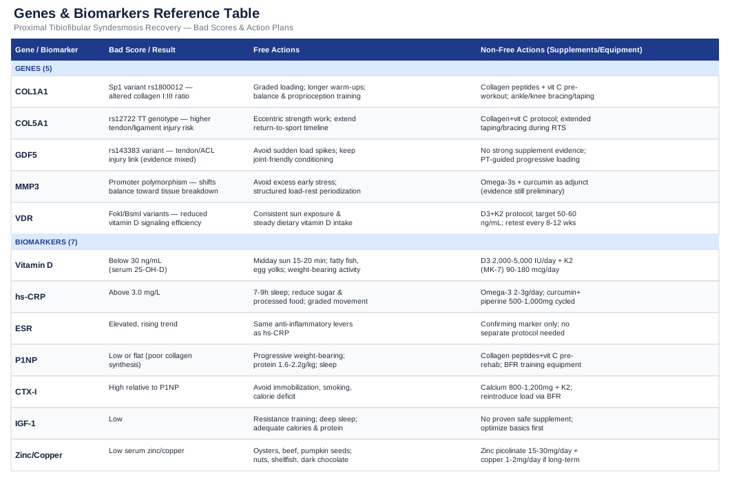
Before the deep dive: this article ranks seven blood biomarkers — inflammatory tone, three separate collagen-building nutrients, a cross-linking mineral, a glycation marker, and a local repair growth factor — that shape how well a stretched or torn proximal tibiofibular ligament rebuilds itself, with a measurement method, a cost range, and a two-track correction plan (lifestyle-only, then supplement- or equipment-assisted) for each one. It then turns to four genes tied to collagen structure and joint development, explains what a "risk" variant actually changes mechanically, and what can realistically be done about it. A separate section distills ten research-backed lessons on loading, collagen timing, and hormone effects from the tendon-ligament research of Dr. Keith Baar, and a final section evaluates which complementary therapies — laser, massage, balance-based biofeedback — have genuine evidence for joint and ligament sprains rather than just marketing claims. If you have been told to "just give it time," what follows is the more specific version of that advice.
7 Biomarkers That Reveal How Well Your Ligaments Can Heal
The proximal tibiofibular joint is stabilized mainly by the anterior and posterior tibiofibular ligaments, reinforced by the biceps femoris tendon and the arcuate ligament complex. When these structures are overstretched — commonly from a twisting knee injury, an inversion ankle sprain that transmits force up the leg, or direct trauma — recovery depends on the same biology that governs every ligament: fibroblasts laying down new type I and type III collagen, cross-linking enzymes stabilizing that collagen into load-bearing fibers, and an inflammatory response that clears debris without dragging on so long that it stalls repair. A systematic review of proximal tibiofibular joint instability and a more recent report describing it as an underrecognized cause of lateral knee pain both note that conservative management outcomes are inconsistent — which is exactly the kind of gap where individual biology, not just brace choice, may explain who recovers well and who does not.
The seven biomarkers below are the ones with the most direct, mechanistic link to that repair process. None of them is specific to the tibiofibular joint on its own — no lab has studied this joint in isolation — but each has documented human evidence for a ligament, tendon, or connective tissue role, which makes it a reasonable and practical proxy.
High-sensitivity C-reactive protein (hs-CRP)
Hs-CRP is the standard blood marker of systemic low-grade inflammation. Acute inflammation right after a sprain is necessary — it recruits the cells that clear damaged tissue and kick off repair — but a persistently elevated hs-CRP weeks after the injury suggests the inflammatory phase is not resolving on schedule, which tends to slow the transition into the remodeling phase where new collagen is actually organized into strong fibers. A recent review specifically reframes CRP as more than an acute-phase bystander in tissue injury and repair, with a direct role in shaping how repair proceeds.
How to measure it
Hs-CRP is a standard blood draw available through any primary care visit or direct-to-consumer lab panel. Cost typically runs from around $10 to $35 in the US when ordered outside insurance, and it is included in most comprehensive longevity or "inflammation" panels (Quest, LabCorp, Function Health, or a physician order). A useful range for a joint injury context is under 1.0 mg/L; between 1 and 3 mg/L is moderate; above 3 mg/L (in the absence of an active infection) suggests meaningful background inflammation worth addressing before your next hard training phase.If the score is bad, the plan without supplements
Prioritize sleep consistency (7 to 9 hours, stable wake time), since short sleep reliably raises inflammatory markers within days. Reduce ultra-processed food and added sugar intake, which independently elevates hs-CRP in cohort studies. Keep total training load and injury-site loading progressive rather than aggressive during the first 6 to 8 weeks post-sprain — a joint that is still inflamed does not benefit from being pushed. Recheck hs-CRP every 4 to 6 weeks; it should trend down as the joint stabilizes.If the score is bad, the plan with supplements or equipment
Omega-3 fatty acids (1.5 to 3 g/day combined EPA/DHA, with food, cycling on for 8 to 12 weeks then reassessing) have the most consistent human evidence for modestly lowering hs-CRP. Curcumin with piperine (500 to 1000 mg/day) is a reasonable second-line option with weaker but positive evidence. Side effects are generally mild — fishy aftertaste or loose stools with omega-3s, occasional GI upset with curcumin — and both should be paused a week or so before any surgical procedure due to a theoretical bleeding-risk interaction. Contrast therapy (alternating hot and cold) or a sauna practice 2 to 3 times weekly can also nudge inflammatory tone down, though the ligament-specific evidence for this is indirect.25-hydroxyvitamin D
Vitamin D receptors are present on tenocytes and fibroblasts, and vitamin D status has been linked to how well tendons remodel extracellular matrix, control local inflammation, and proliferate new repair cells. A 2025 scoping review of vitamin D and postoperative tendon healing found consistent epidemiological association between deficiency and poorer healing outcomes, while flagging that trial quality in this specific area is still limited — an honest caveat rather than a reason to ignore it.
How to measure it
A standard 25-OH-D blood test costs roughly $20 to $50 out of pocket, or is often bundled into annual physicals. Aim for 30 to 50 ng/mL; below 20 ng/mL is classified as deficient and is the range most associated with musculoskeletal complications.If the score is bad, the plan without supplements
Ten to twenty minutes of midday sun exposure on bare arms and legs several times a week, weather and skin tone permitting, meaningfully raises vitamin D synthesis over 6 to 8 weeks. Fatty fish (salmon, mackerel, sardines) two to three times weekly adds a modest dietary contribution.If the score is bad, the plan with supplements or equipment
Vitamin D3, 2000 to 4000 IU daily with a meal containing fat for absorption, is the typical correction dose for a moderate deficiency; more severe deficiency (under 12 ng/mL) is often dosed higher short-term under physician supervision. Retest at 8 to 12 weeks rather than continuing indefinitely at a high dose, since fat-soluble vitamin D can accumulate; true toxicity is rare but has been reported above sustained daily doses in the tens of thousands of IU. Pairing with vitamin K2 (90 to 120 mcg/day) is a common practice to support calcium handling, though this is more relevant to bone than ligament specifically.Plasma vitamin C (ascorbate)
Vitamin C is a required cofactor for the prolyl and lysyl hydroxylase enzymes that stabilize the collagen triple helix — without adequate vitamin C, newly synthesized collagen is structurally weaker. This is not theoretical: a well-known human trial found that vitamin C-enriched gelatin taken before intermittent exercise roughly doubled a blood marker of collagen synthesis compared with placebo, directly demonstrating the vitamin C-collagen link in living tissue.
How to measure it
Plasma vitamin C is a less commonly ordered test (roughly $40 to $80 through specialty or functional-medicine labs) and is not part of most routine panels — you may need to specifically request it. Because levels swing quickly with recent intake, it is a rough proxy at best; a nutrition history (do you regularly eat vitamin-C-rich produce) is almost as informative.If the score is bad, the plan without supplements
Two to three daily servings of citrus, bell peppers, kiwi, or broccoli reliably cover baseline requirements (roughly 75 to 90 mg/day) for most adults. Smokers and people under high physical stress have measurably higher vitamin C turnover and need more from diet or supplementation.If the score is bad, the plan with supplements or equipment
For active collagen loading around rehab sessions, the protocol used in the human trial above is 15 g of gelatin or hydrolyzed collagen with roughly 50 to 250 mg of vitamin C, taken 30 to 60 minutes before a rehab or loading session, on training days only (not a daily standing supplement). Side effects are minimal at these doses; above 2000 mg/day of vitamin C can cause GI upset or, in susceptible individuals, contribute to kidney stone risk, so there is no benefit to exceeding the studied range.Serum copper and ceruloplasmin
Copper is the required cofactor for lysyl oxidase, the enzyme that cross-links collagen and elastin fibers into a mechanically strong matrix — without adequate copper, collagen is produced but stays structurally loose. Classic work established that copper is directly required for elastin and collagen synthesis, and ceruloplasmin, the copper-carrying blood protein, is the standard clinical proxy for copper status, as detailed in the NCBI biochemistry review of ceruloplasmin.
How to measure it
Serum ceruloplasmin runs about $30 to $60; serum copper is often ordered alongside it for around $25 to $50. Normal ceruloplasmin is roughly 20 to 40 mg/dL. Note that inflammation and oral contraceptives raise ceruloplasmin independent of true copper status, which can mask a real deficiency — pair with hs-CRP for context.If the score is bad, the plan without supplements
Shellfish (especially oysters), organ meats, nuts, seeds, and dark chocolate are the richest dietary copper sources; most people who are not restricting these foods rarely become deficient from diet alone.If the score is bad, the plan with supplements or equipment
True dietary copper deficiency is uncommon outside of malabsorption conditions, bariatric surgery, or excessive zinc supplementation (zinc competes with copper for absorption). If confirmed low, 1 to 2 mg/day of copper for 8 to 12 weeks, then retest, is a standard correction; sustained high-dose copper supplementation is not advisable without medical supervision since chronic excess copper is hepatotoxic. If you are also taking zinc for wound healing, keep the ratio around 8 to 15 parts zinc to 1 part copper to avoid inducing a deficiency.Homocysteine
Elevated homocysteine does more than raise cardiovascular risk — it actively interferes with collagen cross-linking. Research shows homocysteine suppresses expression of lysyl oxidase, the same cross-linking enzyme that depends on copper, through an inflammatory and epigenetic pathway — meaning a high homocysteine level can undercut ligament repair through a second, independent mechanism.
How to measure it
A homocysteine blood test costs roughly $30 to $70 and is widely available. A healthy target is under 9 to 10 micromol/L; above 15 is considered elevated and worth actively correcting.If the score is bad, the plan without supplements
Homocysteine is primarily driven by B-vitamin status (B6, B9/folate, B12), so increasing leafy greens, legumes, eggs, and animal protein covers most mild elevations. Reducing alcohol intake also helps, since alcohol interferes with folate metabolism.If the score is bad, the plan with supplements or equipment
A B-complex containing methylfolate (400 to 800 mcg), B12 (500 to 1000 mcg, methylcobalamin form if you carry an MTHFR variant), and B6 (25 to 50 mg) daily for 8 to 12 weeks typically normalizes mild-to-moderate elevations; retest after that window rather than supplementing indefinitely at high doses, since long-term high-dose B6 (above 100 mg/day for extended periods) has been associated with peripheral neuropathy in rare cases.HbA1c and fasting glucose
Chronically elevated blood sugar accelerates the formation of advanced glycation end-products (AGEs), which create abnormal, non-enzymatic cross-links in collagen. Direct tissue work shows AGEs measurably reduce tendon collagen fiber sliding, stiffening tissue in ways that impair normal, elastic ligament function rather than strengthening it usefully.
How to measure it
HbA1c is a standard $15 to $40 blood test, often included in annual labs. Fasting glucose is usually run alongside it for a similar cost. Target HbA1c under 5.6 percent; 5.7 to 6.4 percent is prediabetic range and is where AGE-related tissue effects start becoming relevant even without a diabetes diagnosis.If the score is bad, the plan without supplements
Walking 10 to 15 minutes after meals, prioritizing protein and fiber earlier in meals, and reducing refined carbohydrate and added sugar intake are the highest-leverage, no-cost interventions, typically improving HbA1c measurably within 3 months.If the score is bad, the plan with supplements or equipment
A continuous glucose monitor (roughly $50 to $100 per month) can identify which specific foods spike your glucose, which is often more actionable than a single HbA1c number. Berberine (500 mg, two to three times daily with meals, cycled 8 to 12 weeks on and a few weeks off) has reasonable evidence for improving glycemic markers; it can cause GI upset and should be discussed with a physician if you take other glucose-lowering medication, due to additive effect.IGF-1 (insulin-like growth factor 1)
IGF-1 is produced both systemically by the liver and locally within connective tissue, where it supports fibroblast proliferation and collagen synthesis during repair. A recent review describes IGF-1's role in empowering tendon regenerative therapies, while also noting that clinical trial results have been mixed — a fair example of promising mechanism outpacing proven clinical benefit so far.
How to measure it
Serum IGF-1 costs roughly $60 to $120 and is not part of routine panels; it is most useful if you suspect a broader growth-hormone-axis issue (poor sleep, low protein intake, aging) rather than as a routine post-sprain check. Reference ranges are age- and sex-specific, so interpret relative to your lab's stated range.If the score is bad, the plan without supplements
Adequate total protein intake (roughly 1.6 to 2.2 g/kg body weight per day for someone actively rehabbing), resistance training, and deep sleep are the three biggest levers for healthy systemic IGF-1, all without any supplement cost.If the score is bad, the plan with supplements or equipment
There is no well-supported, safe supplement that meaningfully and reliably raises IGF-1 for ligament repair specifically; approaches marketed this way (colostrum, deer antler velvet) have thin human evidence. The more defensible route is addressing the upstream drivers — sleep, protein, resistance training — and, for a confirmed clinical deficiency unrelated to the sprain itself, referral to an endocrinologist rather than self-supplementation.4 Genes Linked to Ligament Strength and Injury Risk
Blood biomarkers are more actionable, but genetics explains part of why two people load-match on paper yet respond to the same sprain differently. This is the kind of terrain that geneticist Ali Torkamani and biohacking communicator Gary Brecka both point to when they talk about testing collagen and connective-tissue genes before assuming an injury is simply "bad luck." The evidence here is real but generally epidemiological — association studies in athletic cohorts, not proof that a single variant determines your outcome — so treat it as a modifier of risk, not a diagnosis.
COL5A1
COL5A1 encodes a component of type V collagen, which regulates the diameter and organization of the type I collagen fibrils that make up ligaments and tendons. A meta-analysis of COL5A1 polymorphisms and tendon-ligament injury risk found the rs12722 "T" variant associated with increased susceptibility to soft tissue injury, plausibly because it shifts the type V to type I collagen ratio toward thinner, less tensile-strength fibrils.
If the gene is bad, the plan without supplements
Since this variant is associated with more compliant (less stiff) connective tissue, emphasize slow eccentric and isometric loading progressions during rehab rather than rushing back to high-speed cutting or pivoting activity, and extend your return-to-play timeline modestly beyond generic protocols.If the score is bad, the plan with supplements or equipment
Pair collagen-supportive nutrition (the vitamin C and copper strategies above) with a stability brace or kinesiology taping during early return to activity, and consider a proprioceptive balance-board program (see the complementary section below) for at least 8 to 12 weeks, since neuromuscular control can partly offset inherently more compliant ligament tissue.COL1A1
COL1A1 encodes the primary chain of type I collagen, the dominant structural protein in ligaments, tendons, and bone. Variants in this gene are more classically tied to bone density and rupture risk in tendon injury cohorts, and it is frequently discussed alongside COL5A1 in the same review of the genetics of sports injuries and athletic performance, which summarizes multiple collagen-gene associations with soft tissue injury susceptibility.
If the gene is bad, the plan without supplements
Build a longer, more gradual strength progression before reintroducing high-impact or high-torque knee movements, and prioritize consistent, moderate loading over sporadic high-intensity sessions, which appears to matter more for collagen remodeling quality than raw training volume.If the score is bad, the plan with supplements or equipment
The gelatin-plus-vitamin-C pre-loading protocol described earlier is specifically designed around this collagen-synthesis pathway; combining it with a structured resistance program (2 to 3 sessions weekly) gives the clearest, most studied combination for anyone with a collagen-related risk variant.GDF5
GDF5 (growth and differentiation factor 5) is essential for normal joint and intra-articular ligament development; mice lacking the gene show failed formation of intra-articular ligaments. In humans, a well-replicated regulatory polymorphism in GDF5 is associated with joint remodeling capacity and osteoarthritis risk, suggesting the same gene that shapes joint development also influences how well a joint repairs and remodels after injury later in life.
If the gene is bad, the plan without supplements
Since GDF5-linked risk is more about long-term joint remodeling capacity than acute ligament strength, focus on maintaining full, pain-free range of motion and consistent low-impact conditioning (cycling, swimming) between rehab phases to protect the joint surfaces while the ligament heals.If the score is bad, the plan with supplements or equipment
There is no supplement that meaningfully alters GDF5 expression in humans with current evidence. The more realistic accommodation is longer-term joint monitoring — periodic imaging or orthopedic follow-up if clicking, catching, or swelling persist beyond a normal healing window — since this variant is more about cumulative joint wear than a single fixable deficiency.TNC (Tenascin-C)
Tenascin-C is an extracellular matrix protein expressed at tendon and ligament attachment sites during mechanical loading and repair; certain TNC variants have been associated in athletic cohorts with altered soft tissue injury risk, discussed within the same sports genetics review referenced above. Evidence here is earlier-stage than for the collagen genes, so treat any single TNC result cautiously.
If the gene is bad, the plan without supplements
Because tenascin-C responds to mechanical loading itself, a structured, progressive loading program — rather than prolonged immobilization — is the most defensible way to support whatever tenascin-C-mediated repair signaling your tissue is doing naturally.If the score is bad, the plan with supplements or equipment
No supplement currently has credible human evidence for modifying tenascin-C-related injury risk; the practical response is the same graded-loading approach used above, potentially guided by a physical therapist experienced in ligament rehabilitation rather than a generic timeline.10 Lessons From Keith Baar's Research on Loading, Collagen, and Ligament Repair
Much of the most useful, clinically translatable research on tendon and ligament repair over the last decade comes from Dr. Keith Baar's lab at UC Davis, summarized at length in a wide-ranging conversation on The Tim Ferriss Show. Baar's work is a useful counterweight to older "rest and ice" thinking, and several of his findings challenge default rehab assumptions directly.
1. Ligament and tendon tissue remodels far slower than muscle
Muscle protein turns over in days; collagen-based tissue turns over over months to years. This is the core reason ligament rehab timelines feel painfully slow compared with muscle recovery — it is not that you are healing "wrong," it is that the tissue biology is inherently slower.2. Short, specific loading bouts stimulate more collagen synthesis than continuous loading
Baar's research found that tenocytes become refractory to further mechanical stimulus after roughly 10 minutes of load, meaning longer sessions do not proportionally add benefit and may simply add fatigue and injury risk without added repair signal.3. The "load, then rest 6 to 8 hours, then load again" pattern roughly doubles collagen synthesis
Spacing two shorter loading bouts across a day, rather than one long session, appears to more effectively stimulate collagen production — a practical, free adjustment to rehab scheduling that most standard physical therapy plans do not use.4. Vitamin C-enriched collagen before loading nearly doubles a marker of collagen synthesis
This is the same human trial cited in the biomarker section above — 15 g of gelatin or hydrolyzed collagen with vitamin C, taken 30 to 60 minutes before the loading session, timed specifically around the workout rather than as a general daily supplement.5. Isometric holds may prime the ligament-tendon interface better than dynamic movement alone
Sustained, moderate-intensity isometric contractions appear to load connective tissue in a way that prepares it for higher-speed activity, which is why many modern ligament and tendon rehab protocols now front-load isometric work before reintroducing dynamic loading.6. Reflexive anti-inflammatory use right after injury may blunt the repair signal
Baar's "anti-RICE" argument is that the inflammatory phase is a genuine repair signal, and routinely suppressing it with NSAIDs in the first 48 to 72 hours may reduce the very stimulus needed for collagen remodeling — a genuinely contrarian point relative to reflexive over-the-counter pain management, worth discussing with a physician rather than adopting blindly, especially if pain is severe.7. Local IGF-1 exposure matters more for tendon-ligament repair than systemic growth hormone
This reinforces why chasing higher growth hormone through supplements is largely beside the point for ligament healing — what matters is the tissue's local exposure to IGF-1 during and after loading, which is influenced far more by mechanical stimulus and protein availability than by circulating hormone levels.8. Estrogen fluctuations measurably change ligament stiffness and injury risk
A systematic review of menstrual cycle phase and ACL injury risk surrogates found moderate evidence for increased injury risk around ovulation, when estrogen peaks and appears to reduce fibroblast collagen synthesis — a genuinely underused piece of context for timing rehab loading intensity in naturally cycling women.9. Heat before loading changes tendon stiffness gains measurably
Warming tissue before a loading session appears to improve the mechanical adaptation response compared with loading cold tissue, supporting a genuine physiological rationale for warm-ups beyond generic injury-prevention folklore.10. Genetics sets the range, but the loading pattern decides where you land in it
Even with a less favorable collagen genotype, the training and nutritional pattern used during rehab has a large, well-documented influence on the outcome — which is the central, practical reason this article treats genetics as context rather than destiny.Complementary Approaches Worth Considering
A small number of complementary modalities have genuine, if sometimes mixed, human evidence relevant to ligament sprain recovery. This is not an exhaustive wellness list — it is a short, deliberately filtered set, leaving out anything without a real evidence trail for connective tissue or joint sprain recovery.
Low-level laser therapy (photobiomodulation)
Photobiomodulation is proposed to accelerate soft tissue repair by stimulating mitochondrial activity in fibroblasts, potentially speeding collagen production during the early healing phase after a ligament sprain. For a joint like the proximal tibiofibular joint, where the injured ligaments sit close to the skin surface, this is at least a plausible mechanism worth understanding rather than dismissing outright.
The evidence, however, is genuinely mixed rather than clearly positive. A large, well-designed randomized clinical trial of low-level laser therapy in ankle sprains found no benefit over placebo across 217 patients, with the placebo group actually reporting better function at some follow-up points — a result that should temper expectations considerably. More recent, smaller trials combining laser with active functional exercise have shown more promising results, suggesting any benefit may depend heavily on pairing it with active rehab rather than using it alone.
Realistically, if you try photobiomodulation for a tibiofibular ligament sprain, treat it as a possible adjunct to active rehab, not a substitute for it, and do not expect it to meaningfully change your timeline on its own based on the strongest available trial evidence.
Biofeedback-based balance and proprioceptive training
Proprioceptive training uses real-time balance feedback — typically a wobble or balance board — to retrain the neuromuscular control that keeps a joint stable under load, which is directly relevant after any ligament sprain since the mechanoreceptors within the ligament itself are damaged alongside the fibers.
The evidence here is unusually strong for a complementary approach. A large randomized controlled trial of home-based proprioceptive training after ankle sprain found the recurrence rate dropped from 33 percent in controls to 22 percent in the training group over one year, and a separate controlled trial of balance-board training in athletes found significantly fewer sprains in the intervention group. While these trials focused on the ankle rather than the proximal tibiofibular joint specifically, the underlying neuromuscular mechanism — retraining joint position sense after ligament damage — applies directly.
A realistic protocol is 10 to 15 minutes of single-leg balance or wobble-board work, 4 to 5 times weekly, beginning once acute pain and swelling have settled, and continuing for at least 8 to 12 weeks even after the joint feels stable, since recurrence prevention is the main benefit shown in these trials.
Massage therapy
Massage is commonly used during ligament and soft tissue rehab to reduce local stiffness, support circulation, and manage compensatory muscle tension around an injured joint, particularly in the muscles crossing the knee and proximal fibula that often tighten protectively after a sprain.
Evidence for massage in soft tissue recovery is moderate and somewhat inconsistent depending on what outcome is measured. A review of sports massage for recovery of skeletal muscle from strenuous exercise found it more reliably improves perceived soreness and short-term range of motion than objective measures of tissue repair speed — a useful, honest distinction between feeling better and healing faster.
For a proximal tibiofibular ligament sprain, massage focused on the surrounding musculature (biceps femoris, peroneals, lateral gastrocnemius) once acute swelling has resolved, at a frequency of once or twice weekly for several weeks, is a reasonable comfort- and mobility-focused addition alongside — not instead of — the active loading and proprioceptive work described above.
Conclusion
A proximal tibiofibular ligament sprain heals according to the same underlying biology as any connective tissue injury, but how well and how fast depends on details a standard rehab handout never asks about: your inflammatory tone, your collagen-building nutrient status, your blood sugar control, and, to a smaller but real degree, the collagen and joint-development genes you carry. Tracking hs-CRP, vitamin D, vitamin C, copper and ceruloplasmin, homocysteine, HbA1c, and IGF-1 gives you a concrete, correctable picture of your repair capacity rather than a guess, and understanding your COL5A1, COL1A1, GDF5, and TNC status adds useful context for how aggressive or conservative your return-to-activity timeline should be.
None of this replaces a proper orthopedic evaluation if instability, locking, or persistent lateral knee pain continues. The next practical step is a simple one: ask your physician for the panel above at your next visit, log how your symptoms track against your training load for a few weeks, and use both to have a more specific conversation about your rehab plan than "wait and see."
Musculoskeletal: Joint Conditions Sports Injuries