This article contains AI generated content.
Proximal Tibiofibular Syndesmosis Injury: 5 Genes And 7 Biomarkers To Track
Introduction
If you're dealing with a proximal tibiofibular syndesmosis injury, you already know the frustrating part isn't the diagnosis — it's what comes after. You've likely been told to rest it, brace it, do some physio, and "give it time." That advice isn't wrong, but it's not really answering the question you actually have: why does this joint still feel loose, or still ache, when someone else with a similar sprain was back to normal in half the time?
Generic recovery advice treats every ligament injury as roughly the same problem with the same timeline. In reality, the proximal tibiofibular joint is a small, often overlooked structure, and how well it heals depends on things a standard six-week physio plan doesn't measure — how efficiently your body is laying down new collagen, how well-controlled your inflammatory response is, whether you have the raw nutrient cofactors on hand to rebuild tissue, and to some extent, the connective tissue tendencies you were born with.
This article goes past the generic version. Instead of another list of stretches, it looks at the measurable biology behind ligament repair: blood-based biomarkers that reflect what's actually happening in your tissue right now, and the genetic tendencies that shape your baseline connective tissue resilience. Both are covered in enough depth to actually act on, not just read about.
None of this is about miracle fixes — a syndesmosis injury still needs time, load management, and often professional guidance. But better information changes the decisions you make along the way: what to test, what to ask your physio or doctor about, and where the leverage actually is. This article walks through the biomarkers worth tracking, what your genes may be telling you, a science-based look at pain and healing from one of today's most-cited health podcasts, and a short review of complementary approaches with real evidence behind them for this kind of injury.
Summary
Here's the short version of what's ahead. Seven blood biomarkers — from vitamin D to collagen turnover markers like P1NP and CTX-I — can tell you whether your body is actually in an active repair state or quietly stuck in a catabolic holding pattern, and each one comes with a practical plan, with and without supplements, to move it in the right direction. Underneath that, five genes (COL1A1, COL5A1, GDF5, MMP3, and VDR) help explain why some people are more prone to syndesmosis and other ligament injuries in the first place, and why the same rehab program doesn't work identically for everyone. Beyond the lab work, a closer look at the neuroscience of pain and healing — drawn from one of Andrew Huberman's most detailed episodes — challenges a few habits many people default to without thinking, like reaching for ice or anti-inflammatories on day one. And for the complementary side, a short, evidence-checked review covers massage therapy, photobiomodulation, biofeedback, and tai chi, all of which have real, condition-relevant human research behind them. Taken together, these sections aren't meant to replace your care team — they're meant to make the conversation with them a lot more specific.
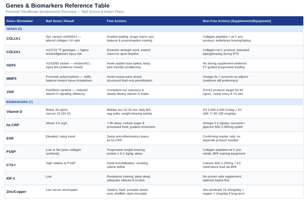
The 7 Biomarkers Worth Tracking After a Proximal Tibiofibular Syndesmosis Injury
The proximal tibiofibular joint relies on a fairly small, densely packed set of ligaments to keep the fibular head stable against the tibia during every step, pivot, and load transfer through the lower leg. When that syndesmosis is sprained or partially torn, healing isn't just a matter of "time passing" — it's a biological process of clearing damaged tissue, laying down new collagen, and remodeling it into something with real tensile strength. The biomarkers below are the ones with the clearest, most actionable link to that process. None of them are specific to this one joint — no lab test is — but together they give a much more precise picture of your healing environment than symptoms alone.
Vitamin D (25-hydroxyvitamin D)
Vitamin D does more for connective tissue than most people realize. Beyond bone, it acts through the vitamin D receptor in ligament and tendon tissue, and low levels have been associated with a higher rate of ligament and soft-tissue injuries, along with slower functional recovery after reconstruction surgery. A systematic review on vitamin D and ACL injury rates found lower vitamin D status linked to both higher injury incidence and worse post-reconstruction outcomes, and a separate study of young patients with lower-extremity musculoskeletal complaints found ligament and cartilage injuries were associated with the lowest vitamin D levels of any complaint category (systematic review on vitamin D and ACL injury, vitamin D status in young patients with lower-extremity complaints).
How to measure it
A basic serum 25(OH)D blood draw, typically $30–$50 out of pocket (often covered if ordered by a physician). Conventional labs flag sufficiency above 30 ng/mL; many sports-medicine and longevity-focused clinicians aim for 40–60 ng/mL during active tissue repair.If the score is bad, the plan without supplements
Get 15–20 minutes of midday sun exposure on bare skin several times a week (adjusted for skin tone and latitude), eat fatty fish, egg yolks, and fortified dairy regularly, and keep doing weight-bearing activity within your rehab limits, since mechanical loading interacts with vitamin D signaling in bone and connective tissue.If the score is bad, the plan with supplements or equipment
Vitamin D3, 2,000–5,000 IU daily with a meal containing fat, paired with vitamin K2 (MK-7, 90–180 mcg) to support proper calcium utilization. Retest in 8–12 weeks, then again at 6 months; there's no need to cycle it off entirely, but dosing should be adjusted based on retesting rather than left on autopilot. Avoid doses above 10,000 IU/day without physician supervision — sustained megadosing risks hypercalcemia. A UVB sunlamp (roughly $100–150) is a reasonable equipment option for those in low-sunlight climates.High-sensitivity C-reactive protein (hs-CRP)
CRP rises within hours of tissue injury and is one of the most sensitive general markers of inflammation in circulation, capable of increasing up to 1,000-fold in acute injury or infection (CRP as a prognostic inflammatory indicator). In the early days after a syndesmosis sprain, some elevation is expected and even necessary — it reflects immune cells arriving to clear damaged tissue. What matters more for tracking recovery is the trend: hs-CRP that stays elevated well past the acute phase can suggest the inflammatory process hasn't resolved, which tends to correlate with slower, less complete healing.
How to measure it
A standard hs-CRP blood test, usually $15–$30, often bundled into broader metabolic panels. Peter Attia and other longevity-focused physicians commonly use hs-CRP under 1.0 mg/L as an optimal target, with 1–3 mg/L considered average and above 3 mg/L flagged for follow-up.If the score is bad, the plan without supplements
Prioritize 7–9 hours of sleep, reduce ultra-processed food and added sugar, eat fatty fish 2–3 times weekly, and resume graded movement rather than prolonged full immobilization — disuse itself can prolong low-grade inflammation in and around the joint.If the score is bad, the plan with supplements or equipment
Omega-3 fish oil, 2–3 g combined EPA/DHA daily for 8–12 weeks, then reassess hs-CRP; watch for mild GI upset and use caution if you're on blood thinners. Curcumin with piperine, 500–1,000 mg daily, cycled 8 weeks on and 2 weeks off, can modestly support inflammation resolution but may cause GI discomfort and should be avoided with a history of gallstones.Erythrocyte sedimentation rate (ESR)
ESR moves more slowly than CRP, which actually makes it useful as a confirming, longer-arc marker of inflammation over weeks rather than hours. Used alongside CRP, it helps distinguish a genuinely resolving inflammatory picture from one that's fluctuating or stuck. ESR studied alongside CRP in soft-tissue and inflammatory conditions consistently shows the two moving together, but on different timescales, which is exactly why pairing them is more informative than either alone.
How to measure it
A simple blood draw, usually $10–$20, frequently ordered together with hs-CRP for a combined cost under $40.If the score is bad, the plan without supplements
Same anti-inflammatory lifestyle levers as hs-CRP — sleep, whole-food diet, gradual reloading. ESR isn't really a separate target to chase; treat it as a second data point confirming (or questioning) what hs-CRP is telling you.P1NP (procollagen type I N-terminal propeptide)
P1NP is released when osteoblasts and fibroblasts are actively synthesizing new type I collagen — the dominant structural protein in ligaments. A rising or adequate P1NP level is a reasonably direct signal that your body is in an active building phase, not just idling (reference ranges for P1NP and CTX bone turnover markers).
How to measure it
A specialty blood test, typically $50–$100, more often ordered through sports-medicine or endocrinology labs than a standard primary care panel.If the score is bad, the plan without supplements
Progressive, controlled weight-bearing is the strongest known stimulus for new collagen synthesis — this is where working closely with a physical therapist on graded loading matters more than almost anything else on this list. Pair it with 1.6–2.2 g/kg/day of protein and consistent sleep, since growth hormone pulses during deep sleep support tissue synthesis.If the score is bad, the plan with supplements or equipment
15 g of hydrolyzed collagen peptides with roughly 50 mg vitamin C, taken 30–60 minutes before rehab exercises, is a protocol drawn from tendon-collagen synthesis research and reasonable to cycle through the active rehab phase (8–12 weeks). Blood flow restriction (BFR) training equipment, used under professional supervision, can allow effective loading at lower joint stress — a relevant option early in syndesmosis rehab when full-range loading isn't yet appropriate.CTX-I (C-terminal telopeptide of type I collagen)
CTX-I is the mirror image of P1NP — it reflects collagen breakdown rather than formation. Looking at CTX-I relative to P1NP gives a rough sense of whether your tissue is in net building mode or net breakdown mode, which is far more informative than either number alone (same reference-range study covering both markers).
How to measure it
Blood or urine test, roughly $50–$90, usually ordered alongside P1NP for direct comparison.If the score is bad, the plan without supplements
Avoid prolonged full immobilization — disuse increases resorption markers — and avoid smoking and heavy alcohol use, both of which are independently linked to poorer collagen remodeling. Make sure you're not in a significant calorie deficit during active healing, since undereating pushes the body toward net tissue breakdown.If the score is bad, the plan with supplements or equipment
800–1,200 mg/day of calcium, ideally from diet, plus vitamin K2 (MK-7, 90–180 mcg/day) to direct calcium appropriately. Reintroducing weight-bearing load, potentially via BFR equipment if full loading isn't tolerated yet, is the main lever. Avoid high-dose calcium supplementation if you have a history of kidney stones or hypercalcemia.IGF-1 (insulin-like growth factor 1)
IGF-1 is active almost immediately after ligament injury and stays elevated through much of the healing timeline, driving fibroblast proliferation and new collagen matrix synthesis (IGF-1's role in tendon regenerative therapies). It's one of the more direct biological links between "recovery basics" like sleep and training, and actual tissue repair.
How to measure it
Serum IGF-1 test, roughly $80–$120, typically ordered through an endocrinology or sports-performance lab.If the score is bad, the plan without supplements
Resistance training (as tolerated within rehab limits), consistent deep sleep, adequate total calories, and sufficient protein are the primary natural drivers of IGF-1. Chronic under-eating or overtraining will suppress it.If the score is bad, the plan with supplements or equipment
There isn't a well-evidenced, safe over-the-counter supplement that reliably raises IGF-1 for ligament repair. Growth-hormone secretagogues and peptides like BPC-157 are sometimes discussed online for this purpose, but they're unregulated for this use, human safety data is limited, and they should not be self-administered without direct physician oversight. The higher-value move is optimizing sleep, training, and nutrition first — that's where the actual evidence sits.Zinc and copper status
Zinc and copper are unglamorous but essential — both are cofactors for enzymes directly involved in collagen synthesis and cross-linking, including lysyl oxidase, which stabilizes new collagen fibers. Deficiency in either has been shown to impair wound and connective tissue healing (factors affecting wound healing, including zinc and copper, tissue zinc, copper, and magnesium across wound healing phases).
How to measure it
Serum zinc, roughly $30–$50; serum copper or ceruloplasmin, roughly $40–$60. Neither is part of a standard panel by default, so they usually need to be requested specifically.If the score is bad, the plan without supplements
Zinc-rich foods include oysters, beef, and pumpkin seeds; copper-rich foods include nuts, seeds, shellfish, and dark chocolate. For most people eating a varied diet, this alone closes small gaps.If the score is bad, the plan with supplements or equipment
Zinc picolinate, 15–30 mg/day for 8–12 weeks, taken with food to avoid nausea. Because sustained zinc supplementation can deplete copper, pair it with 1–2 mg/day of copper if using zinc beyond a few months, and retest both every 8–12 weeks rather than supplementing indefinitely without checking levels.Read together, these seven markers tell a more complete story than any single one: whether inflammation is resolving on schedule, whether collagen is actually being rebuilt faster than it's breaking down, and whether the nutrient cofactors needed for that rebuilding are actually available. That's a very different, more actionable picture than "how does it feel today."
What Your Genes May Be Telling You About Ligament Strength and Repair
Biomarkers show what's happening right now; genetics offer context for why your baseline connective tissue behaves the way it does — and why two people with an identical syndesmosis sprain can have meaningfully different injury risk and recovery trajectories. A useful way to think about this, one popularized by researchers like Ali Torkamani and health data communicators like Gary Brecka, is that a gene variant isn't a verdict — it's a lever you now know exists, one you can compensate for with training, nutrition, or monitoring even if you can't change the underlying DNA. A broader narrative review on tendon and ligament genetics lays out this same framing across the field (tendon and ligament genetics: how do they contribute to disease and injury).
COL1A1 — the main structural collagen of ligaments
COL1A1 encodes the alpha-1 chain of type I collagen, the dominant structural protein in ligaments, tendons, and bone (COL1A1 gene reference, NCBI). A well-studied variant in the gene's regulatory region (the Sp1 binding site, rs1800012) is associated with an altered ratio of collagen I to collagen III in connective tissue, which affects overall tissue stiffness versus laxity.
If the gene is bad, the plan without supplements
Favor graded, progressive loading over abrupt increases in training volume or intensity, and build in longer, more thorough warm-ups before activities that stress the lower leg and ankle complex. Proprioceptive and balance training helps compensate for tissue laxity by improving neuromuscular control around the joint.If the score is bad, the plan with supplements or equipment
Collagen peptides (15 g) with vitamin C, timed before loading sessions, cycled through active rehab and return-to-sport phases. An ankle-knee bracing or taping system during higher-risk activities (cutting, pivoting sports) adds a mechanical safety margin while the tissue adapts.COL5A1 — fibril assembly and soft-tissue injury risk
COL5A1 regulates collagen V, which nucleates the assembly of collagen fibrils and influences their diameter and strength (COL5A1 gene reference, NCBI). The rs12722 TT genotype has been associated with a meaningfully higher risk of tendon and ligament injury in Caucasian populations across multiple meta-analyses, while certain protective variants are overrepresented in elite athletes with lower soft-tissue injury rates (meta-analysis of COL5A1 polymorphisms and tendon-ligament injury risk).
If the gene is bad, the plan without supplements
Emphasize eccentric strengthening for the muscles crossing the knee and ankle, extend return-to-sport timelines rather than rushing back at the first sign of stability, and prioritize gradual, structured loading progressions over "testing it" impulsively.If the score is bad, the plan with supplements or equipment
Same collagen-and-vitamin-C protocol as above, plus longer use of supportive taping or bracing during the transition back to unrestricted activity, since this variant is specifically linked to soft-tissue injury recurrence.GDF5 — joint and ligament development signaling
GDF5 (growth differentiation factor 5) plays a role in cartilage, tendon, and ligament development. A variant in the gene's 5' untranslated region (rs143383) has been associated with Achilles tendon pathology and studied for its relationship to ACL injury risk, though findings for ligament injury specifically remain mixed and need larger studies to confirm (GDF5 gene and anterior cruciate ligament rupture).
If the gene is bad, the plan without supplements
Avoid sudden spikes in high-impact training volume, and keep joint-friendly conditioning (cycling, swimming, controlled strength work) as a larger share of your training base while the syndesmosis heals.If the score is bad, the plan with supplements or equipment
There's no strong supplement evidence specific to GDF5 variants — the higher-value approach here is a physiotherapy-guided, carefully progressed loading program with regular reassessment, rather than reaching for a product.MMP3 — the remodeling and breakdown enzyme
MMP3 (matrix metalloproteinase 3) breaks down and remodels collagen and proteoglycans in the extracellular matrix. Promoter polymorphisms affecting MMP3 expression can shift the balance between healthy remodeling and excess tissue breakdown, which matters directly during the weeks a syndesmosis sprain is rebuilding.
If the gene is bad, the plan without supplements
Avoid excessive mechanical stress during the acute and early sub-acute phases, and follow a structured load-rest periodization rather than pushing through discomfort daily.If the score is bad, the plan with supplements or equipment
Omega-3s and curcumin (same dosing as in the biomarker section) are sometimes used to modulate excess matrix metalloproteinase activity, though the evidence here is still preliminary; treat this as a reasonable adjunct, cycled 8–12 weeks, not a primary strategy.VDR — how efficiently your tissue uses vitamin D
VDR (vitamin D receptor) polymorphisms, such as FokI and BsmI variants, affect how efficiently vitamin D signaling functions at the tissue level, including bone-ligament entheses (VDR gene reference, NCBI). This directly connects back to the vitamin D biomarker above — a less efficient VDR variant means the same blood level of vitamin D may produce a weaker tissue effect.
If the gene is bad, the plan without supplements
Maintain especially consistent sun exposure and dietary vitamin D intake rather than relying on occasional high doses.If the score is bad, the plan with supplements or equipment
Some clinicians target the higher end of the sufficiency range (50–60 ng/mL) for people with less efficient VDR variants, using the same D3 plus K2 protocol described earlier, with more frequent retesting (every 8–12 weeks initially) to confirm the higher dose is actually translating into better serum levels.Genetic testing for these variants — through consumer raw-data services paired with third-party interpretation tools, or a clinical genetic panel — typically runs $100–$200. None of this is deterministic; it's context that helps you and your physical therapist decide where to be more conservative and where extra monitoring is worth the effort.
What The Neuroscience Of Pain And Healing Adds To The Picture
Biomarkers and genetics explain the biology of tissue repair, but how you manage the injury day to day — what you reach for when it hurts, how you sleep, whether you move or stay still — also shapes the outcome. One of the more detailed, evidence-grounded explorations of this comes from neuroscientist Andrew Huberman's episode "Essentials: Control Pain & Heal Faster With Your Brain" (Huberman Lab: Control Pain & Heal Faster With Your Brain), which challenges a few habits many people default to automatically. Here are the ten most useful points from it.
1. Pain is a belief system, not just a raw signal
Pain perception is shaped by context, emotion, and expectation as much as by tissue damage itself. That doesn't mean the injury isn't real — it means your mental framing of the injury (catastrophizing versus calm problem-solving) measurably influences how much pain you experience and, in some cases, how quickly you recover function.2. Ice numbs pain but can slow the cleanup process
Ice reduces pain largely through numbing and vasoconstriction, but that same vasoconstriction can cause sludging in blood and lymphatic flow at the injury site — which slows the arrival of the immune cells needed to clear damaged tissue and begin repair.3. Inflammation in the first days is doing necessary work
The inflammatory response recruits immune cells that clean up damaged tissue and kick off the repair cascade. Suppressing it too aggressively, too early, can work against the very process you're trying to speed up.4. Heat often outperforms ice for early recovery
Heat improves tissue fluid clearance and perfusion, which supports the cleanup and remodeling process more directly than ice does — worth discussing with your physical therapist as an alternative or complement to icing, particularly past the first day or two.5. NSAID timing matters more than most people assume
Anti-inflammatories can be important for staying functional and mobile, but taken aggressively in the earliest window after injury, they may blunt the same inflammatory signaling that drives initial repair — a tradeoff worth discussing with a physician rather than defaulting to maximum dosing immediately.6. Sleep position and duration affect tissue clearance
Consistent, sufficient sleep (around 8 hours) supports clearance processes the body relies on for recovery; simple adjustments like sleeping with the injured limb slightly elevated can support fluid drainage away from the joint.7. Gentle movement beats total immobilization
Where safe and appropriate, using the injured limb in a controlled, restricted way — rather than avoiding it entirely — helps preserve motor pathways and tissue quality, which is consistent with modern graded-loading rehab approaches for ligament injuries.8. Visualization and motor imagery can genuinely help
Mental rehearsal of movement, even without physically performing it, has been shown to reduce pain and help preserve neural motor pathways connected to the injured area — a low-cost, no-downside addition to a rehab routine.9. Sunlight and light aerobic activity support the body's repair systems
Regular moderate activity, such as brisk walking a few times a week (as tolerated), alongside natural light exposure, supports broader physiological systems involved in recovery — tying back directly to the vitamin D and inflammation biomarkers discussed earlier.10. Peptide therapies show promise but carry real uncertainty
Compounds like BPC-157 are discussed for their potential to promote tissue repair, but they remain unregulated for this use with limited human safety data. This is a case where enthusiasm has outpaced evidence — worth knowing about, not worth self-administering outside physician supervision.Taken together, these points reinforce a theme that shows up throughout the biomarker and genetics sections too: recovery isn't just about waiting it out, and it isn't about maximally suppressing every symptom either — it's about supporting the specific biological processes doing the actual repair work.
Complementary Approaches Worth Considering
Alongside standard physical therapy, a handful of complementary approaches have real, condition-relevant human evidence behind them — specifically for lower-limb ligament stability, proprioception, and soft-tissue recovery, all of which are directly relevant to a syndesmosis injury.
Massage therapy
Massage and soft-tissue mobilization are relevant here because much of syndesmosis rehab isn't just about the ligament itself, but about the surrounding musculature and fascia that compensate for it during healing — tight, guarded tissue around the knee and lower leg can slow the return of normal movement patterns. A systematic review and meta-analysis on sports massage found consistent benefits for perceived recovery and reduced muscle soreness, supporting its use as a recovery adjunct around training and rehab sessions (sports massage effects on performance and recovery: systematic review and meta-analysis). In practice, this means scheduling light-to-moderate massage of the calf, peroneal, and quadriceps muscles once or twice weekly during the rehab phase — avoiding direct deep pressure over the injured joint itself, and coordinating timing with your physical therapist so it complements rather than interferes with loading protocols.Low-level laser therapy (photobiomodulation)
Photobiomodulation uses red or near-infrared light to stimulate cellular activity, and it's specifically relevant to ligament and tendon injuries because it's been studied for its effects on local blood flow, collagen production, and pain modulation in soft tissue. A systematic review and meta-analysis of randomized controlled trials found benefit for lower-extremity tendinopathy, while also noting that overall evidence quality is still described as moderate rather than definitive (low-level laser therapy for lower-extremity tendinopathy: systematic review and meta-analysis). A realistic approach is to use it as an adjunct to (not a replacement for) standard rehab, typically 2–3 sessions weekly for several weeks under the guidance of a physical therapist or sports medicine provider who has the appropriate device and dosing parameters, rather than attempting to replicate clinical dosing with an inexpensive consumer device.Biofeedback
Biofeedback is particularly relevant for a joint like the proximal tibiofibular syndesmosis, where the underlying problem after injury is often as much about neuromuscular control and proprioception as it is about raw ligament strength. A three-arm randomized controlled trial found that adding neurofeedback training to standard neuromuscular training improved postural control and ankle proprioception more than neuromuscular training alone in people with chronic ankle instability, and a separate meta-analysis found biofeedback measurably alters the biomechanical factors associated with instability (neurofeedback plus neuromuscular training for chronic ankle instability: randomized controlled trial, biofeedback effects on biomechanical factors in chronic ankle instability: meta-analysis). In practice, this typically means working with a physical therapist who uses balance-board or pressure-sensor feedback devices during single-leg stance and dynamic stability drills, a few sessions weekly, progressing as control improves.Tai chi
Tai chi's slow, controlled weight shifts and single-leg balance postures translate reasonably well to rebuilding proprioception and dynamic stability around the ankle-knee-fibula complex after a syndesmosis injury. A randomized controlled trial comparing tai chi to a standard proprioception exercise program in older adults found meaningful improvements in ankle neuromuscular function with tai chi, and related work on functional ankle instability supports its use as a lower-impact balance intervention (tai chi versus proprioception exercise program: effects on ankle neuromuscular function, randomized controlled trial). A realistic starting point once acute pain has settled is two to three 20–30 minute sessions weekly, ideally guided initially by an instructor familiar with modifying stances for people in rehab, since some traditional postures may need adjusting to avoid excessive early load on the joint.Conclusion
A proximal tibiofibular syndesmosis injury responds better to specific information than to generic timelines. Tracking the right biomarkers tells you whether your body is actually rebuilding tissue faster than it's breaking it down, and where the gaps are — inflammation that hasn't resolved, low vitamin D, a mineral cofactor that's quietly missing. Understanding your genetic tendencies adds useful context for why your recovery might need to be more conservative or more closely monitored than someone else's. And the science of pain and recovery, along with a few well-supported complementary approaches, rounds out the picture with practical, low-risk additions to standard care.
None of this replaces your physical therapist or physician — it gives you better questions to bring to them. A sensible next step is choosing two or three of the biomarkers above to check with your doctor, tracking your symptoms and stability alongside them over the next few months, and adjusting your rehab plan based on what the data actually shows rather than guesswork.
Musculoskeletal: Joint Conditions Tendon & Ligament Conditions