This article contains AI generated content.
· UpdatedPseudoachondroplasia Genes and Biomarkers: 3 Key Genes and 6 Biomarkers to Track
Introduction
Living with pseudoachondroplasia, or raising a child who has been diagnosed with it, carries a particular kind of ambiguity. The diagnosis is confirmed, the genetic report is sitting in a folder, and the appointments are scheduled — but the picture of what is actually happening at the cellular level, and what can be meaningfully tracked or influenced, often stays frustratingly blurry. Most rare disease explanations stop at "it is genetic," as if that closes the conversation.
Generic skeletal health information does not apply well here. Advice built around osteoporosis or sports-related cartilage wear was not designed for the biology of pseudoachondroplasia, where the root cause is a misfolded protein trapped inside the cell that made it. The downstream effects — on growth plates, joints, inflammation, and quality of life — are real and measurable, but they require a different lens.
This article approaches pseudoachondroplasia from two complementary angles. The first is genetic: examining the three most relevant genes, what they do in the body, and what the evidence says about influencing their downstream effects even when the mutation itself cannot be changed. The second is biomarker-based: identifying six measurable values that offer a practical, ongoing window into how the body is managing this condition right now.
Neither approach promises a reversal. What they offer is more precise information, and more precise information changes the quality of the decisions you can make — with your doctor, your physio, your nutritionist, and yourself. That shift from generality to specificity is where genuine progress tends to begin.
Summary
This article covers the three genes most central to pseudoachondroplasia — COMP, MATN3, and the ER stress gene network — explaining what goes wrong in each and what evidence exists for influencing the resulting cellular damage. It then maps six practical biomarkers that can be tracked over time to monitor cartilage integrity, inflammation, bone metabolism, and hormonal support. Beyond that, you will find a roundup of the ten most impactful research insights currently reshaping scientific thinking about this condition, as well as an honest look at which complementary therapies have meaningful evidence behind them for people living with pseudoachondroplasia.
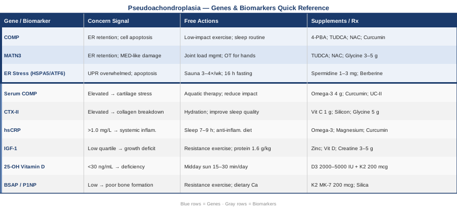
The Genetics of Pseudoachondroplasia: What the Key Genes Reveal and What You Can Do
Pseudoachondroplasia is a rare autosomal dominant skeletal dysplasia. Unlike many complex traits where dozens of genes each contribute a small effect, pseudoachondroplasia is predominantly driven by mutations in a single gene — COMP. That specificity is both a limitation and an advantage: a limitation because the mutation is fixed, and an advantage because the downstream biology is unusually well-mapped and therefore more targetable than in polygenic conditions.
The strategy below is not about reversing the mutation. It is about understanding what the mutation does inside the cell, which pathways it disrupts, and where those pathways offer leverage points that existing and emerging interventions can address.
Gene 1: COMP (Cartilage Oligomeric Matrix Protein)
What it does and why it matters
The COMP gene, located on chromosome 19p13.1, encodes a large pentameric glycoprotein that acts as a structural scaffold in cartilage and tendon extracellular matrix. It cross-links collagen fibrils, interacts with matrilin proteins, and helps maintain the architecture of the growth plate — the active region of bone elongation in children.
In pseudoachondroplasia, mutations in COMP (over 70 have been identified, clustered predominantly in the type III repeats and calmodulin-like domains) produce a protein that misfolds inside the endoplasmic reticulum of chondrocytes. That misfolded protein is not secreted into the extracellular matrix as intended. Instead, it accumulates inside the cell, triggering a cascade of ER stress and ultimately driving chondrocyte death. The growth plate becomes disorganized, endochondral ossification is impaired, and the characteristic short-limbed stature of pseudoachondroplasia results.
NCBI Gene entry for COMP (Gene ID: 1311)
What the mutation actually produces: gain-of-function toxicity
This is a critical distinction. COMP mutations in pseudoachondroplasia are not loss-of-function in the conventional sense. The problem is not simply that COMP is absent. The problem is that the mutant protein is actively harmful — it accumulates in the ER, triggers the unfolded protein response (UPR), induces oxidative stress inside the chondrocyte, and eventually kills the cell through apoptosis. This mechanism is called gain-of-function toxicity, and it changes what therapeutic approaches might be relevant.
If the COMP gene is mutated: the plan without supplements
The first tier of intervention focuses on reducing the burden on chondrocytes through lifestyle and structural support — independently of any supplementation.
Joint load management: Because the cartilage architecture in pseudoachondroplasia is compromised from early development, reducing unnecessary mechanical stress on joints is a genuine biological intervention, not just comfort advice. Low-impact physical activity — swimming, cycling, aquatic physiotherapy — reduces compressive forces on compromised cartilage while maintaining the mechanical stimulation that cartilage needs to remain metabolically active. A consistent routine of 30 to 45 minutes, three to four times per week, appears to strike the right balance. High-impact activity (running on hard surfaces, jumping) should be minimized or avoided, particularly in the presence of joint laxity.
Spinal monitoring and posture: Scoliosis and cervical instability are common complications in pseudoachondroplasia. Regular physiotherapy focused on postural alignment and paraspinal strengthening is not a luxury — it is a way of protecting the spinal cord from the mechanical consequences of ligamentous laxity. A structured programme, ideally supervised initially and then performed independently three times per week, reduces long-term risk of neurological complications.
Sleep and circadian rhythm: Chondrocyte biology is sensitive to circadian disruption. Animal and cell-culture research has shown that circadian clock genes regulate the expression of collagen synthesis genes. Maintaining consistent sleep timing (within a 30-minute window daily), avoiding artificial blue light exposure after 9pm, and prioritizing 7 to 9 hours of sleep may appear minor but represent a genuine signal to the cells that make and maintain cartilage matrix. No specific PSACH trials exist for this, but the upstream biology is solid.
If the COMP gene is mutated: the plan with supplements or targeted interventions
The following are not cures. They are approaches supported by evidence — mostly in cellular models and animal studies, with some early-phase human data for related conditions — that target the same pathways disrupted by COMP mutations.
4-Phenylbutyrate (4-PBA) — chemical chaperone: 4-PBA is an FDA-approved drug used in urea cycle disorders, but its mechanism is relevant here. As a chemical chaperone, it assists in the folding of misfolded proteins inside the ER, reducing the ER stress burden. In cell culture models using COMP mutant chondrocytes, 4-PBA has been shown to reduce COMP retention in the ER and decrease apoptosis. This is early-stage evidence — no clinical trial in pseudoachondroplasia has been completed as of the time of writing — and 4-PBA requires medical supervision. It is not a supplement available over the counter but represents the most scientifically grounded pharmacological target for the underlying pathology.
TUDCA (Tauroursodeoxycholic acid): TUDCA is a bile acid derivative that acts as a mitochondrial and ER protectant. It has been shown to reduce ER stress across multiple cellular models, including those involving protein misfolding. It is available as a supplement (typical dosing in research contexts: 500 to 1,000 mg per day). Side effects are generally mild at lower doses and may include gastrointestinal discomfort. Cycling is prudent (e.g., 8 weeks on, 4 weeks off) given the lack of long-term human data outside liver disease contexts. Use with a medical professional's knowledge, particularly given the hepatic effects at higher doses.
N-acetylcysteine (NAC): ER stress induced by misfolded COMP generates reactive oxygen species inside chondrocytes. NAC is a glutathione precursor and one of the best-studied antioxidants for intracellular oxidative stress. Typical research doses range from 600 to 1,800 mg per day. Side effects at moderate doses include nausea (take with food). Some evidence suggests cycling (e.g., 12 weeks on, 4 off) to avoid blunting of adaptive antioxidant responses. NAC should not be taken alongside nitrate medications.
Curcumin (high-bioavailability formulations): Curcumin inhibits NF-κB, one of the central inflammatory transcription factors activated downstream of ER stress. In skeletal models, curcumin has shown some ability to reduce chondrocyte apoptosis under inflammatory conditions. Bioavailability is the key issue — standard curcumin powder is poorly absorbed. Formulations with piperine or in phospholipid complexes (Meriva, Theracurmin) are preferred. Typical effective doses: 500 to 1,000 mg twice daily of a high-bioavailability form. Side effects include GI sensitivity; avoid in high doses during pregnancy or with anticoagulants.
Omega-3 fatty acids (EPA/DHA): Omega-3s are well-documented anti-inflammatory agents that work in part by reducing downstream inflammatory cytokines (IL-6, TNF-alpha) that are elevated in cartilage pathology. For people with pseudoachondroplasia, managing systemic inflammation may not reverse the core genetic problem, but it may reduce the pace of secondary joint degradation. Typical therapeutic doses: 2 to 4 grams per day of combined EPA+DHA from fish oil or algal sources. No cycling needed; monitor for blood-thinning effects at higher doses.
Gene 2: MATN3 (Matrilin-3)
Why it matters in the PSACH spectrum
MATN3 encodes matrilin-3, a protein that interacts directly with COMP in the cartilage extracellular matrix. Mutations in MATN3 cause a related but distinct condition — multiple epiphyseal dysplasia type 5 (MED) — and the mechanism mirrors PSACH at several levels: matrilin-3 also misfolds in the ER, is retained intracellularly, and generates ER stress-induced chondrocyte death.
NCBI Gene entry for MATN3 (Gene ID: 4148)
For individuals diagnosed with pseudoachondroplasia who receive genetic sequencing, MATN3 variants are worth examining — some cases initially classified as mild PSACH or PSACH-like carry MATN3 mutations rather than COMP mutations, and the distinction matters for family planning and for understanding severity prediction.
If the MATN3 gene shows a variant: the plan without supplements
The structural interventions described for COMP apply equally here: joint load management, spinal monitoring, and sleep consistency. One additional consideration specific to MATN3-related presentations is closer attention to hand and foot joint hypermobility, which tends to be more prominent in MATN3 than in classic COMP-PSACH. Occupational therapy assessment of hand mechanics and grip strength, with specific exercises targeting stabilizer muscles of the small joints, is particularly useful.
If the MATN3 gene score is bad: the plan with supplements or equipment
The supplement strategy overlaps substantially with the COMP approach — TUDCA, NAC, and curcumin all address ER stress, which is the shared pathological mechanism. What differs is the potential role of glycine supplementation, which plays a specific role in collagen synthesis. Matrilin-3 interacts with collagen IX and X in the growth plate; adequate glycine (3 to 5 grams per day, typically from collagen hydrolysate) provides substrate for collagen assembly in the extracellular matrix, supporting whatever matrix-building capacity remains. Side effects are minimal; some individuals report improved sleep quality with glycine at these doses, which is an additional benefit given the circadian-cartilage connection.
Gene 3: The ER Stress Response Network (HSPA5/BiP and ATF6)
The third gene layer: cellular response modifiers
This third category is not a single gene but a network of genes that determines how severely a chondrocyte responds to the accumulation of misfolded COMP or matrilin-3. The two most important mediators are HSPA5 (encoding the ER chaperone BiP/GRP78) and ATF6 (encoding Activating Transcription Factor 6, an ER stress sensor).
BiP is the primary ER chaperone that binds to misfolded proteins, holds them in the ER, and attempts to assist their folding. ATF6 is one of three master sensors of the unfolded protein response (UPR) — it detects ER stress and initiates a transcriptional programme that can either resolve stress or, when overwhelmed, trigger apoptosis.
The key insight here is that the severity of pseudoachondroplasia is not determined solely by which COMP mutation a person carries. It is also shaped by how efficiently that person's ER stress response machinery can compensate. Individuals with more robust BiP expression or more effective UPR activation may experience a milder phenotypic course from the same mutation.
This creates a legitimate epigenetic and environmental target. BiP expression is upregulated by mild ER stress preconditioning — a phenomenon studied in cardiac and neural biology where low-level stress stimuli train cells to mount faster and more effective protective responses. Heat shock (sauna use, warm baths) is the most accessible form of this preconditioning, with growing human evidence for increased heat shock protein expression following regular sauna exposure.
If the ER stress network is underperforming: the plan without supplements
Sauna or heat therapy: Regular exposure to mild thermal stress upregulates heat shock proteins including BiP-family proteins. A protocol of 20 minutes at 80 to 90°C (or comparable dry heat), three to four times per week, has been used in cardiovascular longevity research to induce consistent heat shock protein upregulation. For pseudoachondroplasia specifically, the benefit is speculative but mechanistically grounded: more available ER chaperones may slow the progression of chondrocyte death driven by misfolded COMP accumulation. Important caveat: individuals with cardiovascular concerns or spinal instability should confirm safety with their physician before using high-temperature saunas.
Autophagy support through fasting windows: Autophagy — the cellular process of clearing damaged proteins and organelles — can help degrade misfolded COMP that accumulates in the ER. Mild intermittent fasting (a 14 to 16 hour overnight fast) consistently activates autophagy pathways in humans. This does not require extreme dietary changes and carries a reasonable evidence base for its cellular housekeeping effects.
If the ER stress network is underperforming: the plan with supplements or equipment
Spermidine: A naturally occurring polyamine found in aged cheese, wheat germ, and mushrooms, spermidine is one of the best-studied dietary autophagy inducers. At doses of 1 to 3 mg per day (supplemental form), it has shown autophagy-enhancing effects in human studies. Cycling is not required but periodic breaks (8 weeks on, 2 off) are prudent. No serious adverse effects have been reported at these doses.
Berberine: Berberine activates AMPK and mildly inhibits mTOR, both of which promote autophagy. It also has anti-inflammatory effects downstream of NF-κB. Typical dosing: 500 mg two to three times daily with meals. Side effects include gastrointestinal discomfort; should not be combined with metformin without medical supervision.
Moving from genetics to the practical question of ongoing measurement, the next section covers the six biomarkers that provide a real-time view of how this biology is playing out in the body.
6 Biomarkers to Track in Pseudoachondroplasia
Genetic information tells you what the biological starting condition is. Biomarkers tell you what the body is doing with it right now. For pseudoachondroplasia, the most informative panel is one that tracks cartilage breakdown, inflammation, bone metabolism, and metabolic support simultaneously.
Biomarker 1: Serum COMP
Why it matters
Serum levels of COMP protein reflect cartilage and tendon turnover. In inflammatory arthritis and cartilage degeneration, serum COMP rises because damaged tissue releases the protein into the bloodstream. For pseudoachondroplasia patients, serum COMP may not reflect the standard cartilage degradation signal (since much of the mutant COMP is retained in cells rather than secreted) but it provides a useful longitudinal marker of overall cartilage stress and joint load.
How to measure it
COMP is measured from a blood draw using ELISA assay. It is available through specialized rheumatology or biochemistry laboratories. Cost ranges from approximately $80 to $200 USD depending on the laboratory and country. It is not part of standard panels and typically requires a physician referral specifying the test.
If the score is elevated: the plan without supplements
Elevated serum COMP usually signals increased mechanical stress on cartilage or active joint inflammation. Reviewing activity levels — specifically any high-impact or repetitive loading activities — is the first step. Aquatic therapy or low-impact exercise substitution can reduce mechanical cartilage stress within weeks.
If the score is elevated: the plan with supplements or equipment
Omega-3 fatty acids (2 to 4 g EPA+DHA daily) and curcumin (500 mg twice daily, high-bioavailability) have demonstrated anti-inflammatory effects on cartilage markers. Type II collagen hydrolysate (10 to 40 mg per day of undenatured type II collagen, or UC-II format) has shown some reduction in joint degradation markers including COMP in osteoarthritis trials — evidence is not PSACH-specific but the mechanism is relevant. Frequency: daily, ongoing. Monitor via repeat serum COMP at 3-month intervals.
Biomarker 2: CTX-II (C-Telopeptide of Type II Collagen)
Why it matters
CTX-II is a degradation fragment of type II collagen, the primary structural collagen in articular cartilage. Elevated urinary CTX-II is a well-validated marker of cartilage breakdown and is used in osteoarthritis research as a sensitive indicator of disease progression. In pseudoachondroplasia, where cartilage architecture is compromised, CTX-II offers a measurable proxy for how quickly joint surfaces are degrading over time.
How to measure it
CTX-II is typically measured from a second-morning urine sample, corrected for creatinine. It is available through specialized musculoskeletal research labs and some commercial panels (approximate cost: $100 to $250 USD). Testing annually provides a reasonable longitudinal signal; more frequent testing (every 6 months) is useful when making dietary or therapeutic changes.
If the score is elevated: the plan without supplements
Chronic CTX-II elevation signals ongoing cartilage collagen breakdown. Priority interventions: reduce joint impact loading, ensure adequate hydration (cartilage is 80% water; even mild chronic dehydration concentrates the stress), and review sleep quality (cartilage repair is predominantly nocturnal).
If the score is elevated: the plan with supplements or equipment
Vitamin C (500 to 1,000 mg per day) supports collagen synthesis and is required as a cofactor in procollagen hydroxylation. Silicon (from orthosilicic acid, 5 to 10 mg per day) has early human evidence for supporting collagen cross-linking in connective tissue. Neither is a strong intervention on its own, but both support the collagen-building side of the breakdown/repair equation. Glycine (3 to 5 g per day) directly supplements collagen synthesis substrate. These are low-risk, ongoing daily supplements.
Biomarker 3: High-Sensitivity C-Reactive Protein (hsCRP)
Why it matters
hsCRP is the most accessible and widely available marker of low-grade systemic inflammation. In pseudoachondroplasia, the ER stress caused by misfolded COMP drives an inflammatory cascade that extends beyond the growth plate chondrocyte. Systemically elevated inflammation accelerates cartilage degradation, worsens joint symptoms, and is now understood to impair sleep, mood, and metabolic function through well-characterized neuroimmune pathways.
How to measure it
hsCRP is a standard blood test included in most comprehensive metabolic panels or available as a standalone order. Cost: $10 to $30 USD in most health systems, often covered by insurance. Ideal value: below 0.5 mg/L (Peter Attia's preferred threshold); values above 1.0 mg/L indicate meaningful chronic inflammation warranting intervention.
If the score is elevated: the plan without supplements
Sleep is one of the strongest non-pharmacological reducers of hsCRP. Consistent 7 to 9 hours of high-quality sleep lowers inflammatory markers within weeks. Dietary ultra-processed food reduction, elimination of seed oils high in omega-6, and inclusion of colourful vegetables (polyphenol-rich sources like berries, leafy greens, and olive oil) each independently reduce hsCRP.
If the score is elevated: the plan with supplements or equipment
Omega-3 fatty acids are the most evidence-based supplement for hsCRP reduction. Magnesium glycinate (300 to 400 mg at night) reduces hsCRP and improves sleep simultaneously. Curcumin at therapeutic doses reduces NF-κB-driven CRP production. Frequency: daily. Monitor hsCRP at 3-month intervals when making changes.
Biomarker 4: IGF-1 (Insulin-Like Growth Factor 1)
Why it matters
IGF-1 is the primary mediator of growth hormone's effects on cartilage and bone. It stimulates chondrocyte proliferation and differentiation, and supports extracellular matrix synthesis. In pseudoachondroplasia, the growth plate dysfunction means that downstream IGF-1 signalling is partially disconnected from its normal targets — but maintaining adequate IGF-1 levels ensures that whatever functional chondrocytes remain have the hormonal support they need to perform optimally. IGF-1 also influences muscle mass and bone density, both of which matter significantly for long-term joint protection in this condition.
How to measure it
IGF-1 is a standard blood test available through endocrinologists and many primary care providers. Cost: $30 to $100 USD. Optimal range varies by age — working with a physician to interpret age-adjusted values is essential. Values in the lower quartile of the normal range may warrant investigation, particularly in the context of short stature and metabolic stress.
If the score is low: the plan without supplements
IGF-1 is most powerfully stimulated by resistance exercise and adequate protein intake. Resistance training (bodyweight or adapted equipment, 2 to 3 times per week) is the primary tool. Protein intake should be at least 1.2 to 1.6 g per kilogram of body weight daily, emphasizing leucine-rich sources (meat, fish, eggs, dairy) that most directly stimulate the mTOR-IGF-1 pathway. Sleep quality is the second most important factor — IGF-1 is primarily secreted during slow-wave sleep.
If the score is low: the plan with supplements or equipment
Zinc (15 to 30 mg per day) is an essential cofactor in IGF-1 production and is commonly deficient in Western diets. Vitamin D (see below) also interacts with IGF-1 signalling. Creatine monohydrate (3 to 5 g per day) supports the muscle mass that drives the resistance training stimulus. No special cycling required for any of these.
Biomarker 5: 25-OH Vitamin D
Why it matters
Vitamin D is not just a bone mineral regulator — it is an anti-inflammatory hormone with receptors in cartilage, immune cells, and the spinal cord. In pseudoachondroplasia, adequate vitamin D supports bone density (which matters given the abnormal loading patterns), modulates the inflammatory environment around joints, and influences the expression of several genes involved in chondrocyte survival. Deficiency is extremely common — particularly in people who, due to mobility limitations or pain, spend less time outdoors.
How to measure it
25-OH vitamin D is a standard blood test. Cost: $20 to $60 USD. Optimal target: 40 to 60 ng/mL (100 to 150 nmol/L) in the view of most functional medicine practitioners; above 30 ng/mL is the conventional minimum. Testing twice yearly (before and after winter) is practical.
If the score is low: the plan without supplements
Midday sun exposure (arms and legs, 15 to 30 minutes depending on latitude and skin tone) during summer months is the most efficient source. Dietary sources include fatty fish, egg yolks, and liver — adequate but insufficient alone for therapeutic repletion.
If the score is low: the plan with supplements or equipment
Vitamin D3 supplementation at 2,000 to 5,000 IU per day (with vitamin K2, 100 to 200 mcg, to direct calcium to bone rather than arteries) is the standard approach. Retest at 3 months to adjust dosing. Side effects are rare at these doses but toxicity is possible at very high doses (above 10,000 IU daily long-term) — do not supplement without baseline testing.
Biomarker 6: Bone-Specific Alkaline Phosphatase (BSAP) or P1NP
Why it matters
Bone formation markers — particularly BSAP (bone-specific alkaline phosphatase) or P1NP (procollagen type I N-terminal propeptide) — reflect the activity of osteoblasts, the cells that build bone. In pseudoachondroplasia, abnormal endochondral ossification means that bone formation at the growth plate is disrupted. Tracking bone formation markers over time reveals whether the body's osteoblast activity is adequate to maintain skeletal integrity, particularly as patients age and the risk of early joint degeneration increases.
How to measure it
P1NP is the preferred bone formation marker by most metabolic bone specialists. It is available through clinical labs specializing in endocrinology or bone health. Cost: $50 to $150 USD. It is best interpreted alongside a bone resorption marker (CTX-I from serum) to understand the formation-resorption balance.
If the score is low: the plan without supplements
Resistance exercise directly stimulates osteoblast activity. Even adapted resistance training — seated exercises, resistance bands, water resistance — creates mechanical stimulation that signals bone to form. Adequate dietary calcium from whole food sources (dairy, leafy greens, canned fish with bones) provides raw material. Protein intake supports the collagen scaffold that bone mineralizes onto.
If the score is low: the plan with supplements or equipment
Vitamin K2 (MK-7 form, 100 to 200 mcg per day) activates osteocalcin, the protein that anchors calcium into bone matrix. Silica/orthosilicic acid has early evidence for increasing P1NP in human trials. Both are low-risk, daily, ongoing supplements. Avoid very high-dose calcium supplementation without evidence of deficiency — excess calcium without K2 can increase arterial calcification risk.
10 Research Insights That Are Changing How Scientists Think About Pseudoachondroplasia
The following reflects the most impactful findings from current research into pseudoachondroplasia and related skeletal dysplasias — findings that challenge the resignation often felt after diagnosis and point toward a more actionable biological understanding.
1. The mutation does not fully predict the phenotype
Twin and family studies in pseudoachondroplasia have demonstrated that individuals with identical COMP mutations can have meaningfully different clinical severity. This confirms that genetic background, epigenetic modifications, and environmental factors collectively shape how the mutation expresses. The mutation is not destiny.
2. ER stress is the actual mechanism of damage, not the mutation itself
Chondrocytes die not because the COMP protein is absent from the matrix, but because its accumulation inside the ER overwhelms the cellular stress response. This means that reducing ER stress — even by a fraction — could slow or reduce cell death. This is now an active pharmacological target in skeletal dysplasia research.
3. Chemical chaperones reduce COMP retention in laboratory models
Multiple studies using cell cultures derived from pseudoachondroplasia patients or mouse models have demonstrated that 4-PBA reduces the intracellular accumulation of mutant COMP and decreases chondrocyte apoptosis. This is not a clinical trial result — but it is proof of concept that the mechanism can be pharmacologically addressed.
4. Autophagy enhancement clears misfolded protein load
Research in protein misfolding diseases broadly has established that activating autophagy — the cell's own protein degradation system — can reduce the intracellular burden of aggregated or retained proteins. In PSACH cell models, autophagy induction has shown reduction in ER stress markers. Spermidine, fasting, and rapamycin (in research contexts) are all capable of activating this pathway.
5. Inflammation is not just a consequence — it is an amplifier
Activated UPR pathways in PSACH chondrocytes directly upregulate inflammatory transcription factors including NF-κB. This inflammatory signal extends beyond the cartilage locally and may contribute to systemic low-grade inflammation. This makes anti-inflammatory strategies not cosmetic but structurally relevant to slowing the pace of joint damage.
6. Mouse models have been successfully treated
A transgenic mouse model that recapitulates the COMP mutation of pseudoachondroplasia has been used to test multiple interventions. Results from these studies — including chemical chaperone treatments — have confirmed measurable improvements in chondrocyte survival and growth plate organization. Translating this to human clinical trials is the current frontier.
7. Serum COMP is a feasible monitoring biomarker
Researchers have explored whether serum COMP correlates with joint stress and disease burden in PSACH and related dysplasias. While not yet validated as a clinical standard for PSACH specifically, serum COMP is already in clinical use for rheumatoid arthritis and osteoarthritis monitoring, making its measurement accessible and its interpretation meaningful.
8. Joint laxity and early osteoarthritis are predictable and preventable targets
Longitudinal data from PSACH patient registries shows that early-onset osteoarthritis is nearly universal in pseudoachondroplasia, but its rate of progression varies considerably. Early physiotherapy intervention, joint load management, and muscle strengthening demonstrably slow the progression of joint degeneration — giving a several-decade window of intervention before severe disability develops.
9. Cervical instability requires surveillance, not waiting for symptoms
Research in PSACH has identified that atlantoaxial and subaxial cervical instability can be present without symptoms until neurological compromise occurs. Proactive cervical imaging and specialist monitoring (typically recommended every 3 to 5 years in children and when symptoms develop in adults) changes outcomes significantly. This is a case where knowing the risk changes the action, and the action changes the trajectory.
10. Gene therapy is entering the horizon
While no gene therapy for pseudoachondroplasia has reached clinical trial as of the time of writing, the development of antisense oligonucleotide (ASO) approaches and RNA interference strategies for dominant negative mutations is progressing across the rare skeletal dysplasia field. The PSACH research community is actively engaged in this space. Staying connected to patient advocacy networks such as the Brittle Bone Society, NORD (National Organization for Rare Disorders), and the International Skeletal Dysplasia Society ensures access to emerging trial opportunities.
Complementary Approaches With Meaningful Evidence
Genetic and biomarker strategies address the underlying biology. The following complementary approaches are selected because they have meaningful human clinical evidence for pain management, joint mobility, and quality of life in conditions that share mechanistic features with pseudoachondroplasia — including chronic joint pain, ligamentous laxity, and musculoskeletal impairment.
Yoga (Adapted Practice)
Adapted yoga emphasizes gentle joint mobilization, paraspinal muscle activation, and breathing coordination — all of which address specific challenges in pseudoachondroplasia. Unlike weight-bearing aerobic exercise, a well-designed yoga practice can be entirely non-impact and can be adapted to individuals with significant height and mobility differences. The emphasis on proprioception and postural awareness is particularly relevant given the joint laxity common in PSACH, where proprioceptive training helps reduce fall risk and joint microtrauma.
A randomized controlled trial published in the Annals of Internal Medicine demonstrated that yoga was non-inferior to physical therapy for chronic low back pain over 12 weeks — a highly relevant finding given the prevalence of spinal complications in pseudoachondroplasia. The emphasis on strengthening stabilizing muscles around hypermobile joints maps well to the structural needs of this condition.
Practically, sessions should be conducted with a yoga instructor experienced with hypermobility or skeletal conditions. Two to three sessions per week, 30 to 45 minutes each, focusing on gentle backbends, hip stabilization, and breathing. Poses involving extreme spinal flexion or inversion should be modified or avoided pending cervical stability assessment.
Massage Therapy
In pseudoachondroplasia, the altered biomechanics of a short-limbed body place chronic tension on specific muscle groups — particularly the paraspinal muscles, hip flexors, and calf musculature — that work harder than average to compensate for structural differences. Therapeutic massage reduces muscle hypertonicity, improves local circulation, and has been shown to reduce pain and disability scores in musculoskeletal conditions.
A Cochrane review of massage therapy for low back pain (which shares many features with PSACH-related spinal pain) found short-term improvements in pain and function that were clinically meaningful. While no massage trial exists specifically for pseudoachondroplasia, the musculoskeletal rationale is well-supported.
A practical protocol involves sessions of 45 to 60 minutes every one to two weeks with a therapist familiar with skeletal differences. Techniques should avoid deep pressure directly over the spine given the potential for instability. Focus areas: thoracolumbar junction, hip flexors, and calf-Achilles complex. Inform the therapist of any cervical instability findings before commencing.
Mindfulness Meditation / MBSR
Living with a lifelong genetic condition involves not only physical burden but also a chronic stress response that elevates cortisol and inflammatory cytokines — both of which compound the underlying joint pathology. Mindfulness-Based Stress Reduction (MBSR) is an 8-week structured programme that has demonstrated consistent reductions in chronic pain perception, inflammatory markers, and psychological distress across multiple systematic reviews.
A meta-analysis published in JAMA Internal Medicine found that mindfulness meditation programmes produced moderate evidence of improved pain, depression, and anxiety in patients with chronic medical conditions. For pseudoachondroplasia, where pain is common and emotional exhaustion from lifelong medical management is significant, MBSR offers a grounded, evidence-based tool.
The standard MBSR programme is available in-person through many hospital systems and online through platforms like Palouse MBSR or Mindful Schools. The 8-week commitment requires 30 to 45 minutes of daily practice. After the programme, a maintenance practice of 20 minutes daily is sufficient to sustain benefits. No special equipment is needed.
Breathing-Based Therapies
Controlled breathing techniques — particularly slow diaphragmatic breathing at a rate of around 6 breaths per minute (0.1 Hz resonance breathing) — activate the vagus nerve and shift the autonomic nervous system toward parasympathetic dominance. This directly reduces the systemic inflammatory milieu by lowering sympathetic cytokine signalling. In chronic pain conditions, autonomic dysregulation is a consistent feature, and breathing provides a real-time, cost-free modulation tool.
Research on resonance frequency breathing in heart rate variability biofeedback programmes has shown reductions in inflammatory markers including hsCRP and improvements in pain tolerance scores. The effect is rapid (measurable within four weeks of daily practice) and the mechanism is well understood. For PSACH patients who may also experience sleep-disordered breathing due to spinal or craniofacial anatomy, consultation with a respiratory specialist before beginning intensive breathing protocols is advisable.
Practical application: 10 to 20 minutes of slow-paced diaphragmatic breathing daily, using an app such as Breathwrk, Oxygen Advantage, or a simple metronome set to 6 breaths per minute. Morning practice (before significant activity) and evening practice (30 minutes before sleep) are the most impactful timing windows. No equipment cost is required beyond an optional app subscription.
Conclusion
Pseudoachondroplasia is a genetic condition, and that fact does not disappear. But the biology between the mutation and its long-term consequences is far from fixed. The COMP gene's mechanism of damage — through ER stress, inflammation, and chondrocyte death — creates specific, measurable, and partly addressable targets. The six biomarkers covered here give you and your care team a real-time picture of how those processes are unfolding in the body right now. The complementary approaches offer genuine quality-of-life benefits supported by human evidence, not wishful thinking.
The most useful next step is not the most dramatic one. It is the most specific one: pick one biomarker to baseline, one lifestyle habit to reinforce, and one conversation to have with your treating physician about what monitoring your care plan currently includes and what might be missing. Better decisions follow from better information — and this article has aimed to provide the latter.
Musculoskeletal: Bone Conditions Joint Conditions Spine Conditions
Autoimmune: Inflammatory Conditions Connective Tissue Conditions