This article contains AI generated content.
· UpdatedPyoderma Gangrenosum Genes And Biomarkers — 5 Genes And 6 Biomarkers To Track
Introduction
Pyoderma gangrenosum is one of those conditions that manages to be simultaneously rare enough to be poorly understood and severe enough to upend daily life. The wounds are not just cosmetically distressing — they are deeply painful, slow to heal, prone to worsening with minor trauma, and often connected to systemic diseases that are themselves difficult to manage. If you have spent time in emergency rooms being mistaken for a spider bite victim, or had wound care teams treat your lesions before checking for the underlying inflammatory driver, you are not alone in that experience.
Generic dermatology advice tends to fall short here because pyoderma gangrenosum is not one disease. It is a final common pathway for several distinct immune dysfunctions. In some patients, it is driven by gut inflammation from Crohn's disease or ulcerative colitis. In others, it emerges alongside hematologic disorders or rheumatoid arthritis. In a smaller subset, it is tied directly to inherited mutations in genes that regulate the innate immune response — mutations that make flares nearly inevitable without targeted treatment. Treating every PG patient the same way ignores that biological reality.
What can actually help is getting specific. Specific about which inflammatory markers are elevated in your blood right now. Specific about whether your underlying disease associations are being identified and monitored. Specific about whether your genetics put you in a category where certain biologic therapies are far more likely to work than others. This level of precision is available — it just rarely makes it into a standard consultation.
The goal of this article is to give you a map of that precision. It covers six biomarkers that provide actionable intelligence about what is driving your condition, five genetic factors that can shift how you understand your disease trajectory, a protocol built specifically for autoinflammatory and autoimmune diseases, and evidence-backed complementary strategies worth knowing about. Better information does not replace your medical team — but it gives you something sharper to bring into those conversations.
Summary
Among the six biomarkers covered here, one stands out as consistently overlooked in standard workups: serum IL-6, a cytokine directly upstream of the neutrophilic cascade responsible for PG ulcers, which most dermatologists never order. Another, serum protein electrophoresis, can reveal a hidden blood disorder fueling your flares — a finding that changes treatment completely. You will also learn why fecal calprotectin is worth tracking even if you have no obvious gut symptoms, and how the neutrophil-to-lymphocyte ratio on a standard CBC can serve as a quick-and-cheap proxy for disease activity.
On the genetics side, one gene — PSTPIP1 — is mutated in a defined syndrome that includes PG, and knowing whether you carry it changes which medications your physician should consider first. Two others, MEFV and IL1RN, affect the autoinflammatory cascade in ways that current IL-1 targeting biologics can partially compensate for.
Beyond the biomarkers and genetics sections, you will find a distilled summary of Dirty Genes by Ben Lynch — a framework that explains how common genetic variants maintain an elevated inflammatory set point, and what can be done about it without waiting for medical guidelines to catch up. The article also covers Sarah Ballantyne's Autoimmune Protocol, low-level laser therapy applied to PG wounds, and microbiome-directed approaches that address the gut-skin axis — the one biological connection that may be silently maintaining your condition between flares.
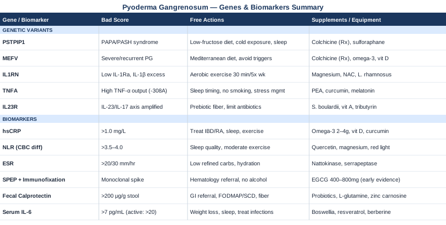
6 Biomarkers to Track If You Have Pyoderma Gangrenosum
Pyoderma gangrenosum sits at the intersection of innate immune dysregulation, neutrophil hyperactivation, and systemic inflammatory disease. The biomarkers that matter most reflect those biological realities — not generic inflammation, but the specific pathways that research has linked to PG pathogenesis, disease severity, and treatment response. These six are worth knowing.
1. High-Sensitivity C-Reactive Protein (hsCRP)
Why It Matters
hsCRP is the most clinically accessible measure of systemic inflammation. In PG, it tends to track with disease activity — rising during active ulceration and falling during remission or successful treatment. It is not specific to PG and cannot distinguish between PG flare and infection (which matters enormously in wound management), but it provides a reliable longitudinal signal. Physicians like Peter Attia have consistently argued that hsCRP should be followed serially rather than checked once, because the trend over time is where the diagnostic value lives. In PG, a persistently elevated hsCRP despite apparent wound healing suggests ongoing systemic inflammatory drive — a reason to look harder at the underlying associated condition.
How to Measure It
Any standard blood draw at most labs. The high-sensitivity version (hsCRP, not standard CRP) detects lower concentrations and is preferable for ongoing monitoring. Cost range: $10–$40 out-of-pocket; often covered under standard inflammatory workup. Target: below 1.0 mg/L for low cardiovascular risk; for PG monitoring, aim to track the trend rather than a fixed threshold. Frequency: at least every 3 months during active disease, or with each clinical review.
If the Score Is Bad: The Plan Without Supplements
The most impactful non-supplement interventions for chronically elevated hsCRP involve addressing the underlying inflammatory driver. If IBD is present, optimizing its treatment is essential — inadequately controlled Crohn's or ulcerative colitis will keep hsCRP elevated regardless of local wound care. Eliminating ultra-processed foods, refined carbohydrates, and vegetable oils high in omega-6 has shown meaningful reductions in hsCRP in multiple clinical trials. Prioritizing sleep (7–9 hours), reducing psychosocial stress through structured practice, and resistance training 2–3 times per week have each shown documented hsCRP reductions. These are not peripheral lifestyle suggestions — they directly affect the inflammatory substrate feeding PG flares.
If the Score Is Bad: The Plan With Supplements or Equipment
Omega-3 fatty acids (EPA+DHA, 2–4g/day) consistently reduce hsCRP in meta-analyses and are well-tolerated at these doses. Start at 2g/day and increase after 4 weeks if tolerated; take with the largest meal to minimize GI side effects. No cycling needed; long-term use is appropriate. Side effect: blood thinning at higher doses — discuss with your physician if on anticoagulants. Vitamin D supplementation (2,000–5,000 IU/day, adjusted to blood level) is consistently associated with lower hsCRP in inflammatory conditions, and deficiency is extremely common in PG patients. Recheck serum 25-OH vitamin D every 3 months while adjusting dose. Curcumin (with piperine, 500–1,000mg/day) has anti-inflammatory evidence at the hsCRP level; cycle off every 8 weeks to assess baseline. References on omega-3 and inflammation are available at PubMed.
2. Complete Blood Count with Differential — Neutrophil-to-Lymphocyte Ratio (NLR)
Why It Matters
PG is a neutrophilic dermatosis — meaning neutrophil dysfunction and dysregulated neutrophil accumulation are central to lesion formation. The neutrophil-to-lymphocyte ratio (NLR), calculated from a standard CBC with differential, captures the balance between innate immune aggression (neutrophils) and adaptive immune regulation (lymphocytes). An elevated NLR signals a systemic state favoring neutrophilic inflammation. Research has validated NLR as a marker of disease severity in inflammatory dermatoses and several systemic inflammatory conditions. In PG, an NLR above 3.5–4.0 during apparent quiescence should prompt investigation into whether systemic inflammation is being adequately controlled. A high NLR also correlates with worse outcomes in associated hematologic malignancies, which affect up to 7% of PG patients. See relevant studies at PubMed.
How to Measure It
Included in any standard CBC with differential — Cost: $15–$50, widely covered. Calculate by dividing the absolute neutrophil count by the absolute lymphocyte count. Normal range: roughly 1.0–3.0 in healthy adults. Optimal for monitoring: check every 3–6 months during stable disease; monthly during active ulceration or treatment changes.
If the Score Is Bad: The Plan Without Supplements
An elevated NLR responds most reliably to addressing the root inflammatory driver — which means ensuring that associated conditions (IBD, RA, myeloid dyscrasias) are being optimally treated, not just acknowledged. From a lifestyle standpoint, poor sleep is one of the most reliable drivers of neutrophilic shift — even a single night of disrupted sleep increases circulating neutrophil counts. Prioritizing sleep quality with consistent timing, light exposure management, and avoiding late alcohol has a measurable effect on NLR over weeks. High-intensity exercise paradoxically spikes NLR acutely but reduces it chronically when training is moderate and consistent.
If the Score Is Bad: The Plan With Supplements or Equipment
Quercetin (500–1,000mg/day with meals) has demonstrated effects on neutrophil activation and NETosis (neutrophil extracellular trap formation) in in vitro and early human studies; clinical evidence is emerging. Take for 8-week cycles with 2-week breaks. Magnesium glycinate (300–400mg at night) supports sleep quality and has mild anti-inflammatory properties that may affect NLR indirectly. Photobiomodulation (red light therapy panels, 630–850nm) applied systemically rather than locally has early evidence for modulating innate immune activation — though PG-specific evidence remains preliminary. Session: 10–20 minutes, 3–5x/week.
3. Erythrocyte Sedimentation Rate (ESR)
Why It Matters
ESR is a slower-moving marker than hsCRP but complementary to it. Because it reflects plasma protein changes rather than CRP production directly, it remains elevated longer after a flare and can signal smoldering inflammation that hsCRP has begun to normalize. In PG with an associated myeloma or paraproteinemia, ESR can be strikingly elevated — sometimes disproportionately so — which serves as a clue to investigate the hematologic angle. Tracking both hsCRP and ESR together gives a more complete picture: when they diverge, the divergence itself is informative.
How to Measure It
Standard blood draw, always included in rheumatologic panels. Cost: $10–$30. Normal: under 20 mm/hr for men, under 30 mm/hr for women (age-adjusted). ESR rises normally with age, so use age-corrected reference ranges (Westergren method). Monitor every 3 months alongside hsCRP; do not track in isolation.
If the Score Is Bad: The Plan Without Supplements
Persistent ESR elevation in PG should prompt a dedicated search for the underlying associated condition, rather than attempts to treat the marker itself. Rheumatology referral is warranted if ESR stays persistently above 50 mm/hr without explanation. Dietary changes that reduce fibrinogen and plasma proteins driving ESR include reducing refined carbohydrate intake, which lowers systemic insulin and the downstream inflammatory signaling that raises fibrinogen. Adequate hydration also normalizes plasma viscosity independently.
If the Score Is Bad: The Plan With Supplements or Equipment
Nattokinase (2,000 FU/day, between meals) has early evidence for reducing fibrinogen, one driver of elevated ESR. Use with caution if on anticoagulants; take in 4-week cycles. Serrapeptase (40,000–120,000 IU/day) is similarly used to reduce plasma protein-driven ESR elevation in inflammatory conditions, though robust clinical evidence in PG specifically is lacking. Both enzymes should be discussed with a physician before use in patients with active wounds or anticoagulation.
4. Serum Protein Electrophoresis (SPEP) and Immunofixation
Why It Matters
SPEP with immunofixation is one of the most diagnostically important but least-ordered tests in PG workups. Up to 10% of PG patients have an underlying hematologic malignancy, with monoclonal gammopathy of undetermined significance (MGUS) and multiple myeloma being the most clinically relevant. A monoclonal paraprotein circulating in the bloodstream creates an immune-activating environment that can drive and sustain PG — and if this is the actual underlying driver, treating only the skin will always fail. Identification of a paraprotein completely changes the treatment algorithm. Some patients with PG and MGUS have responded to treatment directed at the plasma cell clone rather than conventional PG immunosuppression. Evidence linking PG to hematologic disorders is reviewed at PubMed.
How to Measure It
Blood draw, requires a specific order for SPEP + immunofixation. Cost: $50–$200 depending on whether immunofixation is included; most insurance covers this with a physician's referral. Frequency: at least once as part of initial workup; repeat annually if initially normal but PG remains active or recurs. If SPEP shows a monoclonal spike, immediate hematology referral is warranted.
If the Score Is Bad: The Plan Without Supplements
If a paraprotein is identified, the only appropriate response is hematology consultation and, if applicable, plasma cell-directed treatment. This is a case where lifestyle and supplements are entirely secondary. Monitoring the paraprotein over time, assessing for progression to myeloma with LDH, beta-2 microglobulin, and bone marrow biopsy if indicated, is the priority. No lifestyle intervention meaningfully reduces a monoclonal paraprotein — but reducing overall inflammatory burden (sleep, diet, stress) may slow progression in MGUS by reducing the inflammatory environment that supports clone expansion.
If the Score Is Bad: The Plan With Supplements or Equipment
There are no supplements that address paraproteinemia directly. However, some preclinical evidence suggests that EGCG (green tea extract, 400–800mg/day) may reduce light chain production in plasma cell disorders; clinical evidence remains early and this is not a substitute for hematologic care. Discuss with a hematologist before use. Avoiding alcohol entirely is appropriate, as alcohol affects plasma cell behavior and interferes with treatment monitoring.
5. Fecal Calprotectin
Why It Matters
Fecal calprotectin is a protein released by neutrophils in the intestinal wall during active gut inflammation. It is one of the most sensitive non-invasive markers for IBD activity. This matters enormously for PG because 30–50% of PG patients have an associated IBD, most commonly Crohn's disease and ulcerative colitis, and the gut inflammation does not always announce itself with dramatic GI symptoms. A patient with PG and subclinical gut inflammation — elevated fecal calprotectin but no diarrhea, cramping, or bleeding — may be unknowingly maintaining the immune activation that drives their skin ulcers. Treating the skin without addressing active gut inflammation is like mopping the floor while the tap runs. Relevant research is indexed at PubMed.
How to Measure It
Stool sample, collected at home and sent to a lab. Cost: $40–$150 depending on the lab; increasingly covered by insurance with IBD clinical indication. Normal: under 50 µg/g stool. Borderline: 50–200 µg/g. Elevated: above 200 µg/g. Monitor every 3–6 months in PG patients with known IBD; consider once annually in PG patients without confirmed IBD to screen for subclinical gut activity.
If the Score Is Bad: The Plan Without Supplements
Elevated fecal calprotectin in a PG patient without a confirmed IBD diagnosis should prompt gastroenterology referral and likely colonoscopy. If IBD is confirmed, optimizing IBD treatment — escalating to biologics if topical or aminosalicylate therapy is inadequate — frequently improves PG concurrently. Dietary approaches proven to reduce intestinal calprotectin include the specific carbohydrate diet, the low-FODMAP diet for symptom management, and exclusion of known dietary irritants (gluten in cases of concurrent sensitivity). Increasing soluble fiber intake supports a microbiome environment less conducive to intestinal neutrophil activation.
If the Score Is Bad: The Plan With Supplements or Equipment
Probiotics (multi-strain, including Lactobacillus rhamnosus GG and Bifidobacterium longum, 10–50 billion CFU/day) have evidence for reducing fecal calprotectin in IBD, particularly in ulcerative colitis. Take with food; use continuously for at least 8 weeks before assessing effect. L-glutamine (5–10g/day in divided doses on an empty stomach) supports intestinal barrier integrity, reducing the translocation of inflammatory signals from the gut lumen. No cycling needed. Zinc carnosine (75mg twice daily with food) has specific evidence for gut mucosal repair and reducing intestinal permeability; 4–8 week courses are appropriate.
6. Serum Interleukin-6 (IL-6)
Why It Matters
IL-6 is a pleiotropic cytokine that sits at the center of the acute-phase response and is directly upstream of CRP production. In PG, IL-6 is overexpressed in lesional tissue and elevated in the serum of patients with active disease, reflecting the broader systemic autoinflammatory state. What makes IL-6 particularly important is its role as a bridge between the innate and adaptive immune systems — elevated IL-6 perpetuates neutrophil survival, stimulates acute-phase protein production, and amplifies the inflammatory signal that maintains PG ulcers. IL-6 is also the therapeutic target of tocilizumab, a biologic that has shown benefit in refractory PG cases. Knowing your baseline IL-6 level provides a rationale for discussing IL-6 pathway targeting with your dermatologist, especially in cases refractory to corticosteroids and conventional immunosuppression. Relevant literature can be found at PubMed.
How to Measure It
Blood draw, requires specific order for serum IL-6 (often bundled in cytokine panels). Cost: $60–$200 out-of-pocket; less commonly covered by routine insurance but often orderable by rheumatology or immunology. Normal: typically below 7 pg/mL in healthy adults. Monitor at baseline and after treatment changes; every 3–6 months in active disease. Highly elevated IL-6 (above 20–30 pg/mL) with refractory PG is a finding worth discussing specifically with a rheumatologist.
If the Score Is Bad: The Plan Without Supplements
Elevated IL-6 responds to the same upstream interventions that reduce hsCRP: caloric appropriateness (obesity drives IL-6 production from adipose tissue), sleep optimization, and treatment of the underlying associated disease. Visceral adipose tissue is particularly active in IL-6 production, meaning even modest reductions in central adiposity through diet and exercise have a measurable effect. Intermittent fasting protocols (16:8 time-restricted eating) have shown specific reductions in circulating IL-6 in overweight individuals in multiple trials. Resolving infections — including subclinical dental infections, which are frequently overlooked — can lower IL-6 meaningfully.
If the Score Is Bad: The Plan With Supplements or Equipment
Boswellia serrata extract (standardized to 65% boswellic acids, 300–500mg three times daily with meals) has documented IL-6 inhibitory properties in human trials; 8-week on / 2-week off cycling is reasonable. Resveratrol (150–500mg/day with food) inhibits IL-6 gene transcription at the NF-κB level in human cell studies; evidence in inflammatory conditions is growing though not yet PG-specific. Berberine (500mg two to three times daily before meals) reduces IL-6 in metabolic and inflammatory conditions; take in 8-week cycles. Note: berberine can interact with several medications including metformin and cyclosporine — review with a physician before use.
The six biomarkers above give a layered picture of your inflammatory state, gut involvement, and potential hematologic contributions. Together, they transform PG monitoring from reactive wound management into proactive immune tracking. The genetic layer adds a complementary dimension — explaining why some patients' immune systems are wired toward this kind of dysregulation in the first place.
The Genetics Behind Pyoderma Gangrenosum: 5 Key Genes to Know
The genetic architecture of pyoderma gangrenosum is still being mapped. Unlike conditions with a single causal mutation, PG represents a spectrum of autoinflammatory susceptibility shaped by multiple gene variants that lower the threshold for innate immune activation. Several of these variants are now well enough characterized that genetic testing — through commercial panels or research protocols — can meaningfully inform clinical decision-making.
1. PSTPIP1 — The Syndromic PG Gene
What This Gene Does and Why It Matters for PG
PSTPIP1 (Proline-Serine-Threonine Phosphatase Interacting Protein 1) encodes a scaffolding protein that regulates pyrin, a central component of the inflammasome. Gain-of-function mutations in PSTPIP1 cause PAPA syndrome (Pyogenic Arthritis, Pyoderma Gangrenosum, and Acne) and are also implicated in PASH syndrome (Pyoderma Gangrenosum, Acne, and Suppurative Hidradenitis). In these syndromes, PSTPIP1 mutations lead to constitutive IL-1β secretion through unregulated inflammasome activation — meaning the inflammatory signal never fully turns off. Even in patients without full syndromic presentation, PSTPIP1 variants appear in a subset of PG cases, suggesting they are not confined to classical PAPA/PASH. Identifying this mutation changes the treatment conversation: IL-1 blocking biologics (anakinra, canakinumab) are specifically effective in PSTPIP1-driven disease. Studies on PSTPIP1 and autoinflammatory conditions are indexed at PubMed.
If the Gene Is Bad: The Plan Without Supplements
If PSTPIP1 mutations are identified, the first step is clinical reassessment — specifically, whether a full syndromic workup has been done (joint evaluation, skin assessment, family history). Because PSTPIP1 drives IL-1β overproduction, dietary and lifestyle strategies that reduce inflammasome activation become relevant. These include eliminating dietary triggers of NLRP3 inflammasome activation: processed fructose (high-fructose corn syrup), uric acid-raising purines in excess, and saturated fats in large amounts. Cold exposure (short cold showers, 2 minutes at end of shower) has emerging evidence for modulating IL-1β signaling through TRPM8-pathway effects. Consistent sleep, which regulates circadian inflammasome gating, is non-negotiable.
If the Gene Is Bad: The Plan With Supplements or Equipment
Colchicine (0.5–1.5mg/day) is a prescribed anti-inflammatory that directly inhibits inflammasome formation and has documented efficacy in PSTPIP1-associated autoinflammatory disease — this is a medication discussion, not a supplement, but worth raising explicitly with your physician. On the supplement side, MCC950-equivalent natural approaches — meaning compounds that reduce NLRP3 inflammasome priming — include sulforaphane (from broccoli sprout extract, 25–50mg/day), which has shown NLRP3-suppressing effects in human cell studies. Take continuously; no cycling needed at this dose. Cryotherapy panels or whole-body cold immersion (10–15°C for 5–10 minutes, 3–4x/week) may reduce systemic IL-1β levels over time, though direct PSTPIP1 evidence is extrapolated from inflammation biology generally.
2. MEFV — The Mediterranean Fever Gene
What This Gene Does and Why It Matters for PG
MEFV encodes pyrin, the very protein that PSTPIP1 regulates. Mutations in MEFV cause familial Mediterranean fever (FMF), but heterozygous variants — carrying just one mutated copy — significantly lower the threshold for autoinflammatory episodes without producing the full FMF picture. These carriers show increased susceptibility to a range of neutrophilic dermatoses, including PG. MEFV is relevant to PG because it sits at the junction of neutrophil activation and IL-18 release — two of the key pathological events in PG lesion formation. Carrying heterozygous MEFV variants is more common than generally recognized (up to 1 in 3–5 in some Mediterranean populations) and may explain why some patients have disproportionately severe or recurrent PG compared to others with similar disease associations. Colchicine, which is pyrin's primary modulating therapy, is specifically effective in these patients. Related research is indexed at PubMed.
If the Gene Is Bad: The Plan Without Supplements
A Mediterranean diet pattern — high in olive oil, fish, legumes, vegetables, and low in refined carbohydrates — is mechanistically relevant for MEFV carriers beyond its general benefits. Anti-inflammatory fatty acid ratios in this diet specifically affect the lipid mediators that modulate pyrin activation. Avoiding known MEFV flare triggers — physical stress, surgery, infections — with proactive management (ensuring vaccination coverage, prompt antibiotic treatment of infections) reduces the incidence of autoinflammatory episodes that may precipitate PG flares. If you carry MEFV mutations, fever-like episodes preceding PG flares are a diagnostic clue worth documenting.
If the Gene Is Bad: The Plan With Supplements or Equipment
Colchicine is the specific MEFV-pathway intervention and is available by prescription at low doses (0.5mg once or twice daily); discuss this with a rheumatologist. Beyond prescription options, omega-3 EPA in higher doses (3–4g/day) affects the lipid mediator environment that pyrin-driven inflammation depends on. Vitamin D supplementation to the higher-normal range (60–80 ng/mL serum level) specifically reduces pyrin-related IL-18 in observational studies. Frequency: daily, no cycling needed. The infrared sauna (20–30 min, 2–3x/week at 70–80°C) has been used anecdotally in FMF management and may reduce the frequency of autoinflammatory episodes through heat-shock protein modulation; evidence is limited but the safety profile is favorable.
3. IL1RN — The Interleukin-1 Receptor Antagonist Gene
What This Gene Does and Why It Matters for PG
IL1RN encodes the interleukin-1 receptor antagonist (IL-1Ra), which is the body's natural brake on IL-1β signaling. Variants in IL1RN that reduce IL-1Ra production or activity leave IL-1β signaling chronically unblocked — a state of subclinical autoinflammatory priming that makes PG more likely in susceptible individuals and harder to resolve once established. Anakinra (Kineret), the biologic that has shown increasing evidence for refractory PG, is actually a recombinant form of IL-1Ra — which means patients with IL1RN loss-of-function variants may be deficient in exactly the protein that anakinra replaces. Several polymorphisms in IL1RN have been associated with inflammatory skin diseases and with the severity of neutrophilic dermatoses. Understanding this mechanistically argues for discussing anakinra specifically with your dermatologist, not just as a general immunosuppressant. Relevant studies are indexed at PubMed.
If the Gene Is Bad: The Plan Without Supplements
Boosting endogenous IL-1Ra production through non-pharmacological means is a legitimate biological goal. Aerobic exercise — even a single 30-minute session — acutely increases circulating IL-1Ra in proportion to the IL-1β that exercise simultaneously generates. This self-regulatory response means regular moderate aerobic activity (4–5x/week, 30–45 minutes) is mechanistically supportive for IL1RN variant carriers. Reducing sedentary time, particularly prolonged sitting which elevates IL-1β without the compensatory IL-1Ra increase of exercise, matters independently. Caloric balance is also relevant: obesity reduces IL-1Ra relative to IL-1β, meaning adipose tissue mass has a direct effect on IL-1Ra/IL-1β ratio.
If the Gene Is Bad: The Plan With Supplements or Equipment
Magnesium (400mg glycinate or malate form daily at night) affects IL-1β processing indirectly through NLRP3 inflammasome inhibition; deficiency is pro-inflammatory at the IL-1 level. N-acetylcysteine (NAC) (600mg twice daily, between meals) reduces oxidative stress that amplifies IL-1β production and may support the IL-1Ra/IL-1β balance; take in 3-month cycles with 1-month breaks to assess benefit. Side effect: slight GI discomfort initially, resolves with food. High-dose probiotic therapy (particularly L. rhamnosus strains) has shown IL-1Ra-upregulating effects at the gut-mucosal level in human studies, supporting the microbiome connection.
4. TNFA — Tumor Necrosis Factor Alpha Promoter Variants
What This Gene Does and Why It Matters for PG
TNF-alpha is one of the most central cytokines in PG pathogenesis, and the clinical success of anti-TNF biologics (infliximab, adalimumab, etanercept) in PG confirms its importance. Several polymorphisms in the TNFA promoter region — particularly the -308G/A single nucleotide polymorphism — increase TNF-alpha transcription, resulting in higher baseline and stimulated TNF output. Patients carrying the -308A variant produce significantly more TNF-alpha in response to inflammatory stimuli, which translates to a biologically lower threshold for sustained inflammatory cascades of the type seen in PG. Interestingly, TNFA promoter genotype may also predict anti-TNF treatment response — though this relationship is complex and not yet used clinically to select biologic therapy. Research at PubMed provides the supporting literature.
If the Gene Is Bad: The Plan Without Supplements
TNF-alpha is tightly regulated by the circadian clock — TNF production peaks in the early morning hours, which may explain why some PG patients experience their worst pain overnight. Aligning sleep timing with circadian biology (consistent sleep and wake times, avoiding late-night light exposure) reduces the amplitude of this TNF morning peak. Psychosocial stress is among the most potent activators of TNFA transcription — there is a well-documented pathway from perceived threat through sympathetic nervous system activation to NF-κB-driven TNF gene transcription. Structured stress management (mindfulness, consistent exercise, social connection) is therefore biologically rational for TNFA variant carriers. Smoking dramatically amplifies TNF-alpha output and should be eliminated entirely.
If the Gene Is Bad: The Plan With Supplements or Equipment
Palmitoylethanolamide (PEA) (600mg twice daily with meals) is a fatty acid amide with documented TNF-alpha inhibitory effects in human trials for inflammatory conditions. No cycling needed; safe for long-term use. Curcumin (high-bioavailability form with piperine or lipid complexed, 500–1,000mg/day) directly inhibits NF-κB, the transcription factor through which TNFA polymorphisms exert their amplifying effect. Melatonin (0.5–3mg at lights-out, not higher) is a potent NF-κB inhibitor at physiologic doses; in addition to supporting sleep, it directly reduces TNF-alpha transcription overnight. Use continuously at low dose; avoid doses above 5mg as they may paradoxically suppress endogenous production. Side effect: morning grogginess at higher doses.
5. IL23R — Interleukin-23 Receptor Variants
What This Gene Does and Why It Matters for PG
The IL-23/IL-17 axis has emerged as a central pathway in several autoinflammatory skin and gut conditions. IL23R encodes the receptor for IL-23, a cytokine that drives Th17 cell differentiation and sustains chronic inflammation. Gain-of-function IL23R variants have been associated with psoriasis, IBD, and ankylosing spondylitis — all conditions that share significant overlap with PG. A subset of PG patients, particularly those with associated IBD, carry IL23R variants that amplify the IL-17/IL-23 signaling loop. Biologics targeting IL-12/23 (ustekinumab) and IL-17 (secukinumab) have shown emerging efficacy in PG, and IL23R genotype may eventually inform which biologic pathway to target first. Research on this axis in PG is indexed at PubMed.
If the Gene Is Bad: The Plan Without Supplements
The IL-23/IL-17 axis is particularly sensitive to the gut microbiome. Segmented filamentous bacteria and certain Clostridiales species drive Th17 differentiation in the gut-associated lymphoid tissue — meaning the composition of your microbiome directly affects IL-17 output in a way that IL23R variants amplify. A diet high in soluble prebiotic fiber (inulin, FOS, resistant starch) favors Lactobacillus and Bifidobacterium species that counter-regulate Th17 responses. Avoiding broad-spectrum antibiotic use except when medically necessary preserves the microbiome balance that buffers against IL-23/IL-17 overactivation. Regular moderate exercise has been specifically shown to shift microbiome composition in an anti-Th17 direction.
If the Gene Is Bad: The Plan With Supplements or Equipment
Saccharomyces boulardii (500mg twice daily, 8-week cycles) reduces intestinal IL-17 production in IBD patients and may buffer the gut-level IL-23 amplification driven by IL23R variants. Vitamin A (as preformed retinol, 2,500–5,000 IU/day from cod liver oil) supports Treg/Th17 balance — regulatory T cells antagonize the Th17 cells driven by IL-23, and vitamin A is essential for this counterbalance. Do not exceed 10,000 IU/day as retinol; avoid in pregnancy. Tributyrin (a butyrate supplement, 600mg twice daily) provides the short-chain fatty acid that inhibits Th17 differentiation in the gut lamina propria, directly buffering the IL-23/IL-17 pathway. No cycling needed; take with food.
Genetics offers a map of susceptibility — it explains the terrain. Biomarkers tell you where you currently are on that terrain. The next section shifts from biology to practice, offering a framework that has helped patients with autoinflammatory conditions change their biological baseline through targeted, evidence-informed protocols.
The Book That May Change Your Approach to Pyoderma Gangrenosum
Dirty Genes by Ben Lynch (2018) makes an argument that challenges everything most patients have been told about genetic variants: that your genes are not your destiny, and that specific lifestyle, dietary, and targeted supplement strategies can change how those genes express themselves — even in adulthood. For PG patients whose condition has a genetic foundation, this reframe is practically important.
Lynch — a naturopathic physician and researcher with a background in epigenomics — focuses on seven key "dirty genes" (genes that are either mutated or functionally impaired by environment), but the principles apply broadly to the inflammatory gene variants described above. Here are the ten most impactful ideas from the book for anyone dealing with an autoinflammatory condition:
1. Genes Are Not Destiny — They Are Tendencies
Lynch opens with what he calls the central insight of epigenomics: gene variants create biological predispositions, not fixed outcomes. MTHFR doesn't cause disease; it lowers the threshold for it when the environment is hostile. The same applies to PSTPIP1, IL1RN, and TNFA — they describe risk, not fate.
2. The MTHFR Gene Affects Inflammation Everywhere
MTHFR (methylenetetrahydrofolate reductase) variants impair the methylation cycle, which governs gene expression, neurotransmitter synthesis, and — critically — the production of glutathione, the body's master antioxidant. Poor methylation leaves inflammatory genes less effectively silenced. Lynch argues that methylation support (via methylated B vitamins, not folic acid) is foundational for nearly every inflammatory condition.
3. Dirty Genes Stack — That Is Where Disease Happens
No single variant causes PG. Lynch's key insight is that problems arise when multiple genes are simultaneously "dirty" — functionally impaired by a combination of genetic variants and poor environmental inputs. PG patients who carry PSTPIP1 variants AND have suboptimal vitamin D AND carry TNFA amplifier polymorphisms face a compounding biological burden. Addressing one without the others leaves the stacking effect intact.
4. The COMT Gene Controls Stress Hormone Clearance
COMT variants slow the breakdown of catecholamines — adrenaline, dopamine, norepinephrine. Poor COMT function means stress responses last longer and amplify NF-κB signaling, which controls transcription of TNF-alpha and IL-1β. Lynch describes COMT as one of the most clinically impactful genes for inflammatory disease management because it connects psychological stress directly to molecular inflammation.
5. The DAO Gene and Histamine Intolerance Amplify Skin Reactions
DAO (diamine oxidase) is the enzyme that breaks down dietary histamine. Variants that reduce DAO activity allow histamine to accumulate, which degranulates mast cells and amplifies skin inflammation. Lynch argues that in inflammatory skin conditions, DAO testing and a low-histamine diet trial should be attempted earlier than they currently are. Fermented foods, aged cheeses, and wine are major histamine sources that may worsen inflammatory skin conditions in DAO variant carriers.
6. Glutathione Status Is Foundational
GST/GPX genes govern glutathione S-transferase and glutathione peroxidase activity. Poor antioxidant clearance means oxidative stress accumulates in inflammatory tissue — including PG lesions. Lynch recommends supporting glutathione production through NAC, glycine, and liposomal glutathione rather than passive antioxidant supplementation.
7. NOS3 Affects Blood Flow to Wound Sites
NOS3 (endothelial nitric oxide synthase) variants reduce nitric oxide production, impairing vascular function and wound-site perfusion. Lynch connects poor NOS3 function to impaired wound healing — directly relevant to PG's notoriously slow ulcer closure. L-arginine, L-citrulline, and regular aerobic exercise support NOS3 function and improve local blood flow.
8. Epigenetic Switches Can Be Flipped Within Weeks
One of Lynch's most cited claims: epigenetic changes — methylation marks on DNA that silence or activate genes — can shift meaningfully within 8–12 weeks of consistent behavioral change. Diet, sleep quality, and stress reduction are not slow background interventions. They are active epigenetic modulators that change which genes your cells express at any given moment.
9. Testing Informs Strategy; Guessing Wastes Time
Lynch consistently argues for genetic testing (23andMe raw data interpreted through tools like StrateGene) combined with functional testing (organic acids, methylation markers, glutathione levels) rather than empirical supplementation. For PG patients, knowing which "dirty genes" are in play — among those relevant to inflammation, wound healing, and immune regulation — should precede any supplementation protocol.
10. Slow and Sequential Protocol Changes Prevent Overloading
Lynch warns against changing everything at once. Each gene affects a biological pathway; changing multiple pathways simultaneously makes it impossible to identify what helped and what caused a reaction. He recommends what he calls a "clean" protocol — removing the worst dietary and environmental inputs first, then adding targeted supplements one at a time over weeks. For PG patients whose immune systems are already reactive, this sequential approach reduces the risk of inadvertent flare.
Complementary Approaches With Clinical Evidence
The strategies below are not replacements for medical treatment. They are biologically plausible, have at least some relevant clinical evidence, and address aspects of PG pathophysiology that standard treatment alone does not always reach.
The Autoimmune Protocol (AIP) — Sarah Ballantyne
The Autoimmune Protocol developed by Sarah Ballantyne (detailed in The Paleo Approach, 2014) is a structured dietary elimination and reintroduction framework specifically designed for autoimmune and autoinflammatory conditions. It is based on a substantial body of research into intestinal permeability, the gut microbiome, nutrient density, and immune regulation. Ballantyne, a research scientist, compiled the evidence connecting gut permeability to immune dysregulation and developed a protocol that addresses each of those connections systematically. For PG patients — especially those with associated IBD — the AIP is mechanistically well-aligned with the biology that drives the condition.
The protocol eliminates grains, legumes, dairy, eggs, nightshades, nuts, seeds, alcohol, and refined sugars during a 30–90 day elimination phase. It simultaneously emphasizes organ meats, wild-caught seafood, non-starchy vegetables, bone broth, and fermented foods. The clinical evidence is strongest in IBD: a 2017 pilot study published in Inflammatory Bowel Diseases showed clinical remission in 73% of IBD participants following the AIP after 6 weeks, with endoscopic improvement confirmed. Because PG in IBD patients tracks with gut inflammatory burden, protocols that reduce intestinal permeability and shift gut microbiome composition can indirectly reduce PG activity. The study is indexed at PubMed.
Practically, starting the AIP requires planning but not perfection. The first two weeks are the most challenging; preparing foods in advance and accepting that cravings normalize by week 3–4 helps adherence. Do not attempt AIP during an active, severe PG flare requiring immunosuppressive treatment escalation — the elimination phase can stress the system when inflammatory burden is already high. Instead, begin during a stable or low-activity phase, with gastroenterology input if IBD is co-present. After the elimination phase, systematic reintroduction (one food category per week) identifies specific triggers. For PG, nightshades (which affect the TRPV1 pathway) and dairy (which affects gut permeability markers) are the categories worth paying closest attention to during reintroduction.
Low-Level Laser Therapy / Photobiomodulation
Low-level laser therapy (LLLT), also called photobiomodulation, uses specific wavelengths of red and near-infrared light (typically 630–850nm) to stimulate mitochondrial cytochrome c oxidase, improving cellular energy production and reducing oxidative stress in tissue. For PG, the relevant application is not systemic anti-inflammatory — it is local wound healing support. PG ulcers are notoriously slow to close even after the underlying immune attack is controlled, partly because the tissue damage is deep and vascularity to the wound site is often compromised. LLLT has documented effects on fibroblast proliferation, collagen synthesis, angiogenesis, and neutrophil-to-resolution signaling in wounds.
A 2014 systematic review published in Photomedicine and Laser Surgery found that LLLT significantly accelerated healing in chronic wounds across multiple trial designs, with effects on wound area reduction that were consistent across light parameters. This evidence base supports cautious use of LLLT as an adjunct to standard PG wound care once the inflammatory phase is adequately suppressed — LLLT should not be applied to actively ulcerating PG tissue that is still expanding, as any trauma-adjacent intervention during active pathergy phases risks worsening. The review is indexed at PubMed.
Practical application for PG: LLLT should be used in the post-inflammatory healing phase, after active ulceration has been controlled with corticosteroids or biologics, and after your dermatologist has confirmed the wound is transitioning toward healing rather than expansion. A tabletop device delivering 630nm and 850nm wavelengths (such as devices from established photobiomodulation brands) can be applied at 3–5cm from the wound margin, 10–15 minutes per session, 5x/week. Never apply directly into an open wound bed. Discuss with your dermatologist before beginning — in PG specifically, the pathergy phenomenon means any wound manipulation carries risk, and clearance from your physician is mandatory before attempting any device-based wound adjunct.
Mindfulness Meditation / MBSR
Mindfulness-Based Stress Reduction (MBSR) addresses something that standard immunosuppressive treatment does not: the bidirectional relationship between psychological stress and immune activation. In PG, this is not a minor issue. Stress is both a trigger for flares and a consequence of living with a painful, disfiguring chronic condition. Psychosocial stress activates the hypothalamic-pituitary-adrenal axis and the sympathetic nervous system, both of which stimulate NF-κB-driven inflammatory gene transcription — directly relevant to TNF-alpha and IL-1β overproduction in PG.
MBSR, an 8-week structured program developed by Jon Kabat-Zinn, has been tested in inflammatory skin conditions including psoriasis, with notable findings. A landmark study published in Psychosomatic Medicine (Kabat-Zinn et al., 1998) found that psoriasis patients practicing mindfulness during phototherapy showed significantly faster skin clearance than those who received phototherapy alone. While PG-specific MBSR trials do not yet exist, the biological mechanisms (stress-to-cytokine pathway modulation) are shared, and MBSR's demonstrated effects on IL-6 and CRP in other populations are relevant. Evidence base is at PubMed.
Starting MBSR practically: the standard 8-week protocol involves a weekly 2.5-hour group session and daily home practice of approximately 45 minutes. For PG patients who cannot tolerate group settings due to wound visibility or pain, multiple validated digital MBSR programs (including those based on the original Kabat-Zinn curriculum) provide equivalent home-based delivery. Begin with a 10-minute daily body scan practice if the full protocol feels overwhelming — even brief consistent mindfulness has shown measurable effects on inflammatory markers. Yoga and MBSR-adapted body awareness exercises that avoid pressure on wound sites can be modified safely.
Microbiome-Directed Therapies
The gut-skin axis is one of the most important and underappreciated dimensions of PG management. The connection is not theoretical: 30–50% of PG patients have IBD, and evidence from IBD research shows that gut microbiome composition directly modulates systemic immune activation, barrier integrity, and the cytokine environment that drives neutrophilic skin disease. Even in PG patients without confirmed IBD, subclinical gut dysbiosis may maintain an inflammatory set point that standard treatment cannot fully overcome.
Clinical evidence supports probiotics specifically in IBD-associated conditions. A Cochrane Review of probiotics in ulcerative colitis found significant benefit from VSL#3 (a high-potency multi-strain probiotic) in maintaining remission, with effects on fecal calprotectin and mucosal healing. For PG patients with IBD, using high-potency probiotics as part of IBD management is supported and may secondarily benefit skin disease. More broadly, fecal microbiota transplantation (FMT) has shown early evidence in IBD remission and is being studied as a modifier of the systemic inflammatory environment; several case reports describe incidental PG improvement following FMT for IBD. The Cochrane evidence base is accessible at PubMed.
Practically, microbiome-directed therapy for PG starts with food: increasing dietary fiber diversity — specifically eating 30 or more distinct plant foods per week, as shown in the American Gut Project data — significantly increases microbiome diversity in ways that reduce Th17-promoting bacterial species. Adding a high-potency probiotic (VSL#3 or equivalent multi-strain product with 100+ billion CFU) is the next step for PG patients with concurrent IBD or elevated fecal calprotectin. Prebiotic fibers (inulin, 5g/day, building up slowly to avoid gas) feed the Bifidobacterium species most associated with intestinal barrier support. These are slow interventions — expect 3–6 months before meaningful change in microbiome composition or calprotectin levels.
Breathing-Based Therapies
Slow, paced breathing activates the vagus nerve and increases heart rate variability (HRV) — a measurable marker of parasympathetic tone. Higher parasympathetic tone reduces systemic inflammation through the cholinergic anti-inflammatory pathway: vagal signals suppress macrophage TNF-alpha production through nicotinic acetylcholine receptors on immune cells. For a condition like PG where TNF-alpha is a central driver, this reflex arc represents a legitimate non-pharmacological anti-inflammatory lever.
A 2019 study published in Frontiers in Immunology demonstrated that slow breathing at 6 breaths per minute significantly increased HRV and reduced TNF-alpha and IL-6 in subjects with elevated inflammatory markers. Wim Hof breathing protocol, while more intense, has shown in a controlled human trial (Kox et al., 2014, PNAS) that voluntary breathing techniques can acutely suppress cytokine responses to endotoxin — a finding that, while primarily showing immune modulation capability, suggests breathing techniques can measurably shift immune cytokine output. Relevant evidence is indexed at PubMed.
For practical application in PG: begin with coherent breathing — inhale for 5.5 seconds, exhale for 5.5 seconds, for 10 minutes daily. This produces approximately 5.5 breaths per minute, which consistently maximizes HRV in most adults. No equipment required, though a HRV biofeedback device (such as an HRV4Training app or a Polar chest strap) allows you to track your progress and confirm that your technique is actually engaging the vagal response. Start with 5 minutes daily and extend to 10–20 minutes over two weeks. For PG patients in significant pain, breathing practice during rest positions that do not put pressure on wound sites is entirely compatible with the protocol. The goal is daily consistency over months, not a dramatic acute response.
Conclusion
Pyoderma gangrenosum is biologically complex but not biologically opaque. The six biomarkers covered here — hsCRP, neutrophil-to-lymphocyte ratio, ESR, SPEP/immunofixation, fecal calprotectin, and IL-6 — collectively map the inflammatory, hematologic, and gut-related drivers that standard workups frequently miss. The five genetic variants — PSTPIP1, MEFV, IL1RN, TNFA, and IL23R — explain why the condition presents differently in different people and why some patients respond to biologics that others do not. Together, these layers of information move the conversation from reactive wound care to mechanistically informed management.
The next smart step depends on where you are in your journey. If you have not had a complete inflammatory workup including fecal calprotectin and SPEP, requesting these from your dermatologist or GP is the most actionable immediate move. If you have had the workup and your biomarkers are persistently elevated despite treatment, bringing the IL-6 panel and the genetic context to a rheumatology consultation may open new treatment avenues. And if you are in a stable phase and want to address the biological underpinning — not just manage symptoms — the dietary, microbiome, and stress regulation strategies in this article give you a structured place to start. None of these replace your medical team. All of them give you more to work with.
Digestive: Intestinal Conditions
Skin: Inflammatory Skin Conditions
Autoimmune: Inflammatory Conditions Autoimmune Digestive Conditions