This article contains AI generated content.
· UpdatedQ Fever Arthritis — 6 Biomarkers and 5 Genes to Track
Introduction
Most people who reach a rheumatologist with Q fever arthritis have already been through a confusing stretch of time. The joint pain appeared weeks or months after what seemed like a bad flu, an unexplained fever, or an exposure to farm animals or soil. The original infection may never have been formally diagnosed. What remains is inflammation that mimics several other conditions — reactive arthritis, early spondyloarthropathy, undifferentiated joint disease — without a clear clinical map and often without a clear treatment path.
The difficulty is that standard anti-inflammatory management ignores the most important question: is Coxiella burnetii, the intracellular bacterium behind Q fever, still driving the immune response, or has the joint inflammation become self-sustaining? These two scenarios require fundamentally different treatments. And yet most patients receive the same NSAIDs and physiotherapy regardless of which is happening. Generic advice is not wrong — it just answers the wrong question.
What changes the picture is specific data. A handful of biomarkers can confirm whether the immune system is still responding to Coxiella, measure systemic inflammation intensity, identify whether joint involvement is shifting toward an autoimmune pattern, and track whether any intervention is working. On top of that, genetic variants explain much of the individual variation in who develops arthritis after Q fever and how severe it becomes — giving both a risk map and a set of actionable targets.
This article is structured around two parallel approaches. The first is a biomarker panel of six targeted tests, with practical action plans for both normal and abnormal results. The second explores five gene variants most relevant to Q fever-related joint inflammation, with strategies for each regardless of whether supplementation is used. Beyond these, there is a summary of a book that reframes the entire landscape of post-infectious autoimmunity, and a selection of complementary modalities with real clinical evidence for inflammatory arthritis.
Summary
If you have been managing Q fever arthritis without this level of detail, you are probably making decisions with incomplete data. This article identifies the single blood test that directly confirms whether your immune system is still reacting to Coxiella, two inflammation markers that together reveal far more than either one alone, a genetic marker that explains why your arthritis became chronic when someone else's resolved, and a cytokine test that catches immune activation before it shows up in standard bloodwork. On the genetics side, five specific gene variants are mapped — each with a concrete plan for what to do if you carry the risk version, both with and without targeted supplementation. The article also covers a major book by a clinician-researcher that challenges the conventional protocol for post-infectious autoimmunity, and five evidence-based modalities with specific relevance to inflammatory joint conditions triggered by infection. If you have been working with blunt instruments, this is a more precise starting point.
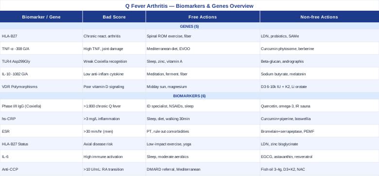
6 Biomarkers Worth Tracking in Q Fever Arthritis
When joint inflammation persists after confirmed or suspected Q fever, a targeted biomarker panel tells you things that a standard CBC and metabolic panel cannot. The six markers below were chosen because each answers a distinct clinical question: Is the infection still driving the response? How intense is systemic inflammation? Is this drifting toward autoimmunity? Is there a genetic risk profile at play? Together they form a monitoring framework rather than a one-time snapshot.
Biomarker 1: Anti-Coxiella Burnetii Phase I and Phase II IgG Antibodies
Why it matters
This is the only test in the panel that is specific to Q fever itself, and it is the one most likely to be skipped once the acute phase has passed. Coxiella burnetii presents in two antigenic phases. Phase II antibodies dominate during acute infection; Phase I IgG titers, when they remain elevated over time, indicate persistent antigenic stimulation — the hallmark of chronic Q fever. In the arthritis context, knowing whether Phase I IgG is still elevated at meaningful levels changes the clinical decision from "manage inflammation" to "this may still be an active infection." According to expert criteria used at Q fever reference centers, a Phase I IgG titer above 1:800 is a major criterion for chronic Q fever diagnosis.
Even in cases of reactive arthritis without true chronic infection, the kinetics of antibody levels matter. Phase II IgG that is rising rather than falling over successive tests indicates the immune system is not winding down. That pattern shifts the clinical priority significantly.
How to measure it
The gold standard is immunofluorescence assay (IFA). Major reference laboratories offer it as a Q fever serology panel including Phase I IgG, Phase II IgG, and Phase II IgM. Cost ranges from approximately $80 to $180 depending on the laboratory and the breadth of the panel ordered. Testing at baseline and then every 3 months is appropriate when monitoring for chronic Q fever or evaluating treatment adequacy.
If the result is abnormal — plan without supplements
Phase I IgG at 1:800 or higher, or rising titers over sequential tests, warrants an infectious disease specialist referral immediately. The standard treatment for confirmed chronic Q fever is long-term combination antibiotic therapy — hydroxychloroquine plus doxycycline for 18 months to 3 years under consensus guidelines established by international Q fever expert groups. Arthritis in this setting is not purely reactive; it has an ongoing infectious driver, and suppressing inflammation without treating that driver can allow the underlying condition to worsen while symptoms are temporarily masked. NSAIDs and low-dose colchicine can manage symptomatic joint pain. Gentle daily range-of-motion exercise during flares, sun exposure for immune support, and sleep optimization (7–9 hours consistently) all support immune competence without pharmacological intervention.
If the result is abnormal — plan with supplements or equipment
While antibiotic therapy remains the cornerstone for elevated Phase I titers, adjunctive support can reduce the inflammatory burden during prolonged treatment courses. Quercetin (500–1000 mg/day with food) has in vitro evidence for inhibiting Coxiella intracellular replication pathways, though human trials specific to Q fever are still limited — consider it supportive rather than therapeutic. Vitamin D3 (4000–6000 IU/day with K2 at 100–200 mcg) is directly relevant because VDR gene polymorphisms — covered in the genetics section — affect immune modulation and are common in reactive arthritis patients; monitor 25-OH-D every 6 months and maintain 40–70 ng/mL. Omega-3 fatty acids (EPA/DHA 2–4 g/day from high-quality fish oil) reduce prostaglandin-mediated joint inflammation and carry the strongest evidence base among supplements for arthritis-associated inflammatory markers. No cycling is required, but monitor LDL at doses above 3 g. Infrared sauna sessions (2–3x per week, 20 minutes at 55–60°C) support lymphatic drainage and have small-trial evidence for reducing inflammatory cytokine burden in arthritic conditions.
Biomarker 2: High-Sensitivity C-Reactive Protein (hs-CRP)
Why it matters
CRP is synthesized by the liver in direct response to IL-6 signaling, making it a downstream readout of systemic inflammatory activity. In Q fever arthritis, its value is primarily as a tracking tool: elevation above 3 mg/L signals active systemic inflammation, and a downward trend over weeks of treatment or lifestyle change confirms that an intervention is having a measurable effect. Peter Attia has consistently placed hs-CRP among the most important routine biomarkers for long-term health, noting that chronic low-grade elevation — even in the 1–3 mg/L range — carries meaningful risk beyond joint disease alone, including cardiovascular consequences that compound over years of uncontrolled inflammatory activity.
In Q fever arthritis specifically, hs-CRP helps distinguish active flares from remission, calibrate the intensity of anti-inflammatory support, and serve as an early warning when elevation returns after apparent improvement.
How to measure it
High-sensitivity CRP — not standard CRP — is the correct version, detecting values in the 0.1–10 mg/L range that are meaningful for chronic inflammation monitoring. It is available at essentially every laboratory at a cost of $10–$30. Draw fasting, away from recent acute illness or vaccination, which transiently spike results. During active treatment adjustments, measure every 6–8 weeks; quarterly once stable.
If the score is bad — plan without supplements
Sleep quality is the most underused lever: consistently short or disrupted sleep independently elevates hs-CRP by measurable amounts across dozens of studies. Eliminating refined carbohydrates and trans fats produces CRP reductions comparable to low-dose statin therapy in some trials. Daily low-intensity exercise — 30 minutes of walking — has demonstrated 25–35% CRP reductions in inflammatory arthritis patients over 12-week periods. In the Q fever context, chronically elevated hs-CRP should also prompt reassessment of whether Coxiella serology warrants retesting, as ongoing antigenic stimulation is one driver that lifestyle changes alone cannot address.
If the score is bad — plan with supplements or equipment
Curcumin with piperine (500 mg curcumin + 5 mg piperine, twice daily with meals) has multiple RCT evidence for hs-CRP reduction in inflammatory arthritis, with some trials showing effects comparable to low-dose NSAIDs. Cycle 8 weeks on, 2 weeks off. GI discomfort is the main side effect at higher doses; avoid combining with blood-thinning medications. Boswellia serrata extract standardized to AKBA (200–400 mg twice daily) has RCT evidence for CRP and joint inflammation reduction in various arthritis types — cycle alongside curcumin. Magnesium glycinate (300–400 mg at night) supports CRP reduction indirectly through sleep quality improvement and NF-kB pathway modulation; continuous use is appropriate. Photobiomodulation (red light at 630–850 nm, 10 minutes directly on affected joints, 3–4x per week) reduces local inflammatory cytokine expression and has growing evidence for reactive arthritis applications.
Biomarker 3: Erythrocyte Sedimentation Rate (ESR)
Why it matters
ESR is older and less specific than CRP, but it captures different aspects of the inflammatory state — particularly fibrinogen levels and immunoglobulin burden — that CRP misses. Used together, the two markers tell different parts of the story. An elevated ESR with normal CRP can indicate a more chronic, immunoglobulin-driven pattern; elevated CRP with normal ESR suggests a more acute, cytokine-mediated process. Rheumatologists use this combination as a diagnostic and monitoring cross-check in reactive arthritis, and the pattern of one normalizing while the other does not is clinically significant.
A persistently elevated ESR above 30 mm/hr in men or above 40 mm/hr in women after apparent symptom resolution in Q fever arthritis should prompt re-evaluation of whether the infection was truly cleared and whether treatment was adequate in duration.
How to measure it
ESR costs $10–$20 and requires no special sample preparation. Draw it alongside hs-CRP and Coxiella serology for maximum interpretive value. Monthly tracking during active flares, then quarterly during maintenance monitoring, is a practical schedule.
If the score is bad — plan without supplements
The same lifestyle interventions that reduce CRP apply here. Additional priorities include excluding concurrent conditions that independently elevate ESR — anaemia, thyroid dysfunction, or kidney disease. In Q fever arthritis, chronic ESR elevation reinforces the need to verify whether antibiotic treatment duration was adequate. Physical therapy focused on joint mobilization reduces ESR in reactive arthritis partly by improving lymphatic drainage and reducing local cytokine concentration in pericapsular tissue.
If the score is bad — plan with supplements or equipment
Proteolytic enzymes — bromelain (500 mg) combined with serrapeptase (120,000 IU) on an empty stomach twice daily — reduce fibrinogen levels and circulating immune complex burden, both of which drive ESR elevation. Take away from meals. Cycle 6 weeks on, 2 weeks off. Do not combine with anticoagulants without medical supervision. Nattokinase (100 mg / 2000 FU, once daily on an empty stomach) similarly reduces fibrinogen with supporting evidence from clinical studies on vascular inflammation; apply the same caution with blood thinners. For local joint inflammation contributing to systemic markers, pulsed electromagnetic field (PEMF) therapy devices (30 minutes daily on affected joints) have clinical evidence for reducing joint-derived inflammatory mediators that contribute to systemic markers like ESR.
Biomarker 4: HLA-B27 Status
Why it matters
HLA-B27 occupies an unusual position in this panel: it is both a genetic marker and a clinical test, and it reframes the entire picture when positive. Between 30% and 60% of patients who develop reactive arthritis after a bacterial trigger carry HLA-B27 — a rate 20 to 30 times higher than background population prevalence. In Q fever arthritis specifically, HLA-B27 positivity is associated with more severe joint involvement, a higher risk of axial (spinal and sacroiliac) disease, and a greater likelihood of chronicity. The NIH Q fever disease overview notes musculoskeletal complications as among the recognized post-Q fever sequelae, and HLA-B27 is the clearest genetic flag for who is at higher risk of that pathway.
Being HLA-B27 negative does not prevent Q fever arthritis, but the clinical course tends toward a shorter duration, less axial involvement, and better spontaneous resolution. Knowing your status calibrates expectations, monitoring intensity, and the threshold for imaging — axial MRI for sacroiliitis is more warranted in HLA-B27 positive patients with any low back or buttock pain component.
How to measure it
HLA-B27 is a one-time genetic test via blood or saliva, performed using flow cytometry or PCR. Cost: $50–$120 at major reference labs. It does not require retesting. The result is binary — positive or negative — sufficient for clinical decision-making, though HLA-B27 subtypes exist that differ in reactive arthritis risk (not routinely reported in standard panels).
If the result is positive — plan without supplements
A positive result is information, not a sentence. It means monitoring for axial disease is warranted, that recurrent reactive arthritis episodes after other infections are more likely, and that achieving and maintaining a low inflammatory burden is particularly important. Daily low-impact exercise — swimming, cycling, yoga with spinal mobility emphasis — is the single most evidence-backed behavioral intervention, as HLA-B27-associated arthritis responds poorly to prolonged immobility. Mediterranean diet adherence is the most RCT-supported dietary framework for reducing reactive arthritis flare frequency and severity.
If the result is positive — plan with supplements or equipment
Low-dose naltrexone (LDN) at 1.5–4.5 mg nightly has accumulating evidence in HLA-B27-associated spondyloarthropathy, acting via opioid growth factor receptor modulation to reduce glial-driven neuroinflammation and inflammatory cytokine production in pericapsular tissue. This requires a physician prescription. Start at 1.5 mg and titrate up over 4–6 weeks; vivid dreams are a common, temporary side effect in the first two weeks. Zinc bisglycinate (25–30 mg/day with food) is specifically relevant: zinc supports metallothionein regulation of immune activation and is frequently deficient in reactive arthritis patients with recurrent flares. Monitor serum zinc every 6 months and balance with copper at 2 mg/day if using doses above 25 mg long-term.
Biomarker 5: Interleukin-6 (IL-6)
Why it matters
IL-6 is the upstream driver of the CRP response, and measuring it directly provides an earlier and more sensitive window into immune activation. In Q fever pathogenesis, Coxiella burnetii has been shown in laboratory studies to actively manipulate IL-6 signaling as part of its immune evasion strategy — making IL-6 monitoring more condition-relevant than it is for most reactive arthritis triggers. Persistently elevated IL-6 is associated with the transition from resolving reactive arthritis to chronic synovitis, partly by driving differentiation of Th17 cells and downstream IL-17 production that perpetuates joint inflammation.
Peter Attia has described IL-6 as one of the most informative cytokine markers in understanding whether inflammation is driven by acute innate immune activation versus smoldering chronic immune dysregulation. In Q fever arthritis, elevated IL-6 with normalized CRP can indicate ongoing deeper immune activity that standard panels miss entirely.
How to measure it
IL-6 is measured via serum ELISA through major reference labs at a cost of $50–$100. Importantly, IL-6 has high diurnal variability and is acutely elevated by intense exercise, stress, and illness. For accurate tracking, draw it fasting in the morning, without strenuous physical activity in the preceding 24 hours. Quarterly measurement during active monitoring is appropriate.
If the score is bad — plan without supplements
IL-6 is acutely sensitive to sleep — even one night of poor sleep produces measurable IL-6 elevation in controlled studies. Stress reduction through structured relaxation or mindfulness practice has direct IL-6-lowering effects at 30 minutes daily. Moderate aerobic exercise (but not high-intensity sessions, which transiently spike IL-6) reduces chronic IL-6 over 8-12 week periods consistently. In the Q fever context, IL-6 elevation alongside elevated Phase I IgG titers is a strong signal that persistent Coxiella activity — not residual autoimmune inflammation — may be the driver, and infectious disease specialist input becomes urgent.
If the score is bad — plan with supplements or equipment
EGCG from green tea extract (400–800 mg/day standardized to 45% EGCG, taken with food) has consistent evidence for IL-6 reduction across inflammatory conditions. Cycle 8 weeks on, 2 weeks off. Avoid on an empty stomach; do not combine with high-dose iron supplementation. Astaxanthin (12–24 mg/day with a fat-containing meal) has RCT evidence for significant IL-6 reduction in inflammatory conditions; continuous use is appropriate. Resveratrol (500 mg/day with a fat-containing meal) inhibits NF-kB-driven IL-6 gene transcription; cycle 8 weeks on, 2 weeks off to avoid estrogenic pathway adaptation. Cold exposure (cold shower or immersion at 10–15°C for 3–5 minutes, 3–4x per week) modulates IL-6 in a hormetic direction — paradoxically anti-inflammatory over time through adaptation. HLA-B27 positive patients should introduce this cautiously and monitor joint stiffness in cold conditions.
Biomarker 6: Anti-CCP Antibodies (Anti-Cyclic Citrullinated Peptide)
Why it matters
Anti-CCP is the most specific marker for rheumatoid arthritis, with specificity above 95%. In Q fever arthritis, it is expected to be negative — and that is precisely its value. When a patient with post-Q fever joint inflammation tests positive for anti-CCP, a critical question arises: did the Coxiella infection trigger an autoimmune transition toward true rheumatoid disease? This is not theoretical. There is documented evidence that severe intracellular bacterial infections can trigger loss of immune tolerance to citrullinated self-antigens in genetically susceptible individuals, particularly those with certain HLA-DRB1 alleles.
A positive anti-CCP result changes treatment intensity, DMARD thresholds, monitoring frequency, and long-term prognosis in ways that purely reactive arthritis management does not account for. A negative result, conversely, provides meaningful reassurance that the arthritis is likely following a post-infectious course with time-limited duration. Thomas Dayspring and rheumatology consensus guidelines both cite anti-CCP as one of the most diagnostically powerful single biomarkers available in arthritis differential diagnosis.
How to measure it
The second-generation assay (anti-CCP2) is standard. Cost: $50–$100. It is generally a one-time test unless there is diagnostic uncertainty. Order it alongside rheumatoid factor (RF) for maximum information: double seropositivity (both anti-CCP and RF positive) in a post-Q fever patient carries major implications and warrants immediate rheumatology referral.
If the result is positive — plan without supplements
Rheumatology specialist referral to determine DMARD candidacy is the first priority. Concurrently, reassessing Coxiella serology is critical — ongoing antigenic stimulation may be fueling the autoimmune transition, and treating any residual infection may reduce the burden on the adaptive immune system. Joint protection strategies become more important: ergonomic support, activity modification, and avoidance of high-load joint use during flares. High-adherence Mediterranean diet with omega-3 and polyphenol emphasis has RCT evidence for improving early RA clinical scores independently of drug therapy.
If the result is positive — plan with supplements or equipment
Fish oil (EPA+DHA, 3–4 g/day) has RCT evidence for reducing anti-CCP titers and clinical scores in early seropositive RA — this is the strongest supplement evidence in this specific context. Vitamin D3 at 5000–6000 IU/day with K2 200 mcg reduces Th17 activity that drives citrullination-driven autoimmunity; maintain 25-OH-D at 50–70 ng/mL. N-Acetylcysteine (NAC) (600–1200 mg/day with food) reduces oxidative stress-driven protein citrullination and has in vitro evidence for RA-relevant autoimmune pathways; cycle 8 weeks on, 2 weeks off. Whole-body vibration platforms (10 minutes, 3–4x per week at 25–50 Hz) have a growing, if small, evidence base for improving joint function and reducing inflammatory markers in early rheumatoid conditions.
What Genetics Reveal: 5 Key Variants Behind Q Fever Arthritis Susceptibility
Not everyone who contracts Q fever develops arthritis. Not everyone with Q fever arthritis develops a chronic form. Much of this variation is genetic. Understanding which immune gene variants you carry helps explain why your response followed the path it did — and opens concrete options for compensating, whether or not you choose to test formally. The five genes below represent the clearest genetic risk architecture for Q fever-related reactive and post-infectious arthritis. Evidence draws from both Q fever-specific immunogenetics research and the broader reactive arthritis genetic literature.
Gene 1: HLA-B27 — The Reactive Arthritis Gateway
HLA-B27 is a class I MHC molecule that presents intracellular peptide fragments to cytotoxic T cells. In people carrying this variant, the immune response to intracellular bacterial antigens — including those derived from Coxiella burnetii — appears to cross-react with self-peptides expressed in joint tissue. This "molecular mimicry" mechanism is the dominant model for why HLA-B27 positive individuals are so disproportionately susceptible to reactive arthritis after intracellular bacterial infections. Population frequency is roughly 8% in Northern Europeans, corresponding to dramatically different background rates of reactive arthritis across ethnic groups. In the context of the 2007–2010 Dutch Q fever epidemic — the largest documented outbreak in history — HLA-B27 positivity was significantly enriched among patients who developed musculoskeletal complications compared to those who recovered without sequelae.
If the gene is bad — plan without supplements
Preventing secondary triggering infections matters most in HLA-B27 positive individuals, who are at high risk for repeated reactive arthritis episodes from any new bacterial exposure — Salmonella, Campylobacter, Chlamydia, and Yersinia are the most common non-Q fever triggers. Maintaining gut microbiome diversity through dietary fiber variety and avoidance of unnecessary antibiotics limits the enteric bacterial reservoir. Daily range-of-motion exercise for the spine and sacroiliac joints is the most evidence-backed behavioral intervention for preventing the axial stiffening that HLA-B27 positive individuals are particularly prone to.
If the gene is bad — plan with supplements or equipment
Low-dose naltrexone (LDN, 1.5–4.5 mg nightly) has the most specific evidence in HLA-B27-associated spondyloarthritis — physician prescription required. Probiotic supplementation (Lactobacillus rhamnosus GG plus Bifidobacterium longum, 10–50 billion CFU daily) reduces gut-derived antigenic triggering in HLA-B27 positive reactive arthritis patients; use continuously. SAMe (S-adenosylmethionine) at 400–800 mg/day on an empty stomach has been studied in spondyloarthropathy with modest benefit on joint pain and morning stiffness; cycle 12 weeks on, 4 weeks off, and monitor mood since SAMe is a methyl donor with mood-active properties.
Gene 2: TNF-Alpha -308 G/A — Amplified Inflammatory Response
The TNF-alpha gene contains a key promoter polymorphism at position -308 (rs1800629) where a G-to-A substitution produces the "A" allele associated with substantially higher TNF-alpha transcription. Carriers of the A allele produce significantly more TNF-alpha in response to the same infectious stimulus than those with the GG genotype. In Q fever arthritis, this means a heightened and more sustained inflammatory response to Coxiella burnetii. The mechanism extends to joint damage: elevated TNF-alpha increases RANKL activation, osteoclast activity in periarticular bone, and sustained NF-kB-driven synovial inflammation — all factors that worsen joint outcomes over time. Research on post-infectious complications of Q fever has found enrichment of the -308 A allele in patients developing chronic fatigue and musculoskeletal symptoms versus those who recovered fully.
If the gene is bad — plan without supplements
Diet is the most accessible lever for TNF-alpha regulation. The Mediterranean pattern consistently reduces circulating TNF-alpha versus Western dietary patterns across multiple RCTs. Extra-virgin olive oil specifically (40+ g/day) provides oleocanthal, which inhibits TNF-alpha synthesis via mechanisms overlapping with ibuprofen at comparable dietary concentrations. Obesity independently amplifies TNF-alpha production from adipose tissue — weight management to a healthy BMI is directly relevant to TNF-alpha load. Resistance training three times per week reduces adipose-derived TNF-alpha significantly over 12-week periods in clinical trials.
If the gene is bad — plan with supplements or equipment
Curcumin phytosome (Meriva formulation) at 1000 mg/day with food has documented TNF-alpha reduction in arthritis studies with substantially better bioavailability than standard curcumin; cycle 10 weeks on, 2 weeks off. Berberine HCl (500 mg twice daily with meals) inhibits NF-kB-driven TNF-alpha gene expression and has additional joint-protective properties; cycle 8 weeks on, 4 weeks off to prevent gut microbiome adaptation. Pentoxifylline (400 mg three times daily) is a methylxanthine drug with specific TNF-alpha synthesis-inhibiting activity used off-label in inflammatory conditions — requires physician prescription; begin at twice-daily dosing and titrate. Red light photobiomodulation at 830 nm, 10 minutes per joint, 3–4x per week has published evidence for local TNF-alpha reduction in arthritic tissue.
Gene 3: TLR4 Asp299Gly — Blunted Innate Recognition of Coxiella
Toll-like receptor 4 (TLR4) is a primary innate immune receptor for bacterial lipopolysaccharide-like structures. Coxiella burnetii has an atypical LPS that signals through both TLR2 and TLR4, but the TLR4 Asp299Gly variant (rs4986790) reduces receptor signaling capacity. This sounds counterintuitive — wouldn't weaker innate immune response limit the inflammatory damage? The paradox resolves this way: a blunted TLR4 response may allow Coxiella to establish a more persistent intracellular reservoir before adaptive immunity mobilizes, increasing the likelihood of chronic post-infectious inflammation. Evidence from post-Chlamydia and post-Salmonella reactive arthritis studies supports TLR4 Asp299Gly as a susceptibility factor for the post-infectious arthritis phenotype broadly, though Q fever-specific genetic data on this variant remain limited — this should be understood as early-stage evidence.
If the gene is bad — plan without supplements
Supporting innate immune function through non-pharmacological means is the priority here. Sleep quality has a direct and proportional effect on NK cell activity and TLR-mediated innate immune surveillance — this is the single most impactful behavioral intervention for individuals with blunted innate immune patterns. Adequate zinc and vitamin A status are both required cofactors for TLR signaling efficiency and are commonly depleted in chronic inflammatory states. Low-intensity regular exercise (not overtraining) stimulates innate immune surveillance without immunosuppressive effects.
If the gene is bad — plan with supplements or equipment
Beta-1,3/1,6-glucan from Saccharomyces cerevisiae (500 mg/day on an empty stomach) has RCT evidence for priming innate immune response via dectin-1 and TLR co-signaling, partially compensating for blunted TLR4 activity; continuous use is appropriate. Andrographis paniculata extract (400–600 mg/day standardized to andrographolide) has immunomodulatory effects on TLR2/4 pathway support and is relevant to intracellular pathogen responses; cycle 6 weeks on, 2 weeks off and avoid in pregnancy. Zinc picolinate (25 mg/day with food) is essential for TLR signal transduction and NK cell function; monitor serum zinc every 6 months and balance copper levels if using above 25 mg/day for extended periods.
Gene 4: IL-10 -1082 G/A — A Weaker Anti-Inflammatory Brake
IL-10 is the primary anti-inflammatory cytokine responsible for limiting immune activation once an infection has been addressed. The -1082 G/A promoter polymorphism (rs1800896) reduces IL-10 transcription in the "A" allele, leaving the immune system with a weaker brake on post-infectious inflammatory responses. Individuals with the AA genotype produce significantly less IL-10, particularly in response to infection, and the resolution of reactive arthritis depends partly on IL-10 successfully dampening the immune response once Coxiella has been cleared. Reduced IL-10 production prolongs this window of inflammatory activity, increasing the risk that joint inflammation becomes self-sustaining rather than resolving.
Ali Torkamani and others working in functional genomics have highlighted IL-10 pathway variants as among the most clinically actionable immune gene polymorphisms, partly because several interventions — dietary and supplemental — can meaningfully compensate for reduced IL-10 production.
If the gene is bad — plan without supplements
Mindfulness meditation (30 minutes daily) is associated with measurable IL-10 increases in multiple controlled studies in inflammatory conditions — this is one of the clearest behavioral interventions with a specific IL-10 effect. Moderate aerobic exercise (not high-intensity sessions, which transiently suppress IL-10) consistently increases regulatory T cell activity and downstream IL-10 production over 8-week periods. Dietary fermentable fiber from vegetables, legumes, and pulses drives butyrate-producing gut bacteria that directly stimulate IL-10 production from intestinal immune cells — this represents a genuinely important dietary mechanism, not a vague "eat more fiber" recommendation.
If the gene is bad — plan with supplements or equipment
Sodium butyrate (1.5–3 g/day with meals) directly stimulates intestinal regulatory T cells and IL-10 production — one of the most mechanistically direct interventions for IL-10 pathway support; continuous use is appropriate. Melatonin at physiological doses (0.3–1 mg at bedtime, not the pharmacological 5–10 mg doses commonly sold) shifts cytokine balance toward IL-10 production in multiple inflammatory condition studies; this is an underappreciated effect that goes beyond sleep support. Prebiotic fiber from PHGG (partially hydrolyzed guar gum, 5–10 g/day in water) reliably increases short-chain fatty acid production and secondary IL-10 from intestinal regulatory T cells; introduce slowly at 2–3 g/day to avoid GI adaptation symptoms.
Gene 5: VDR Polymorphisms — Immune Regulation at Reduced Capacity
The vitamin D receptor (VDR) gene encodes the intracellular receptor through which active vitamin D (1,25-dihydroxyvitamin D3) exerts its immunomodulatory effects. Common VDR polymorphisms — FokI (rs2228570), BsmI (rs1544410), TaqI (rs731236), and ApaI (rs7975232) — affect receptor binding affinity and transcriptional activity, determining how effectively vitamin D signals in immune cells. VDR signaling is critical for regulatory T cell differentiation, suppression of Th17 inflammatory pathways, reduction of IL-17 and TNF-alpha production, and innate antimicrobial defense. Individuals with VDR variants that reduce signaling efficiency are at significantly higher risk for autoimmune and post-infectious inflammatory conditions. Several studies in reactive arthritis populations have found enrichment of VDR FokI "f" allele and BsmI "b" allele compared to healthy controls.
If the gene is bad — plan without supplements
Sun exposure becomes a first-line intervention rather than a lifestyle bonus. Twenty to thirty minutes of direct midday sun on a large skin surface generates the equivalent of 10,000–20,000 IU of vitamin D precursor. Since VDR variants reduce downstream signaling efficiency, achieving higher serum 25-OH-D levels is necessary to drive the same immune effect. Dietary sources alone (fatty fish, egg yolks) are insufficient for meaningful immune-level effects in VDR-variant individuals but contribute at the margin. Ensuring magnesium sufficiency is equally important: magnesium is required for vitamin D activation in the liver and kidneys and is commonly depleted in chronic inflammatory states.
If the gene is bad — plan with supplements or equipment
Vitamin D3 at higher-than-standard doses (6000–10,000 IU/day with K2 200 mcg) is specifically appropriate for confirmed VDR polymorphism carriers who need elevated serum levels to drive adequate receptor signaling through a less efficient receptor; monitor 25-OH-D quarterly and maintain 50–80 ng/mL, with annual calcium monitoring at doses above 6000 IU. Lithium orotate at nutritional doses (5 mg/day) has VDR-upregulating properties via GSK-3-beta inhibition — increasing VDR expression even when receptor binding efficiency is genetically reduced; no prescription required at this dose, but discontinue if mood changes occur. Infrared sauna (40–50°C, 20–30 minutes, 3x per week) enhances regulatory T cell differentiation through heat shock protein 70 induction, partially compensating for reduced VDR-mediated Treg support.
The Book That Reframes Post-Infectious Autoimmunity
The Autoimmune Fix by Tom O'Bryan, DC (2016) is one of the more rigorously referenced books in the functional medicine space, citing over 100 peer-reviewed studies to make a case that challenges standard clinical practice on post-infectious immune conditions. O'Bryan's central argument — that autoimmunity develops on a spectrum over years before clinical diagnosis, and that most patients are in the "silent" or "reactive" phase long before they receive a label — is directly applicable to Q fever arthritis. His framework for understanding how infection-triggered immune events escalate into chronic joint disease offers a practical lens that most rheumatology consultations simply do not have time for.
The following are the ten most impactful ideas from the book, reframed for Q fever arthritis specifically.
1. Autoimmunity Is a Spectrum, Not an On/Off Switch
O'Bryan describes three phases: silent (autoantibodies present, no symptoms), reactive (symptoms begin, no tissue destruction yet), and chronic autoimmune disease (ongoing tissue damage). Q fever arthritis may represent the reactive phase — and staying in that phase versus crossing into chronic autoimmune joint disease depends on what happens next. This framing shifts the goal from "managing symptoms" to "preventing phase progression," which is a fundamentally different clinical target.
2. Every Autoimmune Condition Requires Three Simultaneous Factors
O'Bryan draws on Alessio Fasano's three-hit model: genetic susceptibility (HLA variants), a triggering event (in this case, Coxiella burnetii infection), and intestinal permeability ("leaky gut") that allows immune activation to persist. Addressing any one of the three modifies the outcome; addressing two meaningfully shifts prognosis. The genes section above covers the first hit; the triggering event is already established by Q fever history; gut barrier integrity is the most modifiable of the three.
3. Intestinal Permeability Is the Universal Gateway
The research cited in the book documents that increased intestinal permeability is present in virtually all autoimmune conditions and many post-infectious states. Coxiella infection itself can dysregulate gut mucosal immunity; if untreated, this permeability sustains a low-level immune activation that perpetuates systemic inflammation including joint inflammation. Measuring zonulin or lipopolysaccharide-binding protein (LBP) in blood offers an indirect proxy for gut barrier status.
4. Molecular Mimicry Explains Why Infections Trigger Joint Disease
O'Bryan dedicates a chapter to molecular mimicry — the process by which antibodies raised against bacterial peptides cross-react with structurally similar self-proteins in human tissue. Coxiella burnetii outer membrane proteins share structural homology with several human proteins expressed in synovial tissue, and this cross-reactivity is part of the established model for post-Q fever joint inflammation. Understanding this mechanism helps explain why immune suppression without infection control can backfire.
5. Anti-CCP Can Be Elevated Years Before RA Symptoms
Studies cited in the book show anti-CCP antibodies detectable in blood 5–10 years before clinical rheumatoid arthritis. In the context of Q fever arthritis, a positive anti-CCP is not just a diagnostic label — it is a time-sensitive signal that the autoimmune cascade has started. Early intervention at the reactive phase changes long-term outcomes in ways that intervention at the symptomatic phase cannot replicate.
6. Gluten and Molecular Mimicry Compound Each Other
O'Bryan presents considerable evidence that gliadin peptides (from gluten) are among the most potent triggers of intestinal permeability and share molecular mimicry sequences with multiple self-antigens including collagen II (a joint-tissue protein). For Q fever arthritis patients, particularly those with HLA-DQ2 or DQ8 alleles, gluten exposure may be adding a second layer of molecular mimicry on top of the Coxiella-driven cross-reactivity. A strict gluten elimination for 90 days is the diagnostic and therapeutic test O'Bryan recommends.
7. The Elimination-Reintroduction Protocol Is More Powerful Than Any Single Test
Beyond gluten, O'Bryan outlines an elimination protocol covering gluten, dairy, corn, soy, eggs, and nightshades for 30–90 days, with systematic reintroduction. The logic is not that all these foods are problematic for everyone — it is that in the context of a disrupted immune system post-infection, the threshold for dietary immune activation is significantly lowered, and removal provides a baseline from which to identify individual sensitivities.
8. Gut-Derived Endotoxins Sustain Systemic IL-6 Elevation
Studies referenced in the book link intestinal bacterial translocation (small amounts of LPS crossing the gut barrier) to chronically elevated IL-6 even in the absence of active infection. In Q fever arthritis patients with persistently elevated IL-6 and normalized Coxiella serology, gut permeability-driven endotoxemia is a plausible and underappreciated driver. Addressing gut barrier integrity directly targets this source of IL-6 elevation.
9. Fixing the Terrain Matters More Than Targeting the Trigger
O'Bryan argues — with supporting evidence — that the most durable outcomes in post-infectious autoimmune conditions come from repairing the immune terrain (gut barrier, microbiome diversity, regulatory T cell function, nutrient sufficiency) rather than from indefinite focus on the original pathogen. For Q fever arthritis, this means that once infection is confirmed cleared via serology, the focus should shift to terrain repair rather than continued antibiotic pressure.
10. The Healing Sequence Is Non-Negotiable
The book insists on a specific sequence: remove triggers first, then repair tissue and barriers, then restore microbiome and immune balance. Attempting to restore before removing triggers, or repairing before removal, consistently produces incomplete results. For Q fever arthritis this maps directly to: (1) confirm infection cleared, (2) remove dietary and environmental triggers, (3) repair gut barrier and VDR/IL-10 pathway function, (4) restore regulatory immune balance.
Complementary Approaches With Relevant Evidence
The modalities below were selected because each has meaningful human clinical evidence that applies to the type of joint inflammation seen in Q fever arthritis — specifically post-infectious reactive arthritis with systemic inflammatory components. They are not alternatives to medical management but adjuncts that add value at a practical level.
Mindfulness Meditation and MBSR
Mindfulness-based stress reduction (MBSR) is an 8-week structured program developed by Jon Kabat-Zinn that combines breath awareness, body scan, and gentle movement. Its relevance to Q fever arthritis lies in its dual effect: stress reduction directly lowers cortisol dysregulation that sustains inflammatory cytokines including IL-6 and TNF-alpha, and the program has been shown to increase IL-10 production — the anti-inflammatory cytokine that is genetically blunted in individuals with the IL-10 -1082 AA genotype.
A randomized controlled trial published in Annals of the Rheumatic Diseases (2020) examined mindfulness-based intervention in inflammatory arthritis and found significant reductions in disease activity scores, pain catastrophizing, and inflammatory markers at 8 weeks with effects maintained at 6-month follow-up. A separate meta-analysis in JAMA Internal Medicine confirmed clinically meaningful reductions in CRP in chronic inflammatory conditions with MBSR adherence.
For Q fever arthritis specifically, the recommended starting point is a formal 8-week MBSR course (available online or in person), practiced 30–45 minutes daily. Body scan and breath-focused practices are appropriate during acute flares; gentle yoga-integrated MBSR is better suited to remission phases. The evidence is strongest for chronic inflammatory arthritis; in acute post-infectious phases, evidence is more limited, so maintain conventional medical management as the primary intervention.
Tai Chi
Tai chi is a slow-movement mind-body practice with deep roots in Traditional Chinese Medicine that has been extensively studied in Western rheumatology contexts. For Q fever arthritis, it is particularly relevant because it delivers the specific combination most needed: joint-range-of-motion maintenance, proprioception and balance restoration (impaired in reactive arthritis due to joint effusion and guarding), stress reduction, and low-intensity aerobic conditioning — all without high joint loading.
A well-designed RCT published in Arthritis Care & Research (2010) found that 12 weeks of Tai Chi Chuan significantly improved physical function, pain scores, and health-related quality of life in patients with rheumatoid arthritis, with effects comparable to conventional physical therapy. Additional trials in ankylosing spondylitis — which shares the HLA-B27-associated pathology relevant to Q fever arthritis — have shown significant improvements in spinal mobility and morning stiffness with 24-week Tai chi programs.
For Q fever arthritis, the Yang-style short form (24 movements) practiced for 30–45 minutes, 3–5 times weekly, is the most studied protocol. Begin with a supervised class or video-guided program; transition to independent practice after 4–6 weeks of guidance. The evidence is genuinely meaningful for this condition, though most studies are in established inflammatory arthritis rather than specifically post-Q fever reactive arthritis — apply the evidence with that limitation in mind.
The Autoimmune Protocol from Sarah Ballantyne
The Autoimmune Protocol (AIP), developed by Dr. Sarah Ballantyne (PhD) and detailed in The Paleo Approach (2013), is a dietary elimination and lifestyle framework specifically designed for autoimmune and post-infectious immune dysregulation. It removes foods with evidence for promoting gut permeability and immune activation (grains, legumes, nightshades, dairy, eggs, nuts, seeds, coffee, alcohol) while emphasizing nutrient density from organ meats, oily fish, fermented vegetables, and diverse plant matter. For Q fever arthritis, the AIP is directly relevant to the discussion from The Autoimmune Fix above: it addresses intestinal permeability and dietary molecular mimicry simultaneously.
A pilot RCT published in Inflammatory Bowel Diseases (2017) found significant reductions in clinical disease activity and inflammatory markers in Crohn's disease patients following the AIP over 6 weeks — importantly, this was a condition with established gut-immune dysregulation paralleling what is seen in post-infectious arthritis. For reactive arthritis specifically, clinical studies remain limited, but the mechanistic rationale is strong: reducing gut permeability-derived LPS exposure directly reduces the persistent IL-6 elevation common in chronic Q fever arthritis.
The practical protocol: a strict 30-day elimination phase (no exceptions), followed by systematic reintroduction of one food at a time over 2–3 months. This is not intended as a permanent diet but as a diagnostic and therapeutic reset. Ballantyne's approach includes specific lifestyle factors — sleep prioritization, stress management, light movement — that compound the dietary effect and address the neuroimmune dimension of chronic post-infectious inflammation.
Low-Level Laser Therapy (Photobiomodulation)
Low-level laser therapy (LLLT), also known as photobiomodulation, delivers specific wavelengths of red and near-infrared light (typically 630–1000 nm) at non-thermal intensities directly to inflamed tissue. At the cellular level, it stimulates mitochondrial cytochrome c oxidase, increases ATP production, reduces oxidative stress in synovial tissue, and downregulates pro-inflammatory cytokine expression locally. For Q fever arthritis, where joint inflammation has both infectious and autoimmune components, LLLT's ability to reduce local TNF-alpha and IL-1-beta without systemic immunosuppression is particularly attractive.
A Cochrane systematic review (2010) of LLLT in rheumatoid arthritis examined 7 placebo-controlled trials and found significant pain reduction, morning stiffness reduction, and functional improvement compared to sham treatment, with no significant adverse effects. While the evidence base for reactive arthritis specifically is smaller, the cytokine mechanisms are shared across inflammatory arthritis types, and the safety profile makes it a reasonable adjunct.
Practical protocol: 830 nm wavelength, 100 mW/cm², applied directly to affected joints for 10 minutes per joint, 3–4 sessions per week. Home devices (red light therapy panels or targeted wands) are available in the $150–$600 range. Clinical units are more powerful and can be accessed through physiotherapy practices offering photobiomodulation. Evidence is stronger for peripheral joint involvement than for axial disease; spinal application requires professional guidance.
Microbiome-Directed Therapies
The gut microbiome plays an underappreciated role in reactive arthritis pathogenesis. Several enteric pathogens (Salmonella, Campylobacter) are classic triggers for reactive arthritis via gut-immune axis disruption; Q fever follows a respiratory route but affects gut mucosal immunity through systemic immune dysregulation. In HLA-B27 positive individuals specifically, germ-free animal studies have demonstrated that colonization with specific bacteria is necessary to develop spondyloarthropathy — the microbiome is not merely passive in this disease.
A study in Arthritis & Rheumatology (2017) compared fecal microbiome profiles in patients with reactive arthritis versus healthy controls and found significant reduction in short-chain fatty acid-producing bacteria (Faecalibacterium prausnitzii, Roseburia intestinalis) — bacteria that drive IL-10 production and Treg differentiation. Probiotic and prebiotic interventions targeting this deficit have shown preliminary efficacy for reducing arthritis flare frequency.
The practical approach combines high-dose, multi-strain probiotics (containing Lactobacillus and Bifidobacterium species, 50+ billion CFU, taken with meals, continuously), high-fermentable-fiber dietary intake (30+ g/day from diverse plant sources), and 1–2 servings of fermented foods daily (sauerkraut, kefir, kimchi, tempeh). Avoid unnecessary antibiotic use that disrupts the microbiome, recognizing that the long-course doxycycline/hydroxychloroquine therapy for chronic Q fever can itself deplete short-chain fatty acid producers — targeted probiotic support during antibiotic treatment is therefore especially relevant in this context.
Conclusion
Q fever arthritis is a condition where precision matters. The difference between ongoing Coxiella-driven inflammation and self-sustaining post-infectious autoimmunity determines treatment entirely — and the only way to know which you are dealing with is to look. The biomarkers covered here provide that view: Phase I IgG serology to confirm infectious activity, hs-CRP and ESR for inflammation tracking, HLA-B27 for prognostic framing, IL-6 for early immune activation detection, and anti-CCP to monitor for autoimmune transition. The genetics section adds a layer beneath that — explaining susceptibility through TNF-alpha amplification, TLR4 blunting, IL-10 deficits, and VDR inefficiency, each with actionable compensatory strategies.
The next smart step is not to implement everything at once. Start with the biomarker panel — particularly Coxiella serology if you have not had it repeated since acute infection, and hs-CRP if you have not had it measured recently. Bring those results to an infectious disease specialist and a rheumatologist who is familiar with reactive arthritis. Add the lifestyle interventions with the strongest evidence — sleep, Mediterranean diet, daily low-impact movement — before layering in supplements. And consider genetic testing through a platform like Genomic Life or a functional medicine practitioner to understand which of the five gene variants apply to you specifically, so that targeted supplementation is grounded in your actual biology rather than general arthritis protocols.
Better information does not guarantee better outcomes — but it makes better decisions significantly more likely.
Musculoskeletal: Joint Conditions
Autoimmune: Inflammatory Conditions
Infectious: Bacterial Infections