This article contains AI generated content.
· UpdatedQuadriceps Contracture: 5 Genes And 6 Biomarkers To Track
Introduction
If you are dealing with quadriceps contracture — whether it developed after an injury, a series of injections, a surgical procedure, or simply showed up as stiffness and limited knee flexion that refuses to resolve — you have likely received the standard advice: stretch more, do physical therapy, push through it. For some people, that is enough. For others, the condition stubbornly persists, cycles back, or never fully responds to generic protocols.
That gap between standard advice and actual recovery often comes down to individual biology. Quadriceps contracture is, at its core, a fibrotic process: the muscle and connective tissue become progressively stiffer because the tissue architecture itself has changed. That fibrotic process is not identical in every person. Genetic variants, inflammatory tendencies, and measurable biological signals vary widely — and they change what will actually work for a given individual.
This article does not promise a cure. What it does is offer a more informed perspective. By tracking the biomarkers that reflect what is actually happening inside your tissues, and by understanding the genes that influence fibrosis, inflammation, and muscle repair, you can move from guessing to understanding. That shift in information quality changes what interventions make sense, at what dose, and in what order.
The approaches covered here include tracking six key biomarkers — practical, measurable, and directly actionable — and understanding five relevant genetic variants that shape your baseline risk. A section on the stretching and flexibility research that challenges conventional rehabilitation thinking rounds out the picture, alongside complementary approaches supported by human clinical evidence. None of this replaces a qualified clinician, but it gives you something more valuable than a generic protocol: a framework for making decisions based on your own biology.
6 Biomarkers Worth Tracking in Quadriceps Contracture
Biomarkers do not diagnose a contracture, but they reveal the biological terrain where the contracture is happening. Chronic inflammation, impaired muscle regeneration, and an overactive fibrotic pathway are all measurable — and they all respond to targeted interventions. Tracking these six markers over time gives you a before-and-after picture that most rehabilitation plans simply do not capture.
1. TGF-β1 (Transforming Growth Factor Beta 1)
Why it matters: TGF-β1 is the single most important molecular driver of fibrosis across virtually all tissues, including skeletal muscle. When TGF-β1 signaling is chronically elevated, fibroblasts are activated to produce excess collagen — the defining feature of fibrotic contracture. Research in muscle fibrosis consistently places TGF-β1 at the center of the process, with work by Lieber and Ward in the Journal of Hand Surgery and others documenting its central role in pathological muscle stiffening.
How to measure it: Serum or plasma TGF-β1 via ELISA-based testing, available through specialized or functional medicine labs. Standard reference labs may not include it in routine panels; request it specifically. Cost typically ranges from $80–$180 depending on location and provider. Reference ranges vary by lab, but serum TGF-β1 above 5,000 pg/mL is often considered elevated. In active fibrosis, levels well above this are common.
If the score is bad, the plan without supplements
Aerobic exercise has a well-documented suppressive effect on TGF-β1 activity. Zone 2 cardio (moderate-intensity, 45–60 minutes, 4–5 times per week) downregulates fibrotic signaling over time. Contrast hydrotherapy — alternating cold and warm application to the affected area — may also modulate local TGF-β1 activity. Reducing processed fats, particularly linoleic acid from seed oils, reduces the inflammatory substrate feeding TGF-β1 upregulation.
If the score is bad, the plan with supplements or equipment
Curcumin (turmeric extract) at 500–1,000 mg daily with piperine (black pepper extract) is among the most studied natural TGF-β1 inhibitors. Cycle 8 weeks on, 2 weeks off. Side effects at typical doses are rare but may include mild gastrointestinal discomfort. Resveratrol at 250–500 mg daily has shown anti-fibrotic effects via SIRT1 activation; cycle on the same schedule. Vitamin E tocotrienols at 200–400 mg per day have shown anti-fibrotic properties specifically in muscle tissue in several human studies. Red light therapy (photobiomodulation) devices targeting the quadriceps at 660–850 nm wavelength, applied for 10–20 minutes daily, may reduce local TGF-β1 expression; consumer-grade panels are an accessible starting point.
2. hs-CRP (High-Sensitivity C-Reactive Protein)
Why it matters: hs-CRP is the most accessible general marker of systemic inflammation. Chronic low-grade inflammation — even at levels many clinicians consider within normal range — creates and sustains a pro-fibrotic environment. In the context of quadriceps contracture, elevated hs-CRP suggests the inflammatory phase driving fibrosis has not resolved. Practitioners like Peter Attia emphasize hs-CRP as a cornerstone of routine health monitoring precisely because of its wide-ranging tissue implications.
How to measure it: Included in many comprehensive metabolic panels, or orderable separately through virtually all standard labs. Cost: $15–$50. Optimal target is below 1.0 mg/L; levels above 3.0 mg/L signal clinically concerning systemic inflammatory burden. Most standard panels report CRP rather than hs-CRP — ensure you request the high-sensitivity version for accurate interpretation at the lower end of the range.
If the score is bad, the plan without supplements
Sleep quality is one of the strongest determinants of hs-CRP. Prioritizing 7–9 hours of sleep, consistent sleep-wake timing, and a dark, cool, quiet sleep environment can produce meaningful reductions within 4–6 weeks. Removing ultra-processed foods — particularly refined carbohydrates and seed oils — lowers hs-CRP measurably in controlled studies. Regular low-intensity movement throughout the day (standing, short walks every hour) rather than prolonged sedentary periods independently reduces inflammatory load.
If the score is bad, the plan with supplements or equipment
Omega-3 fatty acids (EPA + DHA) at 2–4 g per day is one of the most consistently replicated anti-inflammatory interventions in clinical trials; suitable for long-term use without cycling. Prioritize high-EPA formulations. Magnesium glycinate at 300–400 mg per day reduces inflammatory signaling and supports sleep quality simultaneously. Quercetin at 500 mg per day has shown hs-CRP-lowering effects in meta-analyses; cycle 8 weeks on, 2 weeks off. Infrared sauna sessions (20–30 minutes, 3 times per week) have demonstrated clinically significant hs-CRP reductions in human studies; avoid if cardiovascular complications are present.
3. 25-OH Vitamin D
Why it matters: Vitamin D is not merely a bone mineral. Skeletal muscle tissue is heavily dependent on vitamin D receptor (VDR) signaling for normal contractile function, repair after injury, and regulation of inflammatory cascades. Low vitamin D levels are consistently associated with muscle weakness, impaired recovery, and increased fibrotic activity. In contracture, suboptimal vitamin D removes a natural brake on the pro-fibrotic process — a brake that costs almost nothing to restore.
How to measure it: Standard blood test (25-hydroxyvitamin D), included in many preventive panels or orderable separately. Cost: $30–$80. Optimal range according to functional medicine practitioners including Peter Attia: 40–60 ng/mL (100–150 nmol/L). Most standard labs flag deficiency only below 20 ng/mL — a threshold that is too conservative for musculoskeletal health optimization.
If the score is bad, the plan without supplements
Midday sun exposure (10–30 minutes of direct skin exposure, 3–5 times weekly when UV index allows) is the physiologically natural correction. This approach also provides benefits no supplement replicates, including UV-induced nitric oxide release. Timing is critical: early morning or late afternoon sun produces very little vitamin D due to low UV-B angle. Fatty fish (salmon, mackerel, sardines, 3–4 times weekly) and egg yolks provide modest dietary contributions.
If the score is bad, the plan with supplements or equipment
Vitamin D3 at 3,000–5,000 IU per day paired with Vitamin K2 (MK-7) at 100–200 mcg per day. Supplementing vitamin D above 2,000 IU without K2 is inadvisable, as K2 directs calcium away from soft tissues. Retest at 90 days. Side effects at these doses are rare, but monitor serum calcium if supplementing long-term at higher doses. At-home narrowband UV-B lamps are an option for individuals with limited sun access; use with appropriate protocols to avoid skin damage.
4. IGF-1 (Insulin-Like Growth Factor 1)
Why it matters: IGF-1 is the primary anabolic hormone for muscle repair and regeneration. It also plays a direct anti-fibrotic role: IGF-1 signaling competes with TGF-β1 at the cellular level, reducing fibroblast activation and promoting myocyte repair rather than scar formation. In quadriceps contracture, low IGF-1 tips the balance toward fibrosis rather than regeneration. Thomas Dayspring and other longevity-focused clinicians consistently include IGF-1 in panels assessing tissue repair capacity.
How to measure it: Serum IGF-1, available through standard labs. Cost: $50–$120. Optimal range varies by age and sex but generally 150–300 ng/mL in adults; below 100 ng/mL in non-elderly adults suggests meaningfully impaired regenerative capacity.
If the score is bad, the plan without supplements
Resistance training is the most potent natural IGF-1 stimulator. Three sessions per week involving multi-joint compound movements (squats, leg press, hip hinge variations) elevates IGF-1 significantly, even at moderate intensity. Adequate protein intake (1.6–2.2 g/kg body weight per day) is essential, as amino acid availability directly influences IGF-1 synthesis. Sleep — specifically deep, slow-wave sleep — is when the majority of IGF-1 is secreted; poor sleep chronically suppresses it.
If the score is bad, the plan with supplements or equipment
Zinc at 15–30 mg per day (paired with copper at 1–2 mg per day to maintain balance) is an essential cofactor for IGF-1 synthesis; cycle 8 weeks on, 2 weeks off. Ashwagandha (KSM-66 extract) at 600 mg per day has shown IGF-1 increases alongside cortisol reduction in multiple randomized controlled trials; cycle 8–12 weeks on, 4 weeks off. Blood flow restriction (BFR) training using specialized cuffs can significantly elevate IGF-1 even at very low loads (20–30% of one-repetition maximum), which is useful when full-load resistance training is contraindicated in the affected limb.
5. IL-6 (Interleukin-6)
Why it matters: IL-6 is a dual-function cytokine. Acutely elevated IL-6 from exercise is beneficial for muscle remodeling. But chronically elevated IL-6 — the kind seen in systemic inflammatory conditions, obesity, and unresolved tissue injury — drives fibrosis and impairs muscle healing. In quadriceps contracture, elevated resting IL-6 signals that the inflammatory environment driving the condition remains active, which is both a diagnostic signal and a modifiable target.
How to measure it: Serum IL-6 via ELISA panel, available through specialized or functional medicine labs. Cost: $60–$150. Standard labs may not include it routinely; request specifically. Resting levels above 3–5 pg/mL in a non-exercised state suggest chronic inflammatory activation worth addressing.
If the score is bad, the plan without supplements
Visceral fat is the primary driver of chronic IL-6 elevation in most adults. Caloric deficit combined with consistent aerobic activity is the most effective long-term IL-6 management strategy. Stress reduction through specific practices — 5 minutes of diaphragmatic breathing before sleep, progressive muscle relaxation — measurably lowers IL-6 through cortisol modulation. Reducing alcohol intake, even modestly, also produces meaningful IL-6 reductions within weeks.
If the score is bad, the plan with supplements or equipment
Fish oil (EPA + DHA) at 3–4 g per day is a primary IL-6 suppressor confirmed in multiple meta-analyses. Melatonin at 0.5–1 mg at bedtime — a physiological dose, not the commonly marketed 10 mg — reduces nighttime IL-6 and improves sleep architecture; suitable for long-term use at this dose. Boswellia serrata extract (AKBA) at 500 mg twice daily has consistent evidence for IL-6 reduction in inflammatory conditions; cycle 12 weeks on, 4 weeks off. Time-restricted eating (16:8 or 18:6 protocol) shows meaningful IL-6 reduction in randomized trials when maintained consistently over 8 weeks or more.
6. Serum Creatine Kinase (CK)
Why it matters: Creatine kinase is the classic marker of muscle cell damage. In quadriceps contracture, persistently elevated CK at rest suggests ongoing muscle breakdown rather than repair — the opposite of what rehabilitation requires. CK tracking also helps calibrate exercise intensity: if CK spikes and stays elevated after a session, the tissue is being damaged faster than it can repair, which actively worsens fibrotic remodeling over time.
How to measure it: Included in many standard blood panels (comprehensive metabolic panel or muscle enzyme panel). Cost: $15–$50. Resting CK in most adults: 55–170 U/L in women, 70–200 U/L in men. Persistent elevation above 300 U/L without recent intense exercise suggests ongoing muscle pathology worth addressing before escalating rehabilitation load.
If the score is bad, the plan without supplements
The most important intervention is exercise dosing: reduce intensity and volume of the affected limb's exercise until CK normalizes, then progress slowly. Cold water immersion (10–15 minutes at 10–15°C, 2–3 times per week) is one of the most robust recovery interventions for CK management in athletic populations. Adequate hydration (minimum 35 mL per kg of body weight daily) is underrated; dehydration meaningfully impairs CK clearance.
If the score is bad, the plan with supplements or equipment
Tart cherry concentrate (equivalent to approximately 90–120 cherries per serving, taken twice daily) consistently reduces CK and inflammatory markers after muscle damage in human trials; no cycling required. Magnesium malate at 400–600 mg per day supports ATP recycling and reduces CK elevation in individuals with suboptimal magnesium status. Compression garments applied after exercise reduce CK elevation and accelerate clearance in multiple randomized studies; can be used after every session without concern.
Moving from biomarkers to genetics adds another layer of precision. While biomarkers show what is happening now, genetic variants reveal your baseline tendencies — the inherent risk factors for fibrosis, inflammation, and impaired repair that persistent good habits can significantly mitigate once they are known.
5 Genes That May Shape Your Quadriceps Contracture Risk
Genetic testing for health optimization is still evolving, and this field moves fast. The variants discussed below have meaningful human evidence — some stronger than others — but none are deterministic. A challenging variant increases probability; it does not write destiny. The most useful framing: genetic data shows you where to be more vigilant and where standard doses of interventions may not be enough.
TGFB1 — The Fibrosis Master Switch
Key variants: rs1800470 (codon 10, Leu/Pro), rs1800469 (C-509T)
What it affects: TGFB1 gene variants directly influence how much TGF-β1 protein your cells produce in response to injury or stress. Pro-fibrotic variants lead to exaggerated TGF-β1 responses — meaning that any muscle injury, including microtrauma from repetitive stress or injection-related damage, triggers a stronger fibrotic cascade than in individuals without these variants. This is arguably the highest-yield genetic factor in quadriceps contracture because TGF-β1 is the direct molecular driver of the condition. TGFB1 polymorphisms are consistently associated with fibrotic disease outcomes across multiple tissue types in large human association studies.
If the gene is bad, the plan without supplements
Individuals carrying pro-fibrotic TGFB1 variants should be especially rigorous about preventing unnecessary muscle microtrauma: avoiding repeated intramuscular injections in the same site, managing exercise loading conservatively after any injury, and taking adequate recovery time between sessions. Zone 2 aerobic exercise (150–200 minutes per week) is one of the most accessible TGF-β1 pathway modulators available without supplementation and should be a non-negotiable baseline for these individuals.
If the gene is bad, the plan with supplements or equipment
Curcumin with piperine (as detailed in the biomarker section) is particularly important for individuals with TGFB1 risk variants. Adding prescription losartan — an angiotensin II receptor blocker that also functions as a TGF-β1 inhibitor — has been specifically studied in fibrotic conditions; this is a clinically meaningful option for severe cases requiring physician involvement. Red light therapy at 660–850 nm, 10–20 minutes daily to the affected quadriceps, is an accessible equipment-based option with a growing evidence base in tissue fibrosis.
COL1A1 — The Collagen Structure Gene
Key variant: rs1800012 (Sp1 binding site polymorphism)
What it affects: COL1A1 encodes the primary structural protein of scar tissue and fibrotic connective tissue. The rs1800012 variant alters the ratio and structure of collagen type I versus collagen type III, influencing whether fibrotic tissue remains organized and functional or becomes disorganized and rigid — the kind of inelastic tissue that defines established quadriceps contracture. This variant is well-documented in tendinopathy, joint hypermobility, and soft tissue injury research; the specific implications for muscle fibrosis are mechanistically sound.
If the gene is bad, the plan without supplements
Eccentric loading exercises — controlled lengthening of the muscle under tension — are among the most evidence-supported interventions for remodeling collagen in a more functional orientation. For quadriceps contracture, this means slow, progressive prone knee flexion exercises or weight-bearing eccentric loading. The collagen remodeling response to mechanical loading peaks 48–72 hours after exercise, meaning training every second day is more effective than daily training for collagen quality adaptation.
If the gene is bad, the plan with supplements or equipment
Vitamin C at 500 mg taken 30–60 minutes before collagen-stimulating exercise amplifies collagen synthesis by supporting prolyl hydroxylase activity, an approach studied by Keith Baar's lab at UC Davis and replicated in human tendon trials. Hydrolyzed collagen peptides at 10–15 g per day taken with vitamin C may further support connective tissue remodeling; continuous use is appropriate, reassess every 12 weeks. Specific collagen peptide types (type I and type II) may differ in relevance depending on the predominant tissue involved.
ACTN3 — The Muscle Fiber Type Gene
Key variant: rs1815739 (R577X — arginine to stop codon)
What it affects: ACTN3 determines whether fast-twitch muscle fibers produce the protein alpha-actinin-3. Individuals with the XX genotype (two copies of the 577X variant) produce no functional ACTN3 — this shifts the entire muscle's contractile and metabolic profile toward endurance-type characteristics. For quadriceps contracture, XX genotype individuals may have inherently slower recovery from muscle trauma and a fiber type distribution that responds differently to rehabilitation protocols. Research by Nan Yang and colleagues at the Children's Medical Research Institute has detailed these physiological effects in multiple human cohorts.
If the gene is bad, the plan without supplements
XX genotype individuals tend to respond better to higher-volume, lower-intensity training than to explosive loading. Rehabilitation protocols should therefore emphasize sustained, slow eccentric loading over high-force interventions. Recovery time between sessions should be extended to 48–72 hours minimum for affected muscle groups, as recovery kinetics are meaningfully different from RR genotype individuals.
If the gene is bad, the plan with supplements or equipment
Creatine monohydrate at 3–5 g per day supports phosphocreatine replenishment in the fast-twitch fibers that ACTN3 deficiency leaves less metabolically efficient — one of the most consistently effective, safe, and well-studied supplements in sports medicine. Long-term use is considered safe; no cycling is required. Blood flow restriction (BFR) training equipment is specifically beneficial for XX individuals who cannot tolerate heavy loads on the affected limb, providing a strong anabolic stimulus at 20–30% of one-repetition maximum.
IL6 — The Inflammatory Tendency Gene
Key variant: rs1800795 (G-174C promoter polymorphism)
What it affects: The C allele at rs1800795 is associated with higher baseline IL-6 production, particularly in response to injury and physiological stress. Individuals with CC or GC genotypes may mount larger and more persistent inflammatory responses to muscle trauma — including the kind of minor repeated trauma that initiates or perpetuates quadriceps contracture. This variant has been studied in the context of athletic recovery, post-surgical inflammation, and inflammatory disease susceptibility.
If the gene is bad, the plan without supplements
Cold exposure — 2–3 minutes of cold water at the end of each shower, daily — is one of the most accessible acute modulators of IL-6 response. Managing psychological stress is equally important: the HPA axis and IL-6 are tightly linked, meaning sustained cortisol elevation directly increases IL-6 secretion. Ten minutes of diaphragmatic breathing twice daily is a specific, measurable practice that has demonstrated cortisol and IL-6 reduction in controlled studies.
If the gene is bad, the plan with supplements or equipment
Individuals with IL6 risk variants should prioritize omega-3 supplementation as a long-term strategy (see biomarker section). Boswellia serrata AKBA extract at 500 mg twice daily provides meaningful additional IL-6 suppression; cycle 12 weeks on, 4 weeks off. Cold water immersion post-exercise (15 minutes at 10–12°C) specifically blunts the post-exercise IL-6 spike, which may be disproportionately amplified in C allele carriers compared to the general population.
VDR — The Vitamin D Receptor Gene
Key variant: rs2228570 (FokI — F/f polymorphism)
What it affects: The VDR gene determines how effectively cells respond to vitamin D. Even with adequate serum 25-OH vitamin D levels, individuals with the ff genotype have a shorter, less functional vitamin D receptor protein — meaning the same circulating vitamin D level produces weaker intracellular signaling. In quadriceps contracture, this matters because VDR signaling in muscle tissue directly suppresses TGF-β1 expression and supports myogenic differentiation over fibroblast activation. VDR polymorphisms and their functional consequences are well-characterized in the literature; direct studies in muscle contracture are limited, but the mechanistic pathway is well-supported.
If the gene is bad, the plan without supplements
For ff genotype individuals, the target serum 25-OH vitamin D level should be set meaningfully higher — 60–80 ng/mL rather than the standard 40–60 ng/mL target — to compensate for reduced receptor efficiency. Maximizing non-supplement vitamin D exposure through midday sun and dietary sources is a reasonable first step, though rarely sufficient for ff individuals to reach elevated targets without supplementation.
If the gene is bad, the plan with supplements or equipment
Vitamin D3 at 5,000–8,000 IU per day (with mandatory K2 MK-7 at 200 mcg per day) may be necessary for ff genotype individuals to reach functional serum targets. Magnesium glycinate at 300–400 mg per day is an essential cofactor for vitamin D activation and should accompany any serious vitamin D optimization. Retest 25-OH Vitamin D every 90 days until target is stable, then semi-annually. Avoid reaching above 100 ng/mL without physician monitoring.
Summary Table: Genes and Biomarkers at a Glance
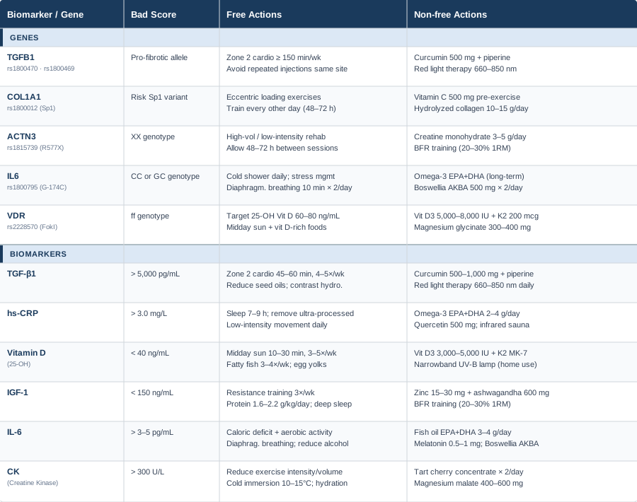
Understanding what the research says about stretching, muscle lengthening, and neurological adaptation opens a practical dimension that biomarker and genetic data alone cannot cover. The work of Andrew Huberman synthesizes a body of science that challenges conventional rehabilitation thinking in ways directly relevant to quadriceps contracture.
What the Science of Flexibility Research May Change About Your Approach
Andrew Huberman's deep examination of stretching physiology — grounded in peer-reviewed research and covered across multiple episodes of the Huberman Lab podcast — challenges several widely held assumptions about how muscle lengthening actually works. For quadriceps contracture, this perspective reshapes the rehabilitation process itself.
1. Flexibility Is Primarily Neural, Not Purely Structural
The most common misconception about limited range of motion is that it is purely mechanical — that the tissue is too short or too stiff to move. Huberman's synthesis of the literature, drawing on work from researchers in kinesiology and neuroscience, emphasizes that in most cases, range of motion limitation is primarily a function of the nervous system's tolerance for length, not the actual tissue architecture. Even as tissue does become fibrotic, the neural component — gamma motor neuron activity, stretch reflex sensitivity — compounds the functional limitation substantially.
Practical implication: static stretching is significantly more effective when the nervous system is calmed first. Techniques that downregulate sympathetic tone before a stretch — 2–3 minutes of diaphragmatic breathing, progressive relaxation of surrounding musculature — can immediately increase effective range of motion even before any structural change occurs.
2. The Optimal Stretching Dose Is Surprisingly Specific
Research synthesized by Huberman suggests that the productive range for neural adaptation in static stretching is 30-second holds, repeated 5 times per muscle per session, performed at least 5 days per week. This specific protocol — approximately 2.5 minutes of cumulative stretch time per muscle per week as a minimum threshold — produced better outcomes than shorter or less frequent approaches in controlled studies. Going significantly beyond this in a single session did not produce proportionally greater adaptation and in some studies produced diminishing returns.
For quadriceps contracture specifically, this means the "stretch more" advice is misdirected not in whether to stretch, but how. Brief, frequent, well-tolerated stretches with deliberate breathing outperform infrequent marathon sessions that the nervous system resists.
3. Heat Before Stretching Produces Mechanical Advantage
For individuals with established contracture, heat applied to the quadriceps before stretching (15–20 minutes of moist heat or a heat pad) mechanically reduces tissue stiffness and lowers the neural resistance to lengthening. This is well-established in basic and clinical tissue mechanics research. Cold applied afterward to manage inflammation does not appear to reverse the thermal priming effect — making a heat-before, cold-after approach physiologically sound for contracture rehabilitation.
4. Eye Position Affects Lower Extremity Flexibility
One of the more counterintuitive findings in this area: the position of your eyes during stretching affects tone in the extremities through vestibulo-spinal and tonic neck reflex pathways. Eyes looking upward during a prone quadriceps stretch modestly increases neural drive to the quadriceps, increasing resistance to the stretch. Eyes looking downward or fixed at a horizontal point reduces this effect. In practical terms: lying face down in a prone stretch with your gaze toward the ground rather than looking up may make the same stretch measurably more effective.
5. PNF Technique Reduces Stretch Reflex Resistance
Proprioceptive neuromuscular facilitation (PNF) is a well-supported tool for temporarily reducing muscle spindle sensitivity and allowing greater range to be achieved. The specific technique relevant to quadriceps contracture: at the end of a passive stretch, isometrically contract the quadriceps against resistance for 6–8 seconds, then deliberately release and deepen the stretch during the relaxation window that follows. Repeat 3–5 times. The post-contraction inhibition window provides a brief period of reduced spindle activity during which the stretch is significantly more effective.
6. Yoga Nidra After Stretching Accelerates Adaptation
Huberman's work on neuroplasticity emphasizes that consolidation of new movement patterns — including increases in usable range of motion — occurs during rest, not during training itself. Twenty minutes of Yoga Nidra or non-sleep deep rest (NSDR) following a stretching session has been shown to accelerate nervous system adaptation. For individuals in rehabilitation for quadriceps contracture, this is a zero-cost, zero-risk addition that meaningfully improves the efficiency of each session.
7. Protein Intake Directly Affects Connective Tissue Adaptation
Huberman frequently notes that flexibility training is a form of tissue stimulus requiring adequate protein for adaptation. Individuals in rehabilitation who are protein deficient — below 1.4–1.6 g per kg body weight per day — will have blunted connective tissue remodeling responses regardless of how consistently they stretch. This is especially relevant for older adults with contracture, where protein intake is often insufficient.
8. Progress Is Non-Linear and Requires Monthly Assessment
Flexibility gains in fibrotic conditions plateau and then seem to jump. Assessing progress week by week leads to premature abandonment of protocols that are actually working on a biological timeline that does not match weekly expectations. Collagen turnover occurs over months, not days. A multi-week plateau is not evidence that the approach has failed; it is often a quiet consolidation phase before the next gain.
9. Aggressive Stretching of Fibrotic Tissue Can Worsen the Condition
Stretching fibrotic tissue with high force past the point of comfortable resistance can cause microtears that trigger further fibrotic repair cycles — actively worsening contracture over time. The evidence strongly supports a "never stretch to pain" principle in established contracture. The goal is the edge of comfortable tension, held there with deliberate breathing, not aggressive mechanical loading through the painful range.
10. Consistency Beats Intensity Across Every Flexibility Study
Across virtually every human study on flexibility adaptation, consistency of practice is the primary predictor of long-term outcomes. Five short sessions per week outperform one long session per week every time. For quadriceps contracture, building a daily 15–20 minute routine — that is manageable enough to actually sustain for months — is far more valuable than an intensive session once or twice a week that the body cannot recover from and the mind resists continuing.
Complementary Approaches With Clinical Support
Each approach below was selected based on meaningful human clinical evidence specifically relevant to muscle fibrosis, contracture management, or rehabilitation of restricted range of motion. None replaces primary medical care or a structured physical therapy program, and evidence quality varies across modalities.
Massage Therapy
Massage therapy is one of the most mechanically direct interventions for conditions involving myofascial restriction. For quadriceps contracture, deep tissue massage and myofascial release techniques address abnormal tissue tension, fascial adhesions, and lymphatic stagnation that accompany fibrotic remodeling. The mechanical deformation of fibrotic tissue under therapeutic pressure — combined with the neurological effects of reducing sympathetic tone in the affected area — makes massage both a structural and neuromodulatory intervention.
A randomized controlled trial by Moyer and colleagues published in the Journal of Orthopaedic & Sports Physical Therapy found that massage therapy significantly reduced passive stiffness and increased range of motion in subjects with restricted lower extremity flexibility compared to controls. For contracture-adjacent conditions, manual therapy applied with sufficient depth — slow, sustained pressure rather than superficial gliding — demonstrated the most meaningful outcomes.
Practically: seek a therapist experienced in myofascial release or deep tissue work specifically for quadriceps and hip flexor complexes. Sessions of 45–60 minutes, once to twice weekly during active rehabilitation, are a reasonable starting point. Aggressive pressure beyond discomfort threshold is counterproductive — communicate this clearly. Home foam rolling between sessions (3–5 minutes daily on the affected quadriceps) maintains gains between appointments.
Low-Level Laser Therapy (Photobiomodulation)
Photobiomodulation uses specific wavelengths of light (typically 630–1100 nm) at non-thermal intensities to stimulate mitochondrial activity, reduce local inflammation, and modulate fibroblast behavior. For quadriceps contracture, it is mechanistically relevant because it acts at several levels simultaneously: reducing local TGF-β1 expression, increasing cellular ATP production in stressed tissues, and modulating the local inflammatory cytokine environment.
A systematic review by Bjordal and colleagues examining LLLT for musculoskeletal conditions found significant benefits in pain reduction and soft tissue healing. Several studies examining LLLT in post-traumatic fibrosis have shown histological evidence of reduced collagen deposition in controlled models, with human evidence growing but more limited for contracture specifically.
Clinically, sessions of 5–15 minutes applied to the affected quadriceps, 3–5 times per week for 4–8 weeks, represent the protocols used in most trials. Consumer-grade red light therapy panels (660 nm + 850 nm) offer an accessible home option at lower irradiance than clinical devices; apply for 10–20 minutes daily at 10–15 cm distance. Side effects are minimal when thermal thresholds are not exceeded.
Yoga
Yoga is not merely stretching. In the context of quadriceps contracture, the combination of sustained hip flexor and quadriceps elongation in poses such as low lunge, prone quad stretch, hero pose, and reclined hero — combined with controlled breathing and progressive proprioceptive challenge — addresses both the structural restriction and the neurological hypersensitivity that compound the condition. The parasympathetic activation achieved during sustained yoga practice also creates the optimal neurological environment for range of motion gains.
A systematic review by Cramer and colleagues published in the Journal of Alternative and Complementary Medicine evaluated yoga's effects on musculoskeletal conditions and found significant improvements in flexibility and functional mobility, with the most benefit observed in consistent practitioners performing 3 or more sessions per week. For conditions with a fibrotic component, yin yoga — passive poses held 3–5 minutes each — is particularly relevant because the extended duration targets deeper fascial layers.
Begin with supported versions of each pose, using blocks, bolsters, or wall support to reduce load on the affected tissues. Avoid any pose creating sharp pain or strong traction sensation in the anterior thigh. A restorative or therapeutic yoga style is more appropriate than power or hot yoga in the active phase of contracture management. Twenty to thirty minutes three times per week of targeted lower body yin practice is clinically meaningful.
Biofeedback
Surface electromyography (sEMG) biofeedback allows patients to see in real time how much electrical activity — tension — is present in the quadriceps at rest and during voluntary relaxation attempts. In many contracture cases, the central nervous system maintains excessive resting tone in the affected muscle as a form of learned guarding, which biofeedback can help unlearn. This is the neurological layer of contracture that purely structural interventions do not address.
Research published in the Archives of Physical Medicine and Rehabilitation demonstrated that EMG biofeedback enhanced neuromuscular control and relaxation in patients with movement disorders affecting lower extremity tone. For quadriceps contracture, biofeedback has been incorporated into post-surgical rehabilitation protocols to help patients both reduce resting tension and selectively activate weakened muscle segments.
Biofeedback sessions are typically conducted by physiotherapists with sEMG equipment. Six to twelve weekly sessions represent a reasonable course. Between clinical sessions, progressive muscle relaxation targeting the quadriceps reinforces the biofeedback learning at home. Consumer-grade wearable EMG devices are now available and can extend biofeedback practice outside clinical settings.
Progressive Muscle Relaxation (PMR)
Progressive muscle relaxation is a structured technique in which muscle groups are deliberately tensed and then fully released in sequence. For quadriceps contracture, PMR addresses the neurologically maintained tension component — the layer of the condition that exists above purely structural fibrotic changes. It requires no equipment, costs nothing, and has an extensive evidence base in musculoskeletal rehabilitation.
A meta-analysis by Conrad and Roth (2007) in the International Journal of Stress Management confirmed that PMR produces significant reductions in muscle tension, sympathetic nervous system activation, and subjective pain compared to control conditions across a range of musculoskeletal presentations. In chronic conditions specifically, PMR practiced consistently reduced resting muscle tension as measured electromyographically and reduced perceived tightness.
A 15–20 minute PMR session daily, with deliberate focus on the affected quadriceps and surrounding hip musculature, is a clinically grounded practice. The standard protocol involves 5–10 seconds of deliberate contraction followed by 20–30 seconds of complete release, moving systematically through the leg. Evening timing — when sympathetic tone is already lower — is optimal. Audio-guided PMR sessions are widely available and improve long-term adherence significantly.
Conclusion
Quadriceps contracture resists generic approaches because it is not a generic condition. The fibrotic process driving it, the inflammatory environment sustaining it, and the neurological patterns compounding it vary from person to person — and the genetic variants and biomarker profiles covered in this article are where those individual differences become actionable.
The most practical next steps: if you have not already, order an hs-CRP and 25-OH vitamin D test — both are inexpensive, widely available, and directly actionable. If genetic testing is accessible to you, TGFB1 and VDR variants are the highest-yield place to start for understanding personal fibrosis risk. Layer in the complementary approaches — massage, photobiomodulation, yoga, or PMR — based on what the evidence suggests is most relevant to your specific presentation and what is realistic to sustain.
None of this replaces working with a qualified physiotherapist or physician, particularly for moderate or severe contracture. But it gives you the information needed to have a more precise conversation and make more targeted choices. Better information is not a cure, but it is consistently the first step toward better outcomes.
Musculoskeletal: Joint Conditions Muscle Conditions
Autoimmune: Inflammatory Conditions Connective Tissue Conditions