This article contains AI generated content.
· UpdatedQuadriceps Tendon Rupture – 6 Genes and 7 Biomarkers to Track
Introduction
A quadriceps tendon rupture rarely arrives without a history. For most people, it is the final event in a much longer process of quiet tissue degradation — collagen losing its architecture, metabolic signals going wrong, inflammation accumulating at a level too low to cause symptoms but high enough to matter structurally. By the time the tendon actually fails, the biology that allowed it to fail has usually been in place for years.
What makes this injury particularly frustrating is that it tends to strike people who feel physically capable. Middle-aged athletes, regular gym-goers, and weekend warriors are caught off guard precisely because they feel strong. Muscle strength and tendon resilience do not always track together. Tendons age differently, adapt more slowly, and carry metabolic vulnerabilities that standard fitness assessments do not capture. The gap between what someone thinks their tendon can handle and what it actually can bears is often where ruptures happen.
Most protocols after a quadriceps tendon rupture are focused entirely on the structural repair: surgical fixation, immobilization, graded loading, return to activity. These steps are necessary and well-evidenced. What they do not address is the underlying biological environment that allowed the rupture to occur in the first place. Without understanding those factors, the same systemic vulnerabilities remain after recovery — which is one reason why re-rupture rates and contralateral tendon injuries are more common than most people expect.
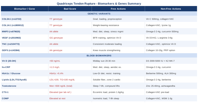
This article takes a more targeted approach. It covers two complementary angles. The first is a set of seven blood biomarkers that, individually and together, can reveal how well your body is supporting tendon collagen, managing inflammation, and maintaining the metabolic conditions that tendons depend on. The second is a look at six genetic variants consistently linked to tendon injury risk and how each can be partially compensated for through targeted interventions. Neither angle provides a complete answer. But both give you specific, actionable information — and that is a meaningful improvement over generic advice about rest, ice, and a standard protein shake.
7 Biomarkers That Reveal Your Tendon Vulnerability
Biomarkers are objective, measurable signals in blood or urine that reflect what is happening at the tissue level. For tendon health, they can tell you whether your collagen environment is anabolic or catabolic, whether inflammation is silently eroding matrix quality, and whether your metabolic and hormonal state supports repair. The seven markers below were selected for their clinical relevance to tendon biology, their measurability in routine lab settings, and — crucially — because each one has a clear action plan when it falls outside the optimal range.
1. 25-OH Vitamin D – The Tendon Repair Foundation
Why it matters: Vitamin D is far more than a bone mineral. It plays a direct role in collagen synthesis, muscle-tendon force transmission, anti-inflammatory gene expression, and immune-mediated tissue repair. Studies in populations with quadriceps, Achilles, and rotator cuff tendon injuries consistently find lower 25-OH vitamin D levels compared to healthy controls. Deficiency at the tissue level impairs the tenocyte's ability to produce new collagen matrix and slows recovery after injury. See related research on PubMed.
What it may reveal: Even sub-optimal levels in the 20–40 ng/mL range — reported as "normal" on many standard lab reports — may be functionally insufficient for tendon repair and regeneration. Chronic deficiency means both collagen maturation and immune-mediated healing are running below capacity.
How to measure it: A 25-OH vitamin D blood test is available at nearly any lab or primary care office. Cost: $30–$80. The optimal functional target is 50–80 ng/mL, not the minimum threshold of 30 ng/mL typically cited on lab reports as "sufficient."
If the score is low, the plan without supplements
Midday sun exposure on large skin surfaces (arms, legs, back) for 20–30 minutes between 10 a.m. and 2 p.m. is the most direct natural source. Dietary sources — fatty fish (salmon, mackerel, sardines), egg yolks, and beef liver — offer modest support but rarely correct a genuine deficiency on their own. Synthesis efficiency drops sharply with age, darker skin tone, and increased latitude, making the sun-only approach unreliable above 40°N in winter months.
If the score is low, the plan with supplements
Vitamin D3 (not D2) at 2,000–5,000 IU per day is appropriate for most adults targeting the 50–80 ng/mL range. For levels below 30 ng/mL, a physician-supervised correction of 5,000–10,000 IU/day for 8–12 weeks is common. Always pair with vitamin K2 as MK-7 (100–200 mcg) to direct calcium appropriately and magnesium glycinate (200–400 mg), which is required to activate vitamin D metabolically — and is itself frequently deficient. Retest at 3 months and adjust dose. Side effects: minimal under 10,000 IU/day; above this threshold, hypercalcemia becomes a real concern — do not exceed without physician monitoring.
2. High-Sensitivity CRP – Your Inflammatory Load
Why it matters: High-sensitivity C-reactive protein (hs-CRP) is one of the most sensitive markers of systemic low-grade inflammation — the kind that operates silently for years while steadily degrading tendon extracellular matrix. Inflammatory cytokines activate matrix metalloproteinases that break down collagen fibrils, impairing both tendon structural integrity and post-injury remodeling. Elevated hs-CRP is associated with tendinopathy in clinical populations and signals an environment where repair is consistently being undermined. See related research on PubMed.
What it may reveal: An hs-CRP above 1 mg/L suggests a proinflammatory state that is working against tendon repair. Values above 3 mg/L warrant deeper investigation: chronic poor sleep, gut dysbiosis, metabolic syndrome, and autoimmune activity are the most common drivers.
How to measure it: Standard blood test, widely available. Cost: $20–$50. Optimal target: below 1.0 mg/L, ideally below 0.5 mg/L. Do not measure within two weeks of acute illness or significant physical stress — both transiently spike CRP in ways unrelated to chronic inflammation.
If the score is elevated, the plan without supplements
The highest-leverage lifestyle interventions are: Mediterranean-style diet (abundant vegetables, olive oil, fatty fish, legumes, minimal ultra-processed foods), sleep optimization targeting 7–9 hours with consistent timing (chronic sleep restriction is among the most potent non-infectious drivers of elevated CRP), and moderate aerobic exercise (30–45 minutes, 4–5 days/week — consistent aerobic exercise has a strong anti-inflammatory effect over time). Time-restricted eating within a 14–16 hour fasting window has also demonstrated measurable anti-inflammatory effects in clinical trials.
If the score is elevated, the plan with supplements
Omega-3 fatty acids (EPA + DHA, 2–4 g/day) from high-quality fish oil or algae-based sources are among the best-evidenced anti-inflammatory interventions available. Bioavailable curcumin (500–1,000 mg/day as Meriva or BCM-95 formulations, which absorb far better than standard curcumin extracts) has robust clinical evidence for lowering CRP. Cycling: 8 weeks on, 2 weeks off for curcumin if taken long-term. Magnesium glycinate (300–400 mg/day) has a modest but consistent anti-inflammatory effect. Side effects: omega-3s at high doses may mildly affect platelet aggregation — relevant if on anticoagulants; curcumin can cause GI discomfort at higher doses, particularly on an empty stomach. Retest at 3 months.
3. HbA1c and Fasting Glucose – The Glycation Problem
Why it matters: This is arguably the most underrecognized risk factor for tendon rupture. Elevated blood glucose — across the full spectrum from occasional high-carb meals to frank diabetes — promotes the formation of advanced glycation end-products (AGEs) that form permanent cross-links between collagen fibers in tendons. This makes tendon tissue progressively stiffer and more brittle, dramatically reducing its ability to absorb dynamic load without tearing. Diabetic patients have a two- to three-fold increased risk of spontaneous tendon rupture. But the risk begins well below the diabetic threshold — pre-diabetic HbA1c levels are sufficient to begin measurably degrading tendon collagen quality. See related research on PubMed.
What it may reveal: HbA1c above 5.5% indicates early-stage collagen glycation load. Fasting glucose consistently above 95 mg/dL, while technically within normal range, warrants attention in the context of tendon health. These numbers should be interpreted together.
How to measure it: Both are standard and inexpensive. HbA1c: $20–$40. Fasting glucose: $10–$20. Optimal targets: HbA1c below 5.4%, fasting glucose 70–90 mg/dL.
If the score is elevated, the plan without supplements
Diet is the most powerful lever here. Reducing refined carbohydrates and added sugar, shifting to a lower-glycemic eating pattern (Mediterranean or low-carbohydrate approaches both work), and eating within a defined daily window (16:8 time-restricted eating) collectively reduce HbA1c, fasting glucose, and AGE formation. Resistance training is the most evidence-backed single intervention for improving insulin sensitivity — 3–4 sessions per week of compound movements (squat patterns, hip hinge, press) measurably lower HbA1c across both diabetic and pre-diabetic populations in well-conducted trials.
If the score is elevated, the plan with supplements
Berberine (500 mg, 2–3 times daily with meals) is the most rigorously studied natural agent for glucose regulation, with multiple meta-analyses showing efficacy comparable to low-dose metformin. Start at 500 mg once daily for the first week to reduce GI adaptation effects. Cycling: 8 weeks on, 4 weeks off, or continuous with the lowest effective dose and periodic breaks. Alpha-lipoic acid (300–600 mg/day) reduces AGE formation directly and improves insulin sensitivity. Magnesium glycinate or malate (300–400 mg/day) is frequently deficient in pre-diabetic populations and impairs glucose metabolism when low. Critical note: berberine should never be combined with metformin or other glucose-lowering medications without physician oversight — hypoglycemia risk is real. Retest HbA1c at 3–4 months.
4. Lipid Panel – LDL-C, Triglycerides, and ApoB
Why it matters: The relationship between lipids and tendon health has two distinct dimensions. First, hypercholesterolemia itself is associated with tendinopathy — cholesterol deposits have been found histologically in degenerated tendons, and patients with familial hypercholesterolemia have markedly elevated rates of tendon xanthomas and spontaneous rupture. Second — and critically underrecognized — statins carry a documented association with tendinopathy and tendon rupture that is real and not trivial. This risk is often dismissed in clinical settings but appears consistently in pharmacovigilance data and case series. If you are on a statin and have had or are recovering from a quadriceps tendon rupture, this connection is worth an explicit conversation with your prescribing physician. See related research on PubMed.
What it may reveal: High LDL-C or ApoB alongside current statin use creates a compounded risk picture for tendon tissue. Elevated triglycerides frequently signal underlying insulin resistance and metabolic dysfunction — the same state that promotes AGE formation in collagen.
How to measure it: Standard lipid panel: $20–$60. Adding ApoB ($30–$80 additional) provides a more accurate measure of atherogenic particle burden than LDL-C alone — a distinction emphasized by Thomas Dayspring and Allan Sniderman in their cardiovascular risk assessment frameworks. Optimal targets: LDL-C below 100 mg/dL, triglycerides below 100 mg/dL, ApoB below 80 mg/dL.
If the score is elevated, the plan without supplements
Increase soluble fiber intake (oats, psyllium, legumes, flaxseed — 10–15 g/day of soluble fiber consistently lowers LDL-C), reduce saturated fat from low-quality sources (processed meats, commercial baked goods), increase zone 2 aerobic exercise (this specifically raises HDL and lowers triglycerides), and eliminate or sharply restrict alcohol, which is a potent driver of elevated triglycerides.
If the score is elevated, the plan with supplements
Omega-3s (EPA + DHA, 2–4 g/day) are highly effective at reducing triglycerides and have a solid safety profile. Plant sterols (1–2 g/day with meals) reduce intestinal cholesterol absorption and are safe for long-term use. Berberine (500 mg 2–3x/day) also lowers LDL-C and TG via PCSK9 inhibition — a mechanism of action supported by a growing number of trials. For those currently using statins and experiencing tendon symptoms, coenzyme Q10 (100–200 mg/day) is widely recommended to address the mitochondrial depletion that statins can cause in both muscle and connective tissue. Side effects: plant sterols are generally well-tolerated; red yeast rice (which contains natural monacolin K, structurally similar to lovastatin) should be used cautiously as it carries equivalent myopathy risk.
5. Total and Free Testosterone – The Anabolic Repair Signal
Why it matters: Testosterone drives collagen synthesis in tendon fibroblasts directly, supports the muscle-tendon interface, and promotes the anabolic state required for daily tissue remodeling. Low testosterone — increasingly common in men over 40 due to aging, chronic sleep debt, elevated cortisol, and metabolic dysfunction — impairs the tendon's ability to repair microdamage that accumulates with physical activity. This is one of the clearest explanations for why spontaneous quadriceps tendon ruptures are predominantly a condition of men in their 40s and 50s. Women are not immune: while their testosterone baseline is much lower, the relative decline with age and metabolic stress carries similar tissue consequences. See related research on PubMed.
What it may reveal: Low free testosterone in particular indicates that anabolic tissue repair signaling is blunted regardless of total testosterone. SHBG (sex hormone-binding globulin) binds testosterone and renders it inactive; high SHBG can make total testosterone look normal while free testosterone is functionally low.
How to measure it: Morning blood draw (testosterone peaks between 7 and 10 a.m.). Total testosterone: $40–$100. Free testosterone: $60–$120. SHBG: often included in broader hormone panels. Optimal for men: total 500–900 ng/dL, free testosterone above 15 pg/mL. For women, reference ranges are considerably lower and must be interpreted relative to age and menstrual status.
If the score is low, the plan without supplements
Sleep quality is the single most powerful lifestyle intervention for testosterone — 70–80% of daily testosterone production occurs during sleep, and chronic restriction below 7 hours measurably reduces levels within days. Resistance training with compound, heavy movements (squat, deadlift, press, pull) 3–5 days/week has a consistent testosterone-elevating effect over time. Reducing visceral body fat (which aromatizes testosterone to estrogen), managing chronic psychological stress (elevated cortisol directly suppresses testosterone), limiting alcohol, and ensuring adequate dietary fat (cholesterol is the direct precursor to steroid hormones) are all meaningful contributors.
If the score is low, the plan with supplements
Zinc (25–40 mg/day with food) is one of the most evidence-based micronutrients for testosterone support — deficiency is common and strongly associated with hypogonadism. Pair with copper (1–2 mg/day) when using zinc long-term to prevent copper depletion. Ashwagandha KSM-66 (300–600 mg/day) has demonstrated significant testosterone increases in randomized trials in men. Tongkat ali (200–400 mg/day) may help by reducing SHBG. Cycling: ashwagandha can be taken continuously; some prefer 12 weeks on, 2–4 weeks off. Tongkat ali: 5 days on, 2 off, or 3 weeks on, 1 week off. Side effects: ashwagandha — rare hepatotoxicity reported at very high doses; start low and monitor; zinc without copper long-term risks copper-deficiency anemia; tongkat ali is generally well tolerated in normal ranges.
6. CTX-1 – Collagen Breakdown Rate
Why it matters: CTX-1 (C-terminal telopeptide of type I collagen) is a direct degradation product of type I collagen — the primary structural protein in tendons, ligaments, and bones. Chronically elevated CTX-1 signals that collagen breakdown is outpacing synthesis, meaning the tendon is operating in a net catabolic state. This is a particularly important marker during tendon recovery, where the balance between breakdown and regeneration determines whether tissue quality improves over time or continues to degrade. See related research on PubMed.
What it may reveal: Elevated CTX-1 in context points to one or more of the following: insufficient anabolic signaling (low vitamin D, low testosterone, low protein intake), excessive mechanical stress without adequate recovery, corticosteroid use (which directly suppresses collagen synthesis), or systemic catabolic states like chronic caloric restriction.
How to measure it: Serum or urine beta-CTX, available at specialty labs and through several direct-to-consumer panels. Cost: $80–$150. A fasting morning sample is required — CTX-1 is diurnal, peaking in the fasting morning state before dropping post-meal. Reference ranges vary by age and sex, so results should be interpreted relative to the lab's reference population.
If the score is elevated, the plan without supplements
Structured mechanical loading is the single most powerful intervention for shifting tendon metabolism from catabolic to anabolic. Eccentric and isometric loading protocols targeting the quadriceps-tendon unit signal tenocytes to increase collagen production and suppress degradative enzyme activity. Adequate dietary protein (1.6–2.2 g/kg body weight/day) is the nutritional foundation — tendons require proline, glycine, lysine, and hydroxyproline as substrates for collagen synthesis, and chronic protein insufficiency is a direct brake on repair. Ensure total caloric intake is not in significant deficit during the recovery period.
If the score is elevated, the plan with supplements
The most scientifically grounded protocol is collagen peptides (10–15 g) with vitamin C (50 mg), taken 30–60 minutes before mechanical loading or physical therapy. This specific timing — established through the work of Keith Baar's lab and published in the American Journal of Clinical Nutrition — creates a priming window during which circulating amino acids from collagen are available when mechanical load stimulates tenocyte collagen synthesis. Glycine (3–5 g/day) is the rate-limiting amino acid for collagen and can be added to any collagen supplementation routine. Vitamin K2 MK-7 (100–200 mcg/day) supports matrix cross-linking quality. No cycling required. Side effects: collagen peptides are exceptionally well-tolerated; vitamin C above 2 g/day may cause loose stools; glycine is among the safest amino acids in human supplementation.
7. COMP – Tendon Matrix Stress Indicator
Why it matters: COMP (Cartilage Oligomeric Matrix Protein) is a non-collagenous structural glycoprotein found abundantly in tendons, ligaments, and cartilage. While CTX-1 measures collagen breakdown products, COMP is released from the extracellular matrix under mechanical stress — elevated serum COMP at rest (not acutely post-exercise) indicates that tendon matrix integrity is compromised and that structural disruption is ongoing. It has been studied as a serum biomarker for Achilles tendinopathy and other connective tissue pathologies, with elevated resting levels correlating with tendon structural abnormalities on imaging. See related research on PubMed.
What it may reveal: Acutely elevated COMP immediately after exercise is normal and transient — it reflects healthy mechanical loading. Chronically elevated COMP measured at rest, however, suggests the extracellular matrix is under persistent structural stress and that repair mechanisms are not keeping pace.
How to measure it: Serum COMP through specialty labs — less routine than the other markers in this list but available through several functional medicine panels. Cost: $100–$250. Most useful in the context of ongoing tendon rehabilitation monitoring rather than general population screening. Measure at rest, avoiding testing within several hours of significant physical activity.
If the score is elevated, the plan without supplements
Reduce excessive mechanical overload while maintaining low-load isometric and eccentric exercises that promote tendon adaptation without exceeding the tissue's current stress threshold. Prioritize sleep quality (7–9 hours) — growth hormone, secreted predominantly during deep sleep stages, is the primary endogenous driver of connective tissue matrix repair. Reduce body mass toward healthy range if applicable; every kilogram of excess body weight adds directly to the compressive and tensile load the quadriceps tendon absorbs with each movement.
If the score is elevated, the plan with supplements
Collagen peptides (10 g/day) with vitamin C (50 mg), 30–60 minutes before loading, remains the cornerstone nutritional protocol for matrix quality across all tendon biomarkers. MSM (methylsulfonylmethane, 1–3 g/day) has shown reductions in serum COMP in osteoarthritis studies, with plausible but less-confirmed extrapolation to tendon tissue. Manganese (2–5 mg/day) is a cofactor for enzymes essential to proteoglycan synthesis in the matrix. These supplements can be taken continuously without cycling. Side effects: MSM is exceptionally well-tolerated long-term; manganese — do not exceed the safe upper intake level (11 mg/day for adults) as neurological toxicity occurs at excess doses.
Taken together, these seven markers give you a working picture of your tendon's metabolic environment. The next layer of insight comes from genetics — not to predict rupture with certainty, but to understand the structural tendencies your biology has built in from the start.
Your Genetic Blueprint for Tendon Resilience
Genetic variants associated with tendon injury risk operate through specific mechanisms: altering the architecture of collagen fibrils, changing the rate at which enzymes degrade the matrix, or impairing the vascular supply that tendons rely on for healing. None of these variants is a sentence. They are predispositions, and most of them can be meaningfully offset through targeted nutritional, loading, and lifestyle strategies. Genetic testing via platforms like 23andMe, AncestryDNA, or dedicated sports genetics panels provides raw data that can be analyzed with tools such as Promethease or interpreted by a sports medicine geneticist.
COL5A1 (rs12722) – Collagen Fibril Architecture
COL5A1 encodes the alpha-1 chain of type V collagen, which regulates the diameter of collagen fibrils during tendon assembly. Smaller, more uniform fibrils tend to be stronger; wider, irregular fibrils are more vulnerable to shear forces. The TT genotype of rs12722 is consistently associated with higher tendon and ligament injury risk across multiple independent studies in different athletic populations. This is one of the best-replicated gene-tendon associations in the literature. See related research on PubMed.
If the gene is unfavorable, the plan without supplements
Emphasize progressive, graduated loading — COL5A1 risk individuals may have less architectural resilience in the fibril structure, making sudden increases in training load disproportionately dangerous. Longer warm-up periods, proprioception and balance training, and deliberate attention to landing mechanics and deceleration patterns reduce the dynamic stresses on the tendon-muscle interface. Avoid abrupt spikes in volume or intensity, especially at the start of a new training phase.
If the gene is unfavorable, the plan with supplements
High-dose vitamin C (500–1,000 mg/day) supports collagen cross-linking and fibril organization. Collagen peptides (10 g/day with 50 mg vitamin C before loading) provide the amino acid substrates needed for fibril remodeling. Proline and lysine (individually 500–1,000 mg/day) are the direct hydroxylated amino acid precursors to collagen formation. These can be taken continuously. Side effects: vitamin C above 2 g/day may cause GI distress; proline and lysine are very well tolerated.
COL1A1 (Sp1 Polymorphism, rs1800012) – Primary Structural Collagen
COL1A1 codes for the alpha-1 chain of type I collagen — the primary structural protein making up roughly 65–80% of tendon dry mass. The TT genotype of the Sp1 polymorphism has been linked to altered collagen fibril morphology and reduced tendon mechanical stiffness in some studies. The same variant is associated with reduced bone mineral density and connective tissue fragility more broadly. See related research on PubMed.
If the gene is unfavorable, the plan without supplements
Weight-bearing exercise — including resistance training and plyometric progressions — mechanically stimulates COL1A1 expression in tenocytes, partially compensating for genetically lower baseline activity. The signal from mechanical load is one of the most direct upregulators of collagen gene expression, making consistent structured exercise non-negotiable for this genotype.
If the gene is unfavorable, the plan with supplements
The same collagen peptide + vitamin C pre-loading protocol applies. Additionally, silicon (as orthosilicic acid, 5–10 mg/day) plays a role in collagen synthesis initiation and has some clinical evidence in connective tissue contexts. Lysine (1,000 mg/day) is particularly important as a precursor to hydroxylysine, the cross-link that gives type I collagen its mechanical strength. Cycle if desired: 12 weeks on, 4 weeks off.
MMP3 (rs679620) – Matrix Remodeling Rate
MMP3 (matrix metalloproteinase-3) is an enzyme that breaks down collagen and other extracellular matrix proteins. It is essential for normal tendon remodeling — but overactive MMP3 variants lead to excessive matrix degradation, particularly under inflammatory conditions. The AA allele of rs679620 is associated with higher MMP3 transcription activity, which in the context of inflammation can tip the balance toward chronic matrix breakdown. This variant has been associated with tendon and ligament injury risk in several genetic association studies. See related research on PubMed.
If the gene is unfavorable, the plan without supplements
Controlling systemic inflammation is the most important compensatory strategy for MMP3 risk — inflammatory cytokines are the primary trigger for MMP3 upregulation. The anti-inflammatory lifestyle protocol described in the hs-CRP section (Mediterranean diet, sleep, aerobic exercise, stress management) directly reduces MMP3 activity. Avoiding excessive fluoroquinolone antibiotic use (which independently upregulates MMPs in tendons) is also critical.
If the gene is unfavorable, the plan with supplements
Omega-3 EPA+DHA (2–4 g/day) and curcumin (500–1,000 mg/day bioavailable form) both demonstrably inhibit MMP activity. Green tea extract (EGCG, 400–600 mg/day) has also shown MMP-3 inhibitory activity in tissue studies. Cycling: curcumin 8 weeks on, 2 weeks off; EGCG can be taken continuously. Side effects: green tea extract on an empty stomach may cause nausea; very high doses of EGCG long-term are associated with rare liver concerns — stay within recommended ranges.
VEGF (rs2010963) – Tendon Vascularization and Healing Capacity
VEGF (vascular endothelial growth factor) regulates the formation of new blood vessels — a process critical for tendon healing, since tendons are relatively avascular structures that depend on new vessel growth to deliver oxygen and repair cells after injury. The GG genotype of rs2010963 is associated with lower VEGF expression, potentially impairing neovascularization during tendon healing. See related research on PubMed.
If the gene is unfavorable, the plan without supplements
Exercise-induced angiogenesis is the most direct way to compensate for genetically lower VEGF expression. Blood flow restriction (BFR) training — low-load exercise with partial arterial occlusion — creates a powerful angiogenic signal and is increasingly used in tendon rehabilitation. Standard aerobic and resistance exercise also upregulates VEGF expression. Additionally, optimizing vitamin D levels (into the 50–80 ng/mL range) supports VEGF expression — vitamin D response elements have been identified upstream of the VEGF gene.
If the gene is unfavorable, the plan with supplements
Vitamin D3 (2,000–5,000 IU/day with K2) is the most directly relevant supplement. L-arginine (3–6 g/day) supports nitric oxide production, which promotes local angiogenesis. Side effects: arginine at high doses (>10 g/day) can cause GI distress and should be avoided in those with herpes simplex history as it may trigger outbreaks; standard therapeutic doses of 3–6 g are generally well-tolerated.
TNC (rs2104772) – Tendon Mechanosensing
Tenascin-C, encoded by the TNC gene, is an extracellular matrix glycoprotein essential for how tendons sense and respond to mechanical stress. It is transiently expressed after tendon loading and plays a key role in the mechanotransduction cascade that drives adaptive remodeling. The AA allele of rs2104772 has been associated with Achilles tendinopathy and other tendon injuries in genetic studies across European and South African populations. This variant may affect how efficiently tendon cells activate the repair process in response to mechanical load.
If the gene is unfavorable, the plan without supplements
Consistent, moderate-intensity mechanical loading is both the stimulus and the compensation. The tendon's ability to upregulate tenascin-C expression depends on regular, dose-appropriate mechanical input. Avoiding sudden large spikes in load (rapid increases in training volume, sport-specific stress without progressive buildup) is particularly important for this genotype, as the mechanosensing machinery may be less efficient at signaling repair when tissue is suddenly overloaded.
If the gene is unfavorable, the plan with supplements
There are no specific supplements that directly modulate TNC expression in humans at this time. The most useful approach remains optimizing the collagen synthesis environment (collagen peptides, vitamin C, vitamin D, protein intake) so that the repair response — once triggered — has all the substrates it needs to execute.
GDF5 (rs143384) – Connective Tissue Development
GDF5 (growth differentiation factor 5) is a signaling protein in the TGF-β superfamily that plays a foundational role in the development of tendons, ligaments, and cartilage. The AA genotype of rs143384 is associated with reduced GDF5 expression and has been linked to increased musculoskeletal injury risk in multiple populations, including elevated risk of Achilles tendinopathy, rotator cuff pathology, and knee ligament injuries. It is also one of the most replicated genetic associations for osteoarthritis.
If the gene is unfavorable, the plan without supplements
Strengthening the full musculature supporting the knee — quadriceps, hamstrings, hip abductors and extensors, calves — reduces absolute load on the quadriceps tendon and compensates mechanically for potentially lower connective tissue development quality. Proprioceptive and neuromuscular control training is especially important to reduce the dynamic stress peaks that predispose structurally weaker tendons to acute failure.
If the gene is unfavorable, the plan with supplements
Collagen peptides (10–15 g/day with vitamin C) and glycine (3–5 g/day) support matrix synthesis broadly. There is emerging interest in PRP (platelet-rich plasma) as an interventional option for those with GDF5-related connective tissue deficits — PRP delivers concentrated growth factors including TGF-β family members locally to the tendon, with evidence from several small randomized trials in tendinopathy. This is a clinical procedure, not a supplement, and requires appropriate medical supervision.
Understanding your genetic predispositions gives you a longer-term lens on vulnerability. The next body of science worth knowing takes a more applied approach — examining specifically how the timing and structure of nutrition around movement can meaningfully alter the biology of tendon repair.
The Collagen Window: 10 Things the Science of Tendons Has Quietly Changed
Much of what is routinely recommended for tendon rehabilitation focuses on physical loading progressions. What gets far less attention is the nutritional and biological environment in which that loading takes place. Over the past decade, a body of research — much of it driven by Keith Baar, PhD, professor of molecular exercise physiology at UC Davis and a leading researcher in musculoskeletal connective tissue biology — has fundamentally reframed how tendons actually adapt. His work, alongside discussions popularized through the Huberman Lab podcast and related science communication, challenges several default assumptions about tendon recovery.
1. Tendons Have a Specific Collagen Synthesis Window
Tendon collagen synthesis does not run continuously throughout the day. It is triggered by mechanical loading and peaks in the hours immediately after. What matters critically is having the right amino acids — particularly glycine, proline, and hydroxyproline — circulating during that window. This is the biochemical basis for taking collagen peptides with vitamin C 30–60 minutes before tendon loading sessions, not after. The timing difference is not subtle: studies from Baar's lab showed collagen synthesis was significantly higher with pre-loading intake versus random timing.
2. Type I Collagen Is the Target, Not Just Any Protein
Whey protein, casein, and plant proteins support muscle protein synthesis effectively but are poor sources of the specific amino acids tendons need: glycine, proline, hydroxyproline, and their hydroxylated forms. Gelatin and hydrolyzed collagen peptides are structurally distinct from standard protein supplements and are the only sources that reliably raise circulating levels of the collagen-specific amino acids required for tendon matrix synthesis. This is not a marketing claim — it is a compositional fact.
3. Vitamin C Is a Non-Negotiable Cofactor
Prolyl hydroxylase and lysyl hydroxylase — the enzymes that stabilize collagen's triple helix structure — are vitamin C-dependent. Without vitamin C, collagen produced by fibroblasts cannot be properly cross-linked and remains structurally weak. Even mild, subclinical vitamin C insufficiency impairs collagen maturation. The dose needed to support tendon collagen synthesis specifically is modest — 50–100 mg alongside collagen peptides before loading is sufficient and well within safe limits.
4. Intermittent, Not Continuous, Loading Is the Optimal Tendon Signal
Contrary to what "more is better" training cultures suggest, tendons adapt best to intermittent loading separated by adequate rest periods between bouts. Continuous loading leads to creep — gradual, time-dependent deformation — and impairs the collagen synthesis response. The ideal protocol appears to involve short bouts of loading (6–10 minutes), rest (6 hours minimum), then a second bout, rather than one long continuous session.
5. Isometric Holds Are the Most Tendon-Protective Form of Loading
Isometric contractions at 70–85% of maximum voluntary contraction, held for 30–45 seconds, provide a strong collagen synthesis stimulus with minimal shear force on the tendon fibers. In early rehabilitation and high-risk phases, isometrics represent the most effective way to load the tendon-muscle unit without provoking further structural damage. This protocol is now well-established in tendinopathy management literature and applies directly to quadriceps tendon rehabilitation.
6. Heat Shock Proteins Protect Tendons From Failure
Heat shock proteins (HSPs) — particularly HSP47, which is involved in collagen folding — are upregulated by thermal stress. Regular sauna exposure (15–20 minutes at 80–90°C, 3–4 times/week) upregulates HSPs throughout the musculoskeletal system. While direct tendon data is limited, the mechanistic case is plausible and the broader cardiovascular and recovery benefits of sauna use are well-supported. Keith Baar has discussed HSP induction as a component of connective tissue resilience in the context of sports injury prevention.
7. Corticosteroid Injections May Accelerate Structural Degeneration
Corticosteroid injections are frequently used for tendon pain management. While they provide effective short-term analgesia, there is consistent evidence that they suppress collagen synthesis and may accelerate tendon structural degeneration over the medium term. For a tendon already compromised — as in the pre-rupture or post-repair state — the biochemical cost of corticosteroid use is worth understanding explicitly. This is not to say they are never appropriate, but the risk-benefit calculation looks different when tendon structural integrity is the primary concern.
8. Mechanical Loading Must Match the Tendon's Current Stiffness
Tendons adapt their stiffness — the resistance to deformation per unit of force — in response to loading history. A tendon that has been immobilized loses stiffness; one that has been chronically overloaded may accumulate microdamage. The rate of load progression in rehabilitation must be calibrated to the tendon's actual stiffness level, not to pain tolerance alone. This is why imaging (ultrasound) during rehabilitation is a more objective guide than symptom perception — a tendon can be significantly compromised structurally before pain becomes limiting.
9. Fluoroquinolone Antibiotics Carry Serious Tendon Risk
Fluoroquinolone antibiotics (ciprofloxacin, levofloxacin, moxifloxacin) have a well-documented, FDA black-boxed association with tendinopathy and tendon rupture through chelation of magnesium and zinc in tendon fibroblasts, inhibition of matrix synthesis, and direct cell toxicity. The risk is highest in those over 60, on concurrent corticosteroids, or with a history of tendon pathology. Anyone in tendon rehabilitation should make any prescribing physician aware of this history and request alternatives when clinically appropriate.
10. Reactive Tendons and Degenerative Tendons Need Different Protocols
Not all tendon pathology is the same. Reactive tendinopathy (acutely overloaded, high pain, structurally still intact) requires load reduction and isometric management. Degenerative tendinopathy (chronic, nodular, structurally disorganized) requires graduated eccentric and heavy slow resistance training to drive matrix reorganization. Applying the wrong protocol to the wrong stage — particularly by loading a reactive tendon heavily — is one of the most common reasons rehabilitation stalls. The continuum model developed by Jill Cook and Craig Purdam remains the most useful clinical framework for categorizing tendon response and matching intervention type accordingly.
Evidence-Supported Complementary Approaches for Tendon Rehabilitation
Beyond biomarkers, genetics, and nutrition, several non-pharmacological modalities have genuine clinical evidence for supporting tendon healing, reducing pain, and improving neuromuscular function in musculoskeletal recovery. The five below were selected for the strength of their evidence and their practical relevance to quadriceps tendon rehabilitation specifically.
Low-Level Laser Therapy (Photobiomodulation)
Low-level laser therapy (LLLT), also known as photobiomodulation, uses specific wavelengths of red and near-infrared light (typically 630–1000 nm) to penetrate tissue and stimulate cellular energy production via cytochrome c oxidase in the mitochondria. In tendons, this promotes tenocyte proliferation, collagen synthesis, and anti-inflammatory signaling. Multiple meta-analyses and systematic reviews support its use in tendinopathy, with effects on pain and function consistently above sham treatment in the short-to-medium term. See related research on PubMed.
The most evidence-supported protocol for tendinopathy uses 808–904 nm wavelengths at a dose of 4–8 J per point, applied 2–3 times per week for 4–8 weeks. A randomized controlled trial published in Physical Therapy in Sport showed significant improvements in pain and functional outcomes in Achilles tendinopathy patients receiving LLLT alongside an eccentric exercise program versus exercise alone. The evidence is strongest for Achilles and patellar tendinopathy; for quadriceps tendon specifically the data is extrapolated rather than direct.
For practical application, clinical LLLT devices are available through physiotherapy clinics and some sports medicine facilities. Consumer-grade red light therapy panels (660 nm and 850 nm) are increasingly available and may provide partial benefit, though they lack the focused tissue penetration of professional devices. Realistic expectations: LLLT works best as an adjunct to a structured loading program, not as a standalone treatment. Start with physiotherapist-guided sessions before committing to home equipment.
Massage Therapy and Manual Therapy
Manual therapy — including deep tissue massage, cross-friction massage, and instrument-assisted soft tissue mobilization (IASTM) — addresses restrictions in the tendon sheath, paratenon, and surrounding musculature that develop after injury and immobilization. Deep friction massage applied directly to the tendon and its junction points is thought to promote matrix reorganization and reduce pathological collagen cross-linking in the healing tendon. Massage of the quadriceps muscle itself also reduces the tension load transmitted to the recovering tendon during rehabilitation loading. See related research on PubMed.
Cross-friction massage (Cyriax method) applied perpendicularly to the tendon fiber direction for 5–10 minutes, 2–3 sessions per week over 4–6 weeks has been studied in several small trials for tendinopathy, with moderate evidence for reducing pain and improving function. A clinical trial published in the Journal of Orthopaedic & Sports Physical Therapy found manual therapy combined with eccentric exercise produced superior outcomes to exercise alone for patellar tendinopathy. Evidence for quadriceps tendon specifically is extrapolated from adjacent tendon research.
The most realistic application for quadriceps tendon rehabilitation is to work with a licensed physiotherapist or sports massage therapist experienced in tendon pathology. Sessions targeting the quadriceps muscle belly (to reduce passive load on the tendon), combined with targeted work on the tendon-bone junction area, fit well within a structured rehabilitation program. Expect to see results over 4–8 weeks of consistent treatment rather than immediate effect.
Biofeedback for Neuromuscular Re-Education
After quadriceps tendon rupture — particularly post-surgical repair — the neuromuscular pathway between the central nervous system and the quadriceps is profoundly disrupted. Patients lose not only strength but voluntary activation efficiency: the brain's ability to recruit the quadriceps fully is impaired by the injury itself and by the protective inhibition that pain and joint effusion impose. Biofeedback addresses this directly by providing real-time auditory or visual signals of muscle electrical activity (via EMG surface electrodes), enabling patients to practice voluntary activation with objective feedback. This accelerates neuromuscular re-education beyond what standard exercise alone achieves. See related research on PubMed.
A systematic review and meta-analysis published in The Journal of Orthopaedic and Sports Physical Therapy found that EMG biofeedback significantly improved quadriceps strength and voluntary activation in patients with ACL injuries, a population whose neuromuscular disruption parallels that of quadriceps tendon repair patients. The protocol typically involves sessions of 20–30 minutes, 3–5 times per week, beginning 2–4 weeks post-surgery as wound healing permits, continuing through the active rehabilitation phase.
For practical implementation, physiotherapy departments offering biofeedback units are the most accessible route. Portable consumer-grade EMG devices are also available and appropriate for home use once the basic technique is learned in clinic. Biofeedback is particularly valuable during the early activation phase when achieving any active quadriceps contraction is the primary goal, and remains useful through the progressive loading phase as symmetry and activation quality are refined.
Mindfulness Meditation and MBSR for Musculoskeletal Pain
Mindfulness-Based Stress Reduction (MBSR) is an 8-week structured program combining mindfulness meditation, body scan, and yoga-based movement. For musculoskeletal injuries, its relevance is primarily through two mechanisms: modulating the pain experience (pain catastrophizing — an amplified, fearful anticipation of pain — is one of the strongest predictors of poor recovery outcomes) and reducing the cortisol burden of chronic stress, which independently suppresses tissue repair. Multiple RCTs support MBSR for reducing pain intensity and improving function across chronic musculoskeletal conditions. See related research on PubMed.
A randomized trial published in Pain found that MBSR produced significantly greater reductions in pain catastrophizing and pain-related functional interference compared to usual care in chronic musculoskeletal pain populations. The standard MBSR protocol involves 45-minute daily practice sessions plus a weekly 2.5-hour group session for 8 weeks. Evidence is strongest for chronic pain contexts rather than acute post-surgical recovery, but the psychological mechanisms — particularly addressing the fear-avoidance beliefs that often derail rehabilitation — are directly applicable to tendon injury recovery.
Realistically, the most accessible starting point is a guided MBSR app (Insight Timer, Waking Up) or community MBSR program. For someone recovering from a significant tendon injury, even 10–20 minutes of daily mindful body scan practice targeting the recovering limb can reduce pain catastrophizing and improve engagement with the rehabilitation process. It works best as a complement to, not a substitute for, structured physiotherapy.
Progressive Muscle Relaxation for Recovery and Sleep Quality
Progressive muscle relaxation (PMR) is a systematic technique involving deliberate tensing and releasing of muscle groups in sequence, producing a generalized physiological relaxation response. In the context of quadriceps tendon rehabilitation, its primary relevance is indirect: improving sleep quality (which directly supports growth hormone secretion and tissue repair), reducing the sympathetic nervous system activation that suppresses healing, and helping patients develop better proprioceptive awareness of the recovering limb. It is one of the most consistently effective non-pharmacological approaches for improving sleep quality in pain populations. See related research on PubMed.
A Cochrane review on psychological interventions for pain management following injury noted significant evidence for relaxation techniques in reducing pain-related distress and improving sleep. For tendon rehabilitation specifically, the mechanism is most plausible through sleep architecture improvement — deep sleep stages are the primary window for growth hormone-driven connective tissue repair, and chronic pain disrupts this architecture significantly. PMR practiced for 20–25 minutes before sleep, daily for 4–8 weeks, produces measurable improvements in sleep onset and quality in musculoskeletal pain populations.
For practical application, guided PMR audio files are widely available for free. The technique requires no equipment and can be performed in the bed immediately before sleep. It is safe, has no adverse effects, and is appropriate to begin immediately post-injury as it does not require physical activity. Its value compounds over time — the relaxation response becomes faster and deeper with practice — so early adoption during the immobilization phase of recovery creates a sustained benefit through the full rehabilitation timeline.
Conclusion
A quadriceps tendon rupture is not a random event, and recovery that ignores its biological context is incomplete. The seven biomarkers covered here — vitamin D, hs-CRP, HbA1c and fasting glucose, lipids, testosterone, CTX-1, and COMP — give you a specific, actionable picture of the metabolic environment your tendon is operating in. The six genetic variants add a layer of structural predisposition that cannot be changed but can meaningfully be compensated for through targeted loading, nutritional, and lifestyle strategies. Together with the evidence around collagen synthesis timing, appropriate loading protocols, and well-supported complementary modalities, these frameworks move the conversation well beyond generic rehabilitation advice.
The most practical next step is to select two or three of the highest-priority biomarkers for your specific situation, arrange testing through your primary care physician or a functional medicine practitioner, and use the results to identify the clearest intervention points. Improving one marker often creates cascading benefits across others. Take the information to a qualified professional — a sports medicine physician, a physiotherapist experienced in tendon pathology, or a functional medicine doctor — and build a recovery and prevention plan grounded in your actual biology, not population averages.
Musculoskeletal Men's Health Endocrine & Metabolic
Musculoskeletal: Muscle Conditions Tendon & Ligament Conditions Sports Injuries
Endocrine & Metabolic: Diabetes & Blood Sugar
Autoimmune: Inflammatory Conditions Connective Tissue Conditions