This article contains AI generated content.
· UpdatedRadiation Dermatitis: 6 Genes and 7 Biomarkers to Track
Introduction
If you are going through radiation therapy — or supporting someone who is — you already know that skin reactions are one of the most visible and uncomfortable side effects. Redness, peeling, pain, and in some cases open sores can appear within weeks and persist long after treatment ends. What most patients are not told is that the severity of these reactions varies enormously between individuals, and that variability is not random. Biology, not bad luck, drives most of it.
Standard oncology protocols treat radiation dermatitis as a predictable consequence of treatment: they apply a grading scale, prescribe barrier creams, and recommend gentle washing. These are reasonable steps. But they do not explain why one patient develops Grade 1 redness while another, receiving the same dose to the same site, develops Grade 3 moist desquamation. That gap — between generic protocol and individual experience — is exactly where deeper investigation becomes useful.
The science of radiation-induced skin injury now points clearly at two layers of personal biology: your inflammatory and oxidative biomarkers, and the genetic variants that shape how your cells respond to ionizing radiation. Neither set of answers is definitive on its own. But together they can give you and your care team a sharper picture of your personal risk profile, what to monitor, and where intervention may actually move the needle.
This article explores both layers. The primary section focuses on the 7 most informative biomarkers you can track — what each one reveals, how to measure it affordably, and what to do when a result is out of range. A shorter but equally useful section covers the 6 key genetic variants most associated with radiation skin toxicity. You will also find a summary of the most impactful research on inflammation and skin recovery, practical complementary approaches with real clinical evidence, and a clear framework for turning information into action.
Summary
Here is what this article covers at a glance:
Biomarkers tracked: TGF-β1, IL-6, TNF-α, hs-CRP, oxidative stress markers (SOD activity / MDA), 8-OHdG, and 25-OH Vitamin D. Each one maps to a specific mechanism behind radiation skin injury and offers a measurable target for intervention.
Genes reviewed: TGFB1, SOD2, XRCC1, ATM, TP53, and VEGF. These six variants collectively cover DNA repair capacity, antioxidant defense, fibrosis risk, and tissue regeneration — all central to how your skin responds to radiation.
What you will also find: A breakdown of an influential Huberman Lab discussion on inflammation and tissue recovery; four complementary modalities with clinical evidence specifically for radiation skin toxicity (low-level laser therapy, mindfulness, microbiome support, and massage); and a practical conclusion on next steps.
The bottom line: Radiation dermatitis is not just a skin surface problem. It is an immune, oxidative, and genetic event. Better information at each of those levels leads to better decisions — before treatment, during it, and after.
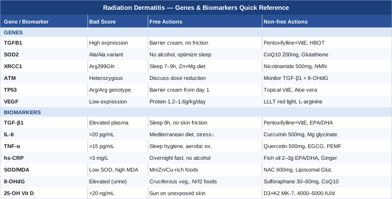
7 Biomarkers to Track for Radiation Dermatitis
Biomarkers are measurable signals in blood, urine, or tissue that reflect what is happening biologically in real time. For radiation dermatitis, the most useful biomarkers cluster around three mechanisms: inflammation, oxidative stress, and tissue repair capacity. Tracking the right combination before, during, and after treatment can reveal why your skin is reacting more severely than expected — and where targeted support may help.
Biomarker 1: TGF-β1 (Transforming Growth Factor Beta 1)
Why it matters: TGF-β1 is the single most studied predictor of late radiation toxicity. It drives the fibrotic cascade that transforms acutely damaged skin into scarred, thickened, or hyperpigmented tissue. Patients with elevated baseline TGF-β1 or a sharp rise during treatment tend to develop more severe dermatitis and are at higher risk for lasting fibrosis. Research in the International Journal of Radiation Oncology Biology Physics consistently identifies plasma TGF-β1 as a clinically meaningful signal — see relevant studies on PubMed.
How to measure it: A standard plasma ELISA test ordered through most specialty labs. Cost ranges from $80–$200 depending on the lab. Some academic medical centers include it in research protocols; ask your oncologist or a functional medicine physician to add it to routine bloodwork. Ideally, test before treatment begins, at week 3–4 of radiotherapy, and 6–8 weeks after completion.
If the score is elevated — plan without supplements: Reduce systemic pro-fibrotic inputs where possible. This includes minimizing processed foods with high advanced glycation end-products (AGEs), protecting irradiated skin from repeated mechanical friction (no tight clothing, no harsh washing), and ensuring adequate sleep (7–9 hours nightly), which independently reduces TGF-β1 signaling. Cold exposure via brief cold showers (1–2 minutes, daily) has shown modest anti-fibrotic signaling effects in pilot data but should only be applied away from the radiation field.
If the score is elevated — plan with supplements or equipment: Pentoxifylline (400 mg, 3x/day) combined with tocopherol (vitamin E, 400–500 IU/day) is the best-studied pharmacological combination for reducing TGF-β1-mediated radiation fibrosis. A meta-analysis in Radiotherapy and Oncology supports this combination for late effects. This should be discussed with your oncologist — not self-prescribed. Omega-3 supplementation (EPA/DHA, 2–3 g/day with food) has secondary anti-fibrotic evidence; cycle on 12 weeks, off 4 weeks. Side effects of pentoxifylline include GI upset and headache; vitamin E at this dose is generally well tolerated.
Biomarker 2: IL-6 (Interleukin-6)
Why it matters: IL-6 is a primary driver of the acute inflammatory cascade that initiates radiation dermatitis within the first 2–3 weeks of treatment. It rises sharply after radiation exposure, recruits immune cells to the skin, and amplifies the local inflammatory environment. Chronically elevated IL-6 between fractions correlates with faster progression from erythema to moist desquamation. See studies on IL-6 and radiation skin toxicity.
How to measure it: Serum IL-6 via ELISA. Cost: $50–$150. Normal range is typically below 7 pg/mL; values above 20 pg/mL during treatment suggest a heightened inflammatory response. Measure at baseline and at the midpoint of radiation treatment.
If the score is elevated — plan without supplements: Anti-inflammatory dietary shifts are the first lever. The Mediterranean-style diet is the best-studied for reducing circulating IL-6 in cancer patients. Specifically: increase oily fish (sardines, mackerel, salmon), reduce refined carbohydrates, and add polyphenol-rich foods (berries, leafy greens, olive oil). Stress reduction matters too — chronic psychological stress is a well-documented driver of IL-6 elevation, and mindfulness-based interventions have shown measurable IL-6 reductions in clinical trials.
If the score is elevated — plan with supplements or equipment: Curcumin (as a standardized extract with piperine or liposomal form, 500–1000 mg/day) has consistent evidence for reducing IL-6. Important caveat: discuss with your oncologist before using curcumin during active radiation treatment, as its antioxidant activity may theoretically interact with treatment mechanisms. Post-treatment use is lower risk. Magnesium glycinate (300–400 mg/day, nightly) is a lower-risk adjunct that supports IL-6 modulation via NF-κB pathway suppression. Cycle curcumin on 8 weeks, off 4 weeks; magnesium can be taken continuously.
Biomarker 3: TNF-α (Tumor Necrosis Factor Alpha)
Why it matters: TNF-α and IL-6 often rise together, but TNF-α plays a distinct role: it activates keratinocyte apoptosis (cell death) in irradiated skin. High TNF-α during radiation correlates with deeper epidermal disruption and slower re-epithelialization. Patients with persistently elevated TNF-α also show a higher risk of secondary infection at wound sites. See PubMed research on TNF-α and radiation skin effects.
How to measure it: Serum TNF-α ELISA. Cost: $60–$150. Values above 15–20 pg/mL are clinically significant in the context of active treatment.
If the score is elevated — plan without supplements: Sleep quality has a disproportionate effect on TNF-α — even one night of fragmented sleep raises it measurably the next morning. Prioritizing sleep hygiene during radiation treatment (consistent schedule, blackout curtains, limiting screens 60 minutes before bed) directly reduces TNF-α amplitude. Moderate aerobic exercise (20–30 minutes, 4–5x/week) is also well-supported for reducing TNF-α in cancer patients, assuming fatigue levels permit.
If the score is elevated — plan with supplements or equipment: Quercetin (500 mg/day with food, 8-week cycles) has shown modest but consistent TNF-α-lowering effects in human trials. Green tea extract (EGCG, 400–800 mg/day as decaffeinated standardized extract) is another option. Both should be paused 3 days before and after each radiation fraction if your oncologist agrees. PEMF (Pulsed Electromagnetic Field) therapy at low-level settings has emerging evidence for reducing TNF-α in soft tissue inflammation, though most data is from osteoarthritis rather than radiation-specific contexts.
Biomarker 4: hs-CRP (High-Sensitivity C-Reactive Protein)
Why it matters: hs-CRP is the most accessible systemic inflammation marker and, while less specific than the cytokines above, its elevation before treatment is a practical red flag for a primed inflammatory state. Patients with pre-treatment hs-CRP above 3 mg/L show higher rates of acute Grade 2–3 dermatitis in observational data. Peter Attia consistently emphasizes hs-CRP as a frontline screening tool precisely because it is cheap, widely available, and interpretable in clinical context.
How to measure it: Standard blood test, included in many metabolic panels. Cost: $15–$40. Target: below 1 mg/L for lowest inflammatory risk. Between 1–3 mg/L is intermediate; above 3 mg/L is high. Measure before treatment and 6–8 weeks post-treatment to track resolution.
If the score is elevated — plan without supplements: Address the upstream drivers: sleep deprivation, sedentary behavior, and dietary ultra-processing all independently elevate hs-CRP. Time-restricted eating (12–16 hour overnight fast) has consistent human evidence for lowering hs-CRP within 4–8 weeks when maintained. Reducing alcohol intake to zero during treatment has a meaningful short-term effect.
If the score is elevated — plan with supplements or equipment: Fish oil (EPA/DHA combined 2–3 g/day with meals) has the strongest evidence for hs-CRP reduction. Thomas Dayspring and Allan Sniderman both reference EPA specifically as a key inflammatory lever. Ginger extract (1–2 g/day standardized) is a lower-cost adjunct with supporting data in clinical populations. Cycle fish oil continuously; reassess hs-CRP at 8–12 weeks to confirm response.
Biomarker 5: Oxidative Stress Markers — SOD Activity and MDA
Why it matters: Radiation generates an immediate and massive burst of reactive oxygen species (ROS) in irradiated tissue. The body's primary defense is superoxide dismutase (SOD), a family of enzymes that neutralize superoxide radicals. When SOD activity is low — whether due to genetic variants (see the SOD2 gene section below) or nutritional deficiencies — ROS accumulate, damaging cell membranes and DNA. Malondialdehyde (MDA) is a byproduct of lipid peroxidation by ROS and serves as a direct indicator of oxidative damage in tissue. Elevated MDA and low SOD activity together are a high-risk profile for severe radiation dermatitis. See PubMed research on SOD and radiation skin oxidative stress.
How to measure it: SOD activity in erythrocytes (red blood cells): specialty labs, $80–$200. Plasma MDA via TBARS assay: $60–$150. These are not routine tests — request them through a functional medicine physician or an integrative oncology center. Baseline measurement before treatment is the most useful reference point.
If the scores are poor — plan without supplements: Maximize dietary antioxidant cofactors: manganese (nuts, whole grains, legumes), zinc (pumpkin seeds, seafood), and copper (organ meats, shellfish) are direct enzymatic cofactors for the three SOD isoforms. A diet chronically low in these minerals will suppress SOD activity even in the absence of any genetic variant. Resistance training (when not contraindicated) upregulates endogenous antioxidant enzyme production over time — this is a longer-term intervention relevant post-treatment.
If the scores are poor — plan with supplements or equipment: Liposomal glutathione (250–500 mg/day) or its precursor N-acetylcysteine (NAC, 600 mg/day) supports endogenous antioxidant capacity, though as with all antioxidants during active radiation, timing matters — pause during treatment fractions and resume the day after, in consultation with your oncologist. Zinc bisglycinate (15–25 mg/day) and manganese (2–3 mg/day, short-term cycles of 8 weeks) address cofactor gaps directly. LLLT/photobiomodulation (see the complementary section) has direct upregulatory effects on SOD activity in irradiated tissue.
Biomarker 6: 8-OHdG (8-hydroxy-2'-deoxyguanosine)
Why it matters: 8-OHdG is one of the most direct markers of DNA oxidative damage available in clinical settings. It is produced when hydroxyl radicals — generated in large quantities by ionizing radiation — attack guanine in DNA. Urinary 8-OHdG correlates with both radiation dose exposure and subsequent skin toxicity severity. High 8-OHdG levels during treatment indicate that oxidative DNA damage is outpacing repair capacity — a state that predicts slower skin recovery and higher infection risk. Research using 8-OHdG in radiation oncology contexts is available at PubMed.
How to measure it: Urinary 8-OHdG via ELISA (first-morning urine, standardized to creatinine). Cost: $80–$180 through specialty labs. Normal urinary values are roughly 0.5–8 ng/mg creatinine in healthy adults; values during radiation treatment will typically be elevated. Use as a trend marker rather than a single-point diagnosis.
If the score is elevated — plan without supplements: Green leafy vegetables and cruciferous vegetables (broccoli, Brussels sprouts) upregulate Nrf2 — the master transcription factor controlling antioxidant gene expression — via sulforaphane and other phytochemicals. This is one of the most evidence-backed dietary interventions for reducing 8-OHdG. Reducing dietary advanced glycation end-products (avoid charred, fried foods at high temperatures) lowers the oxidative substrate burden.
If the score is elevated — plan with supplements or equipment: Sulforaphane (from broccoli seed extract, 30–60 mg/day standardized) has the most direct evidence for Nrf2 activation and 8-OHdG reduction. Coenzyme Q10 (200–400 mg/day as ubiquinol, with fat-containing meal) is a mitochondrial antioxidant with supporting evidence in radiation contexts. Cycle sulforaphane 12 weeks on, 4 weeks off. Ubiquinol can be taken continuously. Both have good safety profiles and low interaction risk in the post-treatment phase.
Biomarker 7: 25-OH Vitamin D
Why it matters: Vitamin D is not conventionally listed as a radiation dermatitis biomarker, but the evidence for its role in skin immune regulation and barrier recovery is strong. Vitamin D receptors (VDR) are expressed on keratinocytes, and vitamin D signaling directly regulates keratinocyte differentiation and the skin barrier function disrupted by radiation. Observational data in breast cancer patients show that pre-treatment 25-OH vitamin D deficiency (below 20 ng/mL) correlates with higher-grade acute skin reactions. See PubMed research.
How to measure it: Standard serum 25-OH vitamin D. Cost: $30–$80, often covered by insurance. Optimal range for skin health and immune function is 40–60 ng/mL (100–150 nmol/L). Below 30 ng/mL represents deficiency.
If the score is low — plan without supplements: Sensible sun exposure to non-irradiated skin — 10–20 minutes of midday sun on arms and legs — is the safest and most physiologically natural way to raise vitamin D. This must avoid the radiation field entirely. Dietary sources are limited but include fatty fish, egg yolks, and UV-exposed mushrooms.
If the score is low — plan with supplements or equipment: Vitamin D3 supplementation with vitamin K2 (MK-7 form): for deficiency, 4,000–5,000 IU/day D3 with 100–200 mcg K2 daily is a well-supported repletion protocol. Retest at 8–12 weeks. At maintenance levels (above 40 ng/mL), 1,500–2,000 IU/day is typically sufficient. K2 is included to support calcium routing and reduce any risk from D3 supplementation in the cardiovascular context. No cycling needed; this is a long-term supplement for most cancer patients, especially those with limited sun exposure.
Genetics and Epigenetics of Radiation Skin Toxicity
Knowing your biomarkers tells you what is happening now. Knowing your genetic variants tells you why — and what your baseline risk architecture looks like before a single radiation fraction is delivered. The six genes below are the most consistently implicated in radiation dermatitis severity across human genetic studies. All are actionable in some form, even if the gene itself cannot be changed.
Gene 1: TGFB1 (TGF-β1 Gene)
What it affects: The TGFB1 gene encodes the TGF-β1 protein covered in the biomarker section. The rs1800469 promoter polymorphism (C>T) alters transcription activity, with certain variants associated with higher TGF-β1 expression and greater fibrotic response to radiation. This is one of the most replicated genetic associations in radiation toxicity research. See relevant studies.
If the gene is suboptimal — plan without supplements: Since TGF-β1 overexpression is the mechanism, the non-supplement protocol focuses on preventing fibrosis triggers: avoid skin trauma to the irradiated field (no friction, no tight straps), prioritize wound hydration with plain barrier emollients (zinc oxide or petroleum-based), and maintain anti-inflammatory nutrition throughout treatment and recovery.
If the gene is suboptimal — plan with supplements or equipment: Pentoxifylline + vitamin E (described in biomarker section) is the most evidence-backed pharmacological strategy for carriers of the high-expression TGFB1 variant. Discuss proactive use with your radiation oncologist. Post-treatment, hyperbaric oxygen therapy (HBOT) has shown the ability to downregulate TGF-β1 in irradiated tissue through repeated sessions of 20–40 dives at 2.0–2.4 atmospheres — a significant investment ($150–$300/session in the US) but with strong data for late radiation fibrosis.
Gene 2: SOD2 (Superoxide Dismutase 2)
What it affects: The SOD2 gene encodes the mitochondrial form of superoxide dismutase. The Val16Ala polymorphism (rs4880) affects how efficiently the SOD2 enzyme is imported into mitochondria. The Ala/Ala genotype is associated with reduced mitochondrial antioxidant capacity and measurably higher oxidative stress under radiation exposure. This variant is common (around 30–40% allele frequency in European populations) and has been associated with increased radiation skin and mucosal toxicity in multiple studies. See PubMed research.
If the gene is suboptimal — plan without supplements: Minimize mitochondrial stressors during treatment: reduce alcohol to zero (alcohol directly depletes mitochondrial antioxidant capacity), avoid high-intensity exercise during active radiation (moderate aerobic exercise is preferable), and optimize sleep, which is when mitochondrial repair and biogenesis are most active.
If the gene is suboptimal — plan with supplements or equipment: Mitochondrial-targeted antioxidants are most relevant here. Ubiquinol CoQ10 (200–300 mg/day), MitoQ (if available through specialty channels, 10 mg/day), and liposomal glutathione (250–500 mg/day, post-treatment) directly support mitochondrial antioxidant defenses. Magnesium malate (400 mg/day) supports mitochondrial function as a cofactor. Cycle CoQ10 and glutathione on 12-week periods with 4-week breaks; reassess with MDA levels.
Gene 3: XRCC1 (X-ray Cross-Complementing Protein 1)
What it affects: XRCC1 coordinates the base excision repair (BER) pathway, the primary mechanism for repairing single-strand DNA breaks caused by ionizing radiation. The Arg399Gln polymorphism (rs25487) reduces BER efficiency in carriers, meaning more unrepaired DNA damage accumulates in skin cells between radiation fractions. This variant has been associated with higher acute dermatitis grades in breast and head-and-neck cancer patients. See related research.
If the gene is suboptimal — plan without supplements: Support DNA repair with adequate sleep (the BER pathway is most active during the rest phase), and ensure adequate nutritional zinc and magnesium, both of which are cofactors for DNA repair enzymes. Minimize competing DNA damage during treatment — this means avoiding other genotoxic exposures such as smoking and excess alcohol.
If the gene is suboptimal — plan with supplements or equipment: Niacin (as nicotinamide, 500 mg twice daily) has some of the most interesting data here: nicotinamide is a direct precursor for NAD+, which powers the PARP-1 enzyme central to BER pathway function. Studies in non-melanoma skin cancer have shown nicotinamide reduces DNA damage markers. NMN or NR (as NAD+ precursors, 250–500 mg/day) are newer alternatives with preliminary supporting evidence. Discuss these with your oncologist as there is active research on their interaction with radiation treatment.
Gene 4: ATM (Ataxia Telangiectasia Mutated)
What it affects: ATM is the master sensor of double-strand DNA breaks (DSBs), the most severe form of radiation-induced DNA damage. Carriers of heterozygous ATM loss-of-function variants (found in roughly 1–2% of the general population, but enriched in cancer patients) show significantly impaired DSB recognition and repair, leading to higher rates of severe acute and late radiation toxicity. Full ATM mutations cause ataxia-telangiectasia; heterozygous carriers face a more subtle but clinically relevant increase in radiation sensitivity. See PubMed literature.
If the gene is suboptimal — plan without supplements: If ATM heterozygosity is identified before treatment, discuss with your radiation oncologist the possibility of slightly reduced fraction doses or modified treatment planning to reduce skin dose. This is the most impactful intervention available and it operates entirely at the treatment protocol level, not through lifestyle.
If the gene is suboptimal — plan with supplements or equipment: The resveratrol-ATM interaction has generated some mechanistic interest: resveratrol activates SIRT1, which in turn interacts with ATM pathway signaling. Evidence in humans is very preliminary and should not override medical decisions. The more pragmatic tool is careful biomarker monitoring throughout treatment — tracking 8-OHdG and TGF-β1 closely in ATM variant carriers gives earlier warning of escalating tissue damage.
Gene 5: TP53 (Tumor Protein p53)
What it affects: The TP53 Pro72Arg polymorphism (rs1042522) alters p53 function — specifically its ability to trigger apoptosis versus cell cycle arrest in response to DNA damage. The Arg/Arg genotype is associated with stronger apoptotic signaling, which in the context of radiation means higher rates of keratinocyte cell death in the irradiated field and potentially more pronounced acute skin breakdown. This is still an area of active research with mixed findings, but the biological rationale is mechanistically strong. See PubMed.
If the gene is suboptimal — plan without supplements: Prioritizing skin barrier protection physically is particularly important for Arg/Arg carriers: start using a medical-grade barrier cream or silicone gel sheet on the treatment field from day one of radiotherapy, rather than waiting for visible dermatitis to appear. Early physical protection reduces the degree of epidermal disruption before it escalates.
If the gene is suboptimal — plan with supplements or equipment: Vitamin E oil (topical tocopherol acetate) applied to the skin margin — never on open wounds — has modest evidence for reducing acute radiation skin damage when used pre-emptively. Aloe vera gel (inner leaf, preservative-free) applied twice daily to the non-broken skin field is well-tolerated and has biological plausibility as a mild anti-inflammatory barrier support, though the human evidence for radiation dermatitis specifically is mixed.
Gene 6: VEGF (Vascular Endothelial Growth Factor)
What it affects: VEGF drives the angiogenic response — new blood vessel formation — that is essential for skin healing after radiation damage. Polymorphisms in the VEGF gene (including rs2010963 and rs3025039) affect VEGF expression levels, and lower VEGF expression is associated with impaired wound healing and prolonged skin recovery after radiation. This is particularly relevant for late-phase recovery (weeks 4–16 post-treatment) rather than acute inflammatory severity. See related studies.
If the gene is suboptimal — plan without supplements: Protein intake is a key non-supplement driver of angiogenesis and collagen remodeling. Cancer patients under treatment frequently undereat protein; targeting 1.2–1.6 g/kg/day of high-quality protein (eggs, fish, legumes) meaningfully supports the tissue repair cascade that VEGF regulates. Moderate aerobic exercise post-treatment also upregulates VEGF expression through hypoxia-inducible factor pathways.
If the gene is suboptimal — plan with supplements or equipment: LLLT/photobiomodulation (red light, 620–680 nm) has direct VEGF-stimulating effects in irradiated skin — the best non-pharmacological tool here. Red light therapy panels used at home (10 minutes/session, daily) are the most practical application. L-arginine (3–5 g/day with food), a precursor to nitric oxide, supports vascular recovery and has modest human evidence in wound healing contexts. Cycle L-arginine 8 weeks on, 4 weeks off. Side effects are rare but include GI discomfort at higher doses.
What the Science on Inflammation and Tissue Recovery Really Reveals
Andrew Huberman's podcast episode on inflammation — "Understanding and Controlling Inflammation" (released in 2023 on the Huberman Lab) — synthesizes a wide body of research that is directly applicable to radiation dermatitis management, even though the episode does not address radiation specifically. The following 10 insights from that discussion and the studies it references are among the most impactful for anyone managing treatment-related tissue injury.
1. Inflammation is necessary before it becomes damaging
The episode opens with a crucial reframing: inflammation is not the enemy. The acute inflammatory phase after injury is the signal that initiates healing. The problem arises when that phase fails to resolve. In radiation dermatitis, this resolution failure — driven by persistent cytokine signaling and impaired clearance — is exactly what separates Grade 1 from Grade 3 outcomes.
2. Sleep is the primary anti-inflammatory tool available to everyone
Deep slow-wave sleep triggers the glymphatic system and downregulates IL-6, TNF-α, and other pro-inflammatory cytokines simultaneously. Huberman references studies showing that even partial sleep restriction (6 hours vs 8 hours for one week) produces measurable increases in circulating inflammatory markers. During radiation treatment, protecting sleep is not a luxury — it is a direct clinical intervention.
3. The omega-6/omega-3 ratio matters more than total fat intake
High dietary omega-6 (from seed oils, processed foods) competes with omega-3 for the same metabolic enzymes, shifting eicosanoid production toward pro-inflammatory prostaglandins. Huberman cites research showing that lowering the omega-6:omega-3 ratio to below 4:1 reduces CRP and IL-6 within 6–8 weeks. Most Western diets run at 15:1 to 20:1.
4. Cold exposure accelerates inflammatory resolution — with caveats
Brief cold exposure (cold shower, 1–3 minutes) acutely reduces prostaglandin synthesis and promotes norepinephrine-mediated anti-inflammatory signaling. The episode explicitly notes this should never be applied to healing wounds or fragile skin. For radiation patients: cold exposure to unexposed body areas (hands, neck, feet) may still deliver systemic benefits without risk to the treated field.
5. Sunlight and circadian rhythm are direct immune regulators
Morning light exposure (10–20 minutes of outdoor light within 1 hour of waking) sets circadian timing and regulates the cortisol awakening response, which has downstream effects on immune modulation. Blunted cortisol rhythms — common in cancer patients — are associated with higher baseline inflammation. This is a zero-cost, zero-risk intervention available to anyone.
6. The gut microbiome is an upstream regulator of systemic inflammation
Huberman references multiple studies showing that dysbiosis (imbalanced gut microbiome) elevates systemic LPS (lipopolysaccharide) leakage, driving IL-6 and CRP. This mechanistic link is increasingly studied in radiation oncology, as pelvic and abdominal radiation directly disrupts intestinal flora. Supporting the microbiome is not separate from managing dermatitis — it is upstream of it.
7. Breathing protocols (nasal, slow, diaphragmatic) reduce inflammatory tone
The episode covers the neuroscience behind respiratory-driven vagal tone modulation. Slow nasal breathing at 5–6 cycles per minute activates the parasympathetic nervous system and measurably reduces cortisol and TNF-α over 4–8 weeks of daily practice. This is actionable for anyone, regardless of fitness or treatment status.
8. Exercise is anti-inflammatory — but dose matters during treatment
Moderate aerobic exercise (zone 2, 60–70% max heart rate) upregulates anti-inflammatory myokines including IL-10 and IL-6RA (the anti-inflammatory IL-6 form). Very high-intensity exercise, by contrast, temporarily spikes inflammatory markers. During radiation treatment, moderate exercise is supported; high-intensity training should be deferred.
9. Dietary polyphenols amplify Nrf2 more than isolated antioxidants
Huberman discusses research showing that whole food polyphenols — from berries, coffee, green tea, dark chocolate — activate Nrf2 more effectively than isolated antioxidant supplements. This has direct relevance to 8-OHdG and oxidative stress markers: the diet is the primary tool, supplements are secondary amplifiers.
10. Resolving inflammation requires specific pro-resolving molecules
The episode covers the specialized pro-resolving mediators (SPMs) — lipoxins, resolvins, protectins — that actively shut off inflammation. These are derived from EPA and DHA. This explains mechanistically why omega-3 supplementation reduces inflammatory biomarkers: it is not just suppressing inflammation but providing the substrate for active resolution. This is the biochemical argument for EPA/DHA at therapeutic doses (2–3 g/day) during and after radiation treatment.
Complementary and Alternative Approaches
Low-Level Laser Therapy (Photobiomodulation)
Photobiomodulation (PBM) uses red and near-infrared light (typically 620–1000 nm) to stimulate cellular energy production through cytochrome c oxidase in the mitochondrial electron transport chain. In the context of radiation dermatitis, PBM is relevant because it both increases ATP production in damaged keratinocytes and has direct anti-inflammatory and anti-fibrotic effects — reducing TGF-β1, upregulating SOD activity, and stimulating VEGF-driven angiogenesis in recovering tissue.
A randomized controlled trial published in Supportive Care in Cancer (Censabella et al., 2016) demonstrated that PBM significantly reduced acute dermatitis severity in breast cancer patients undergoing radiotherapy, with treated patients showing lower RTOG toxicity grades and faster healing compared to controls. Multiple subsequent meta-analyses, summarized in this PubMed search, confirm consistent benefit with no reported adverse events from the light therapy itself.
In practice, apply PBM using a clinical-grade red light device (630–670 nm for superficial skin effects) at 3–4 J/cm² per session. Treatment is typically 3–5 sessions per week during active radiotherapy. Importantly, apply PBM after each radiation fraction, not before. Home red light panels can be used for post-treatment recovery (daily, 10 minutes per session to the healing skin). Avoid applying to open wounds or active moist desquamation until re-epithelialization begins.
Mindfulness Meditation and MBSR
Mindfulness-Based Stress Reduction (MBSR) is an 8-week structured program combining meditation, body scan, and gentle movement. Its relevance to radiation dermatitis extends beyond psychological comfort: MBSR has documented effects on reducing circulating IL-6, cortisol, and TNF-α, directly addressing three of the seven biomarkers in this article. Cancer patients undergoing radiation treatment face compounded psychological stress that amplifies the inflammatory substrate of skin toxicity.
A well-cited randomized trial by Lengacher et al. in breast cancer patients (PMID 22251769) demonstrated significant reductions in fear of recurrence and improvements in immune markers in patients completing MBSR. Subsequent work has shown MBSR specifically reduces IL-6 in cancer populations. The mechanism runs through reduced HPA axis activation: lower chronic stress hormone levels suppress the cytokine cascades that amplify radiation skin damage.
Practically, MBSR is accessible through the University of Massachusetts online program, local hospital-based integrative oncology programs, and app-based adaptations (Insight Timer, Calm). The minimum effective dose appears to be 20 minutes of daily formal practice. Start during the week before radiation begins to establish the habit; maintain through treatment and for at least 8 weeks post-treatment. No side effects; the only risk is inadequate practice frequency reducing benefit.
Microbiome-Directed Therapies
The gut microbiome is now recognized as a central regulator of systemic inflammatory tone, and its disruption during cancer treatment — through antibiotic use, treatment-related GI effects, dietary changes, and radiation itself (especially in pelvic sites) — directly elevates systemic markers including LPS, IL-6, and CRP. Supporting the microbiome during radiotherapy is increasingly studied as an indirect strategy for reducing treatment toxicity, including dermatitis at distant skin sites.
A clinical trial by Delia et al. in rectal cancer patients found that oral Lactobacillus acidophilus and Bifidobacterium longum supplementation significantly reduced radiation-induced intestinal toxicity and systemic inflammatory markers. While most microbiome-radiation trials focus on GI endpoints, the downstream reduction in circulating inflammatory cytokines is mechanistically relevant to skin outcomes. See related trials on PubMed.
Practical protocol: Begin a multi-strain probiotic containing Lactobacillus rhamnosus GG, Bifidobacterium longum, and Lactobacillus acidophilus (minimum 10 billion CFU/day) two weeks before radiotherapy starts. Continue through treatment and for 8 weeks post-treatment. Pair with prebiotic foods (garlic, onions, leeks, oats) to sustain the bacteria. If pelvic radiation is part of your treatment, discuss microbiome support proactively with your oncologist. Probiotics are generally safe during cancer treatment; rare exceptions include severely immunocompromised patients.
Massage Therapy
Massage therapy in the radiation oncology context primarily addresses the lymphedema and tissue fibrosis that develop in the weeks and months after treatment, rather than acute dermatitis itself. Manual lymphatic drainage (MLD) — a specialized form of light-pressure massage — directly moves lymph fluid out of congested, irradiated tissue, reducing the edema and protein accumulation that drives secondary fibrotic remodeling.
A Cochrane systematic review and supporting RCTs in breast cancer patients confirm that MLD reduces limb volume in lymphedema and improves patient-reported skin texture scores in irradiated areas. These findings, available through PubMed searches, support MLD specifically for patients with late radiation fibrosis or lymphedema. Evidence for acute dermatitis specifically is limited; this is a post-treatment tool.
To apply safely, seek a certified lymphedema therapist (CLT) rather than a general massage therapist — the pressure and technique differ significantly. Start MLD no earlier than 4–6 weeks post-treatment once the skin has fully re-epithelialized. Sessions are typically 45–60 minutes, weekly initially then moving to monthly for maintenance. Avoid any massage directly over active radiation burns, open skin, or areas of confirmed infection.
Conclusion
Radiation dermatitis sits at the intersection of physics, biology, and individual genetic makeup. The skin reaction you experience is not simply a function of how much radiation was delivered — it reflects your inflammatory baseline, your oxidative stress capacity, your DNA repair efficiency, and the specific genetic architecture you carry. Each of those layers is partially measurable and partially modifiable.
The most useful next step is not to take every supplement on this list simultaneously. It is to identify your personal weakest link. If you have access to basic bloodwork, starting with hs-CRP and 25-OH vitamin D gives you two highly actionable data points at low cost. If you are preparing for radiation treatment, discussing TGF-β1 baseline testing with your oncologist — or at least reviewing whether your treatment center offers integrative oncology consultation — is a concrete and reasonable ask. If treatment has already concluded and skin recovery is slow, the oxidative stress markers and genetic panels are worth exploring.
The science covered here will not replace your radiation oncologist's care. But it offers a sharper lens on what is happening in your body — and that sharper lens is what transforms generic management into something genuinely tailored. Start with one measurement, one habit, one conversation with your care team. That is the real next smart step.