This article contains AI generated content.
· Updated[TITLE level="1"] tag.
Relapsing Polychondritis: 5 Genes And 6 Biomarkers To Track
Introduction
Living with relapsing polychondritis means navigating a condition that most physicians encounter only a handful of times in their careers. The average diagnostic delay runs between two and four years, and even after diagnosis, the treatment path often feels blunt — corticosteroids calibrated to quiet the most visible flares, with little attention paid to what is actually driving the inflammation beneath the surface. Many patients are left wondering why their disease behaves so differently from the next person with the same label, or why the same drug that stabilizes one person barely moves the needle for them.
The answer frequently lies in individual biological variation. Relapsing polychondritis is not a monolithic disease — it is a spectrum shaped by genetic predispositions, cytokine dynamics, and structural markers that differ from one patient to the next. A person carrying specific HLA variants will generate a different immune response profile than someone without them. A patient whose interleukin-6 remains persistently elevated between flares is under a different biological pressure than someone whose inflammation appears only episodically. Treating these two patients identically makes little biological sense, yet that is the default in most clinical settings.
This article focuses on what you can actually measure and track. Rather than repeating the standard clinical overview of RP, it takes a precision-oriented approach: identifying the biomarkers that reveal your current inflammatory state and the genetic variants that may predispose you to specific immune dysregulation patterns. Neither is a magic answer, but together they create a far more actionable picture than a diagnosis alone.
Two core strategies are explored here. The first covers six key biomarkers — measurable from blood — that can help you and your rheumatologist monitor disease activity, anticipate flares, and assess whether interventions are working. The second maps five genetic variants associated with RP susceptibility and suggests targeted lifestyle and supplement strategies for each. Used together, they represent a meaningful shift from reactive to proactive management.
6 Biomarkers to Track in Relapsing Polychondritis
Most patients with RP receive CRP and ESR results as part of routine monitoring, but the full biomarker picture is richer than that. The six markers below span acute inflammation, cartilage-specific damage, cytokine activity, and immune cell dynamics. Not all are available at every lab, and costs vary widely — but knowing which ones matter and why gives you a real basis for informed decisions.
1. High-Sensitivity C-Reactive Protein (hs-CRP)
Why it matters: CRP is produced by the liver in response to inflammatory cytokines, particularly IL-6. In relapsing polychondritis, hs-CRP correlates closely with disease activity — it rises during auricular, nasal, and laryngeal flares and typically falls during remission. It is one of the most responsive markers for tracking whether treatment is working or a flare is developing. Studies have shown that persistently elevated hs-CRP between flares is associated with a more aggressive disease course and greater cumulative organ damage.
How to Measure It
Standard CRP is available at virtually every lab. High-sensitivity CRP (hs-CRP) is the preferred version because it detects lower-grade chronic inflammation that standard CRP misses. Request hs-CRP specifically when ordering labs. Cost range: $15–$75 depending on your region and insurance coverage. Target: below 1.0 mg/L is low risk; 1.0–3.0 mg/L is borderline; above 3.0 mg/L reflects active systemic inflammation. For RP monitoring, serial measurements (every 6–8 weeks during active disease) are more informative than a single value.
If hs-CRP Is Elevated — Plan Without Supplements
The most powerful non-supplement interventions for CRP reduction are dietary and behavioral. Adopting an anti-inflammatory eating pattern — eliminating refined sugars, seed oils, and ultra-processed foods while increasing omega-3-rich fish, colorful vegetables, and polyphenol sources — can reduce hs-CRP by 30–40% within 8–12 weeks. Sleep is equally critical: getting below seven hours per night reliably drives hs-CRP up. Aerobic exercise at moderate intensity (brisk walking, cycling) three to four times per week also reduces CRP through IL-6-mediated pathways, provided it does not trigger RP joint inflammation. Stress reduction matters too — chronic psychological stress activates the HPA axis and keeps IL-6 elevated. Evidence: PubMed search on CRP and lifestyle interventions.
If hs-CRP Is Elevated — Plan With Supplements or Equipment
Fish oil (EPA + DHA): 2–4 g/day of combined EPA+DHA has demonstrated consistent CRP reduction in multiple randomized trials. Use triglyceride-form fish oil for better absorption. Cycle: can be taken continuously. Side effects: fishy burps (take with meals or use enteric-coated capsules); mild blood thinning at high doses — disclose to your physician if on anticoagulants.
Curcumin (with piperine): 500–1000 mg/day of bioavailable curcumin (phospholipid-complexed or nanoparticle forms preferred). Reduces NF-κB signaling, which drives CRP production. Cycle: 8 weeks on, 2 weeks off is a reasonable approach. Side effects: occasional GI upset; may interact with blood thinners.
Boswellia serrata (AKBA form): 100–200 mg/day of AKBA-enriched extract. Inhibits 5-LOX enzyme involved in cartilage inflammation. Particularly relevant for RP given the cartilage-specific mechanism. Side effects: generally mild; occasional nausea.
2. Erythrocyte Sedimentation Rate (ESR)
Why it matters: ESR measures how quickly red blood cells settle in a tube — the faster they fall, the more inflammatory proteins are present in the blood. In RP, ESR is a secondary but complementary marker to CRP. It is slower to change than CRP (takes days versus hours), making it better for tracking sustained disease activity over weeks rather than acute flares. ESR is also affected by anemia, which is common in RP, so results need to be interpreted alongside a full blood count.
How to Measure It
Included in most standard inflammatory panels. Cost: $5–$35. Normal range: men under 15 mm/hr; women under 20 mm/hr (Westergren method), though these thresholds rise with age. For RP monitoring, an ESR above 40 mm/hr during apparent remission warrants attention. Measure alongside CRP for the most complete picture.
If ESR Is Elevated — Plan Without Supplements
The same lifestyle interventions that lower hs-CRP apply here: anti-inflammatory diet, sleep optimization, stress management, moderate exercise. Addressing underlying anemia is also critical — iron deficiency and anemia of chronic inflammation both artificially elevate ESR. Ensure ferritin, B12, and folate are adequate before interpreting an elevated ESR purely as an inflammatory signal.
If ESR Is Elevated — Plan With Supplements or Equipment
Omega-3 fatty acids: same protocol as for CRP. Fish oil 2–4 g/day continuously. Vitamin D3 + K2: Vitamin D deficiency is strongly linked to elevated inflammatory markers and is disproportionately common in autoimmune patients. Target serum 25-OH vitamin D between 50–80 ng/mL. Dose: 2000–5000 IU/day D3 with 100–200 mcg MK-7 (K2). Cycle: continuous with periodic testing every 3–6 months. Side effects: excessive dosing can cause hypercalcemia — monitor blood levels. Infrared sauna: 20-minute sessions 3x per week have shown reductions in inflammatory markers in several small trials. Use only during remission in RP — heat can theoretically stress inflamed cartilage during flares.
3. Anti-Type II Collagen Antibodies
Why it matters: This is the most RP-specific biomarker on this list. Type II collagen is the dominant structural protein in hyaline cartilage — the tissue targeted in RP. Antibodies against type II collagen have been detected in a significant proportion of RP patients and are considered a marker of active autoimmune cartilage attack. Their presence may help distinguish RP from mimicking conditions, and elevated titers correlate with more aggressive auricular and laryngotracheal involvement. Research on anti-type II collagen in RP.
How to Measure It
Not available at all standard labs — typically requires a reference or academic center lab. Test name varies: anti-collagen II antibodies, anti-CII IgG. Cost: $100–$350. Useful both at diagnosis (to support confirmation) and longitudinally during flares. Not all RP patients will test positive — a negative result does not rule out the diagnosis.
If Anti-Type II Collagen Is Elevated — Plan Without Supplements
Elimination of dietary gluten and dairy: Molecular mimicry — where dietary proteins share structural sequences with self-antigens — has been proposed as a mechanism driving anti-collagen antibody production. While direct evidence in RP is limited, both gluten and casein have been implicated in autoimmune antibody elevation more broadly. A 12-week strict elimination trial is reasonable. Focus also on gut barrier integrity: avoiding NSAIDs when possible (they damage intestinal lining), managing gut dysbiosis, and stress reduction.
If Anti-Type II Collagen Is Elevated — Plan With Supplements or Equipment
Undenatured type II collagen (UC-II): 40 mg/day of undenatured collagen — counterintuitively — has been shown to induce oral tolerance to type II collagen through regulatory T-cell pathways in the gut-associated lymphoid tissue, reducing the immune attack on cartilage. This effect is dose-specific: do not use denatured (hydrolyzed) collagen for this purpose, which works differently. Cycle: 3 months continuous minimum. Side effects: very rare; occasional mild GI symptoms. Quercetin: 500–1000 mg/day. Quercetin acts as a mast cell stabilizer and inhibits antibody-mediated inflammatory cascades. Side effects: minimal; avoid very high doses (>3g/day) due to potential kidney effects with prolonged use.
4. Interleukin-6 (IL-6)
Why it matters: IL-6 is the master upstream driver of both CRP and ESR, but measuring it directly provides earlier and more specific information. In RP, IL-6 rises before CRP does and reflects active synovial and cartilage membrane inflammation. Chronically elevated IL-6 also drives fatigue, anemia, and the systemic symptoms that many RP patients find just as disabling as the structural flares themselves. IL-6 is the biological target of tocilizumab (Actemra), a biologic used in refractory RP — which underscores its centrality to the disease mechanism. PubMed: IL-6 in relapsing polychondritis.
How to Measure It
Available through most larger lab services though not always included in standard panels. Cost: $50–$180. Normal: below 7 pg/mL. During RP flares, levels can reach 50–200 pg/mL. Pair with hs-CRP; if IL-6 is elevated but CRP is not, early-stage or low-grade inflammation may be present.
If IL-6 Is Elevated — Plan Without Supplements
Cold exposure: Cold water immersion (10–15 minutes at 14–16°C) or cold showers have demonstrated measurable reductions in circulating IL-6 in several controlled trials. This works in part through catecholamine release and vagal nerve activation. Start with contrast showers (30s cold alternating with warm) and gradually extend cold exposure. Frequency: 3–5x/week. Moderate aerobic exercise also reduces resting IL-6 over time, though very intense exercise temporarily spikes it. Time-restricted eating (16:8 intermittent fasting) has shown IL-6 reduction in several metabolic trials.
If IL-6 Is Elevated — Plan With Supplements or Equipment
Omega-3 fatty acids: 3–4 g EPA+DHA/day reduces IL-6 gene transcription. Continuous use. Resveratrol: 250–500 mg/day of trans-resveratrol with meals. Inhibits STAT3 signaling downstream of IL-6. Cycle: 8 weeks on, 2 weeks off. Side effects: very high doses (>2g) may disrupt hormonal signaling in women; estrogen-sensitive individuals should use lower doses. Melatonin: 0.5–3 mg at bedtime. Beyond sleep promotion, melatonin has demonstrated IL-6 inhibitory activity in several immune studies. Side effects: grogginess at higher doses; start at 0.5 mg.
5. Cartilage Oligomeric Matrix Protein (COMP)
Why it matters: COMP is a structural glycoprotein found almost exclusively in cartilage, tendons, and ligaments. When cartilage is actively damaged, COMP is released into circulation. In RP, elevated serum COMP reflects the ongoing destruction of auricular, nasal, and articular cartilage even during periods of apparent clinical quiescence. This makes it a particularly valuable marker — it can reveal subclinical cartilage damage before the next visible flare and may predict long-term structural outcomes. Research in rheumatoid arthritis and ankylosing spondylitis has validated COMP as a damage biomarker; its application in RP is increasingly recognized. COMP biomarker research.
How to Measure It
Available through specialty and academic reference labs. Test name: serum COMP or cartilage oligomeric matrix protein. Cost: $80–$250. Not routinely ordered in most rheumatology practices, but can be requested specifically. Useful as a baseline at diagnosis and monitored every 3–6 months. Elevated values (typically above 12 U/L, though reference ranges vary by lab) indicate active cartilage turnover or damage.
If COMP Is Elevated — Plan Without Supplements
Avoid mechanical cartilage stress during flares: High-impact activities that load inflamed cartilage structures (running, heavy lifting, contact sports) accelerate COMP release. Switch to low-impact alternatives during active disease. Ensure adequate sleep — growth hormone released during deep sleep is the primary driver of cartilage matrix regeneration. Address vitamin D status (see ESR section) since deficiency impairs chondrocyte function. Swimming and aquatic exercise allow joint loading and maintenance of muscle mass without percussive cartilage stress.
If COMP Is Elevated — Plan With Supplements or Equipment
Undenatured type II collagen (UC-II): 40 mg/day as noted above — supports cartilage matrix repair through tolerance induction. Cycle: minimum 3 months continuous. Glucosamine sulfate: 1500 mg/day. Provides substrate for glycosaminoglycan synthesis in cartilage. Evidence strongest in osteoarthritis but mechanism directly relevant to matrix repair. Side effects: mild GI upset; not recommended in shellfish allergy (use synthetic forms). Photobiomodulation (red/near-infrared light therapy): 630–850 nm devices applied to affected cartilage areas (ears, nose, joints) for 10–15 minutes per site, 3–5x/week. Evidence in cartilage repair includes several controlled trials showing enhanced chondrocyte metabolism and reduced cartilage breakdown markers.
6. Neutrophil-to-Lymphocyte Ratio (NLR)
Why it matters: NLR is a simple calculation derived from a standard complete blood count: divide absolute neutrophil count by absolute lymphocyte count. It has emerged as a surprisingly robust marker of systemic inflammation and immune dysregulation across numerous conditions. In autoimmune disease, elevated NLR reflects both heightened innate immune activation (neutrophils) and relative lymphocyte suppression — a pattern associated with more severe disease, treatment resistance, and increased infection risk. It is free information already sitting inside the CBC most RP patients get regularly. NLR in autoimmune disease.
How to Measure It
Calculate from any complete blood count (CBC with differential). No extra cost — included in the standard CBC you already receive. Normal NLR: 1.0–2.5. Above 3.0 suggests elevated inflammatory stress; above 5.0 correlates with severe systemic inflammation or infection risk. Track trends over time rather than single values.
If NLR Is Elevated — Plan Without Supplements
Sleep quality is the single most powerful lever for NLR. Even one night of poor sleep significantly elevates neutrophil counts. Prioritize 7–9 hours, consistent timing, cool bedroom temperature (18–20°C), and darkness. Chronic stress elevates cortisol, which directly shifts the neutrophil-lymphocyte balance. Mindfulness-based stress reduction (MBSR), regular walking, and social connection all help. Aerobic exercise at moderate intensity (not exhaustive) trains the immune system toward better lymphocyte resilience over time.
If NLR Is Elevated — Plan With Supplements or Equipment
Ashwagandha (KSM-66 or Sensoril extract): 300–600 mg/day. Adaptogen that reduces cortisol and has demonstrated improvements in immune cell ratios in multiple randomized trials. Cycle: 8–12 weeks on, 4 weeks off. Side effects: rare; avoid in autoimmune thyroid conditions without physician oversight. Vitamin D3: As noted above. Vitamin D directly supports lymphocyte function. Target blood level 50–80 ng/mL. Melatonin: 0.5–3 mg at bedtime. Beyond IL-6 suppression, melatonin supports lymphocyte activity and natural killer cell function during sleep.
The Genetic Picture: 5 Key Variants Worth Knowing
Genetic testing does not diagnose relapsing polychondritis and cannot predict its severity with certainty. What it can do is explain why your immune system is wired toward certain inflammatory patterns and identify where targeted lifestyle or supplementation interventions are most likely to help. Genetic data is best obtained through whole-genome sequencing services or targeted autoimmune gene panels. Consumer platforms like 23andMe provide raw data that can be analyzed through third-party tools.
Gene 1: HLA-DR4 (HLA-DRB1*04)
What it does: HLA-DR4 encodes a cell surface protein that presents antigens to T-helper cells. It is the most consistently replicated genetic association with relapsing polychondritis, found in significantly higher frequency in RP patients compared to healthy controls across multiple studies. HLA-DR4 influences which antigens trigger a T-cell response — in RP, it may facilitate abnormal reactivity to type II collagen peptides presented by cartilage-resident immune cells. Carrying this allele does not cause RP on its own, but it significantly lowers the immune activation threshold for cartilage-targeted autoimmunity. HLA-DR4 and relapsing polychondritis research.
If HLA-DR4 Is Present — Plan Without Supplements
Since HLA-DR4 increases susceptibility to antigen-driven T-cell activation, the most important intervention is minimizing immune provocation from the gut. Intestinal permeability allows partially digested proteins (including wheat gliadins and dairy caseins) to cross the gut barrier and interact with immune cells. In HLA-DR4 carriers, these interactions may be more likely to trigger cross-reactive T-cell responses. Strict adherence to the Autoimmune Protocol (AIP) diet for a minimum of 90 days is the most evidence-informed dietary approach for this variant. Avoid infections where possible — vaccination is advisable — since active infections can trigger molecular mimicry cascades. Prioritize sleep and circadian consistency, which regulate T-regulatory cell function that keeps self-reactive T-cells in check.
If HLA-DR4 Is Present — Plan With Supplements or Equipment
Vitamin D3 (high-normal range): Target serum 60–80 ng/mL. HLA-DR4 carriers with autoimmune conditions benefit particularly from optimized vitamin D status, as vitamin D directly suppresses Th17 cell differentiation — a key driver of cartilage inflammation. 3000–5000 IU/day with K2. Test every 6 months. Side effects: monitor serum calcium if dosing above 5000 IU/day long-term. Omega-3 fatty acids: 3–4 g EPA+DHA/day continuously. Omega-3 metabolites (resolvins, protectins) reduce MHC class II-mediated antigen presentation — directly relevant to HLA-DR4 biology. Undenatured type II collagen (UC-II): 40 mg/day. Oral tolerance induction via gut-associated lymphoid tissue reduces CD4+ T-cell reactivity to cartilage antigens — a direct counter to the HLA-DR4 mechanism.
Gene 2: TNFA -308G>A (rs1800629)
What it does: The rs1800629 variant in the TNF-alpha gene promoter region increases transcriptional activity of the TNFA gene, resulting in higher baseline and stimulated TNF-alpha production. TNF-alpha is a master pro-inflammatory cytokine that drives NF-κB activation, cartilage matrix degradation, and systemic inflammation. This variant is associated with more aggressive courses of multiple autoimmune conditions. In RP, elevated TNF-alpha activity is reflected in the success of TNF blockers (adalimumab, etanercept, infliximab) as salvage therapies in refractory cases.
If TNFA -308G>A Is Present — Plan Without Supplements
Time-restricted eating (16:8 or 18:6): Fasting states reduce NF-κB activity and downstream TNF-alpha gene expression. Implement a daily eating window of 8–10 hours, with the feeding window earlier in the day for maximal circadian benefit. Frequency: daily. Cold exposure (cold showers, cold plunge 3–5x/week) activates norepinephrine release which directly suppresses TNF-alpha production. Mediterranean dietary pattern — high olive oil, fish, vegetables, low refined carbohydrates — has demonstrated TNF-alpha reduction in multiple clinical trials.
If TNFA -308G>A Is Present — Plan With Supplements or Equipment
Curcumin (phospholipid-complexed): 500–1000 mg/day. Curcumin directly inhibits NF-κB nuclear translocation, reducing TNF-alpha gene transcription. Use bioavailable form (Meriva or BCM-95). Cycle: 8 weeks on, 2 weeks off. Side effects: GI upset at high doses; blood thinning effect — disclose to physician. Boswellia serrata (AKBA): 100–200 mg/day of AKBA-standardized extract. Inhibits 5-LOX independent of TNF-alpha but synergistic anti-inflammatory effect. Cycle: continuous. Side effects: rare nausea. Infrared sauna: 20-minute sessions 3x/week reduce circulating TNF-alpha levels. Use during remission only.
Gene 3: IL6 -174G>C (rs1800795)
What it does: The rs1800795 G allele in the IL-6 gene promoter region is associated with higher constitutive IL-6 transcription. As already discussed in the biomarker section, IL-6 is a central driver of RP pathology — it elevates CRP, drives the acute phase response, promotes Th17 differentiation, and contributes to systemic symptoms including fatigue and anemia of chronic inflammation. Carriers of the G allele at this locus tend to have higher baseline IL-6 and greater inflammatory response magnitude to immune stimuli. IL-6 gene polymorphism and autoimmune conditions.
If IL6 -174G>C Is Present — Plan Without Supplements
Regular moderate aerobic exercise paradoxically reduces resting IL-6 levels over time by improving skeletal muscle metabolism and insulin sensitivity, which are upstream regulators of IL-6 secretion. 30–45 minutes of moderate intensity cardio, 4–5 days per week. Avoid sedentary periods: even prolonged sitting elevates IL-6. Sleep consistency (same bedtime and wake time daily) significantly reduces nocturnal IL-6 elevation patterns. Gut microbiome optimization — increasing dietary fiber, fermented foods — reduces intestinal sources of IL-6-stimulating LPS (lipopolysaccharide) from gram-negative bacteria.
If IL6 -174G>C Is Present — Plan With Supplements or Equipment
Fish oil (EPA+DHA 3–4 g/day): EPA reduces IL-6 gene expression through PPAR-gamma activation. Continuous. Side effects: blood thinning at doses above 3g — disclose to physician if on anticoagulants. Resveratrol (trans form): 250–500 mg/day with meals. Inhibits STAT3 signaling downstream of IL-6 receptor activation. Cycle: 8 weeks on, 2 weeks off. Side effects: possible interaction with blood thinners; women with hormone-sensitive conditions should use lower doses. Magnesium glycinate: 300–400 mg/day at bedtime. Magnesium deficiency amplifies inflammatory cytokine signaling including IL-6. Deficiency is common in inflammatory conditions. Side effects: loose stools at high doses — reduce dose if this occurs.
Gene 4: MMP3 (rs3025058, 5A/6A Promoter Polymorphism)
What it does: Matrix metalloproteinase 3 (MMP3) is an enzyme that degrades cartilage matrix components including collagen, aggrecan, and fibronectin. The 5A allele at rs3025058 produces significantly more MMP3 than the 6A allele — studies show 2–3x higher transcriptional activity. In RP, elevated MMP3 activity accelerates cartilage breakdown during inflammatory episodes and may prevent adequate repair between flares. This variant is associated with more rapid cartilage destruction in rheumatoid arthritis and is biologically plausible in RP. MMP3 polymorphism and cartilage studies.
If MMP3 5A/5A Is Present — Plan Without Supplements
Reduce mechanical stress on affected cartilage — MMP3 is upregulated by both inflammatory cytokines and mechanical loading of damaged tissue. Use cervical collars or protective ear padding during periods of elevated disease activity. Maintain adequate protein intake (1.2–1.6 g/kg body weight) to support collagen synthesis which offsets MMP3-driven matrix degradation. Green tea consumption (3–5 cups/day) provides EGCG, which inhibits MMP gene transcription through epigenetic mechanisms — one of the most studied dietary MMP inhibitors with human evidence.
If MMP3 5A/5A Is Present — Plan With Supplements or Equipment
Green tea extract (EGCG, 400–800 mg/day): Equivalent to 6–10 cups of green tea. Directly inhibits MMP3 gene transcription via histone deacetylase pathways. Take with food to reduce GI irritation. Cycle: continuous with breaks every 3 months given caffeine content considerations. Side effects: potential liver stress at very high doses (above 800 mg) — use standardized extracts and avoid combining with alcohol. Vitamin C (liposomal form, 1–2 g/day): Cofactor for collagen synthesis; also inhibits MMPs through hydroxylation of procollagen. Use liposomal form for better absorption. Continuous. Side effects: loose stools at doses above 4g/day. Glycine (3–5 g/day): Direct precursor for collagen synthesis. Inexpensive, well-tolerated. Take in divided doses with meals. Continuous. Side effects: minimal; drowsiness at high doses in some individuals.
Gene 5: TNFAIP3 (A20, rs2230926)
What it does: A20 (encoded by TNFAIP3) is a key negative regulator of NF-κB signaling — it acts as a brake on the inflammatory cascade. Loss-of-function variants in TNFAIP3 result in unchecked NF-κB activation, amplifying TNF-alpha, IL-6, and IL-1beta signaling simultaneously. TNFAIP3 variants have been associated with multiple autoimmune conditions including lupus, rheumatoid arthritis, and psoriatic arthritis. Given RP's overlapping immunological features with these conditions, TNFAIP3 dysfunction is a plausible contributor to amplified flare responses. TNFAIP3 and autoimmune regulation.
If TNFAIP3 Variant Is Present — Plan Without Supplements
Since A20 deficiency means NF-κB turns on too easily and doesn't switch off promptly, avoiding NF-κB activating stimuli is the key strategy: minimize processed food intake (high in advanced glycation end-products that activate NF-κB), manage chronic infections and periodontal disease (bacterial LPS is a potent NF-κB activator), and prioritize consistent circadian rhythm (NF-κB has a diurnal rhythm that gets amplified by circadian disruption). Meditation and mind-body practices have demonstrated NF-κB gene expression reduction in several human trials.
If TNFAIP3 Variant Is Present — Plan With Supplements or Equipment
Quercetin (500–1000 mg/day): One of the most potent natural NF-κB inhibitors, working partly through TNFAIP3 pathway restoration. Take with bromelain for enhanced absorption. Cycle: 8 weeks on, 2 weeks off. Side effects: generally mild; possible kidney effects at sustained very high doses. Curcumin + piperine (500–1000 mg/day): Synergistic with quercetin for NF-κB suppression. See dosing in TNFA section. Photobiomodulation (red light, 630–850 nm): Red light therapy at anti-inflammatory doses (3–10 J/cm²) has been shown to modulate NF-κB through mitochondrial photoreceptor mechanisms. Apply over inflammatory sites 3–5x/week. Side effects: minimal at appropriate doses; do not use on active skin infections.
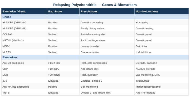
Summary Table: Genes and Biomarkers at a Glance
What a Pioneering Autoimmune Recovery Protocol Can Teach You
In 2010, Dr. Terry Wahls — a clinical professor of medicine at the University of Iowa and an MS patient herself — published the results of her personal recovery protocol and subsequently ran clinical trials testing it in multiple sclerosis patients. Her book The Wahls Protocol is not about MS specifically: it is about the underlying cellular mechanisms driving autoimmune disease and the mitochondrial, microbiome, and dietary leverage points that can modulate them. For RP patients, the framework she built offers one of the most evidence-grounded lifestyle approaches available for any systemic autoimmune condition.
Her approach challenges the standard thinking that lifestyle has only marginal influence on autoimmune disease activity. The clinical trial results — including measurable reductions in fatigue, inflammatory markers, and neurological function scores — suggest the opposite. What follows are the ten most impactful insights from her protocol.
1. Mitochondrial Dysfunction Is at the Root
Wahls argues that mitochondrial failure precedes and amplifies immune dysregulation. When mitochondria in immune cells are impaired, regulatory pathways (including T-regulatory cells) lose function, and inflammatory effector pathways dominate. The first priority is restoring mitochondrial nutrient density: coenzyme Q10, B vitamins, sulfur compounds, and antioxidants — all obtainable from food before supplements.
2. Nine Cups of Vegetables and Fruits Daily
Her protocol requires 3 cups of leafy greens (kale, collards, chard), 3 cups of sulfur-rich vegetables (cabbage, onions, mushrooms), and 3 cups of deeply colored produce daily. This is not arbitrary: each category maps to specific cellular needs. Leafy greens provide folate and vitamins K and B. Sulfur vegetables support glutathione synthesis — the body's primary cellular antioxidant. Colored produce provides antioxidants that suppress NF-κB and reduce CRP.
3. Omega-3 to Omega-6 Ratio Is Critical
The standard Western diet has an omega-6 to omega-3 ratio of approximately 20:1. Inflammatory thresholds drop dramatically as this ratio approaches 4:1. Wahls prescribes wild-caught fatty fish at least 3 times per week, elimination of seed oils, and grass-fed animal products — which have 3–5x more omega-3 content than grain-fed equivalents.
4. Gluten and Dairy Exclusion Is Non-Negotiable in Phase 2
Beyond a preliminary phase, Wahls removes gluten and dairy completely. Gliadin (wheat protein) increases intestinal permeability and can activate innate immune receptors. Casein (dairy protein) shares structural sequences with cartilage-related proteins in some individuals. For RP patients carrying HLA-DR4, this exclusion has particularly strong theoretical support.
5. The Gut Microbiome Shapes Immune Tone
Wahls identifies a low-diversity gut microbiome as a consistent feature of autoimmune patients and prescribes both prebiotic foods (resistant starch, fermentable fiber from the nine cups protocol) and fermented foods (sauerkraut, kimchi, kefir if tolerated). A healthy microbiome produces short-chain fatty acids that support intestinal barrier integrity and regulatory T-cell function — both critical in RP.
6. Electrical Stimulation of Muscles
An unusual element of the Wahls Protocol is e-stim (electrical muscle stimulation), used to maintain mitochondrial density in muscles when exercise tolerance is limited by disease. Wahls used it herself before recovering functional mobility. For RP patients with joint limitations, TENS and e-stim devices offer a non-impact way to maintain muscle mitochondrial health.
7. Stress Activates Immune Pathways Directly
Psychological stress elevates cortisol and catecholamines which activate NF-κB independently of immune triggers. Wahls prescribes a daily mind-body practice as a core therapeutic — not supplementary — component of the protocol. Even 10 minutes of focused breathing or meditation daily produces measurable changes in gene expression patterns in immune cells.
8. Sleep Is When Immune Calibration Happens
During deep sleep, the glymphatic system clears metabolic waste from the brain, T-regulatory cell function is restored, and inflammatory cytokine signaling is reset. Wahls emphasizes sleep as part of the therapeutic protocol — not optional background. She recommends 7–9 hours with consistent timing and blue light elimination after sunset.
9. Toxin Reduction Reduces Immune Burden
Heavy metals (mercury, lead), pesticides, and plasticizers activate immune cells and impair detoxification pathways. Wahls prescribes filtering drinking water, reducing processed food packaging exposure, and consuming sulfur-rich vegetables that support hepatic phase II detoxification. In RP patients, reducing the total immune burden from environmental toxins may lower the threshold for clinical flares.
10. Tracking Function Matters More Than Symptom Diaries
Wahls encourages patients to track functional outcomes — how far they walk, how much they sleep, grip strength, energy at specific times of day — rather than relying exclusively on subjective symptom scores. This creates objective data points that reveal trends invisible in quarterly rheumatology appointments and empowers patients to connect specific inputs (diet changes, sleep improvements) with measurable outcomes.
Complementary and Integrative Approaches
Beyond biomarker optimization and genetic insight, several evidence-grounded complementary approaches deserve attention in RP management. The four modalities below have meaningful support in autoimmune inflammatory conditions, even if direct RP-specific trial data remains limited due to the rarity of the disease.
The Autoimmune Protocol (AIP) by Sarah Ballantyne
The Autoimmune Protocol is a structured dietary and lifestyle framework developed by Dr. Sarah Ballantyne, a PhD research scientist who used it to put her own autoimmune conditions into remission. It eliminates grains, legumes, dairy, eggs, nightshades, nuts, seeds, and processed foods in an elimination phase, then systematically reintroduces foods to identify individual triggers. Beyond diet, the AIP framework addresses sleep, circadian rhythm, stress management, and physical movement as therapeutic pillars — making it one of the most comprehensive lifestyle protocols for autoimmune disease.
A pilot study published in Inflammatory Bowel Diseases found that 73% of participants with IBD achieved clinical remission after following the AIP diet for six weeks, with statistically significant reductions in inflammatory markers. A subsequent trial confirmed these findings with endoscopic evidence. While direct RP trials have not been conducted, the immunological mechanisms targeted — gut barrier integrity, molecular mimicry reduction, microbiome rebalancing — are directly relevant to RP pathophysiology. AIP diet clinical evidence.
For RP patients, the AIP framework provides a structured starting point. Begin with the full elimination phase (minimum 30–60 days), work with a practitioner to monitor inflammatory markers, and reintroduce foods one at a time every 5–7 days. Pay particular attention to nightshades and eggs, which are among the most common reintroduction reactors in autoimmune patients.
Mindfulness-Based Stress Reduction (MBSR)
MBSR is an 8-week structured program developed by Dr. Jon Kabat-Zinn that combines body scan meditation, mindful movement, and breath awareness. Its relevance to RP lies in the well-documented bidirectional relationship between psychological stress and autoimmune disease activity. Stress activates the HPA axis, elevates cortisol chronically, and directly upregulates NF-κB gene expression in immune cells — increasing inflammatory cytokine production and lowering flare thresholds.
A meta-analysis of 18 randomized controlled trials examining MBSR in autoimmune and inflammatory conditions found significant reductions in both CRP and subjective disease activity, with effect sizes particularly strong in conditions involving chronic pain and fatigue components. MBSR and inflammatory markers. These match the symptom burden many RP patients carry between visible flares.
For practical application, the full 8-week MBSR program (available online through the Center for Mindfulness at the University of Massachusetts) is the reference standard. If 8 weeks feels daunting initially, begin with 10 minutes of breath-focused meditation daily, which already demonstrates measurable changes in inflammatory gene expression profiles within 4–8 weeks.
Microbiome-Directed Therapies
Emerging research consistently links gut microbiome composition to autoimmune disease activity. In autoimmune arthritis conditions, lower microbial diversity correlates with higher disease activity scores and worse treatment response. The gut-immune axis is especially relevant in RP because the intestinal mucosa is where early antigen presentation occurs — including the presentation of dietary proteins that may trigger cross-reactive immune responses against cartilage antigens.
Multiple randomized trials have demonstrated that specific probiotic strains — particularly Lactobacillus rhamnosus GG, Bifidobacterium longum, and multi-strain formulas — reduce circulating inflammatory cytokines and improve gut barrier function. Probiotics and autoimmune arthritis inflammation. Fecal microbiota transplant (FMT) is an emerging area showing promise in autoimmune conditions, though it remains experimental outside of Clostridioides difficile treatment.
For practical application: prioritize dietary diversity (aiming for 30+ different plant foods per week as a microbiome diversity target); introduce 1–2 fermented foods daily (kefir, yogurt if tolerated on AIP reintroduction, kimchi, sauerkraut); consider a multi-strain probiotic (10+ billion CFU) for 8–12 week cycles. Evidence is evolving but the risk-to-benefit ratio is strongly favorable.
Breathing-Based Therapies
Diaphragmatic breathing and structured breathwork protocols activate the parasympathetic nervous system through vagal nerve stimulation, directly reducing sympathetic-driven inflammatory signaling. The relevance for RP extends beyond general stress reduction: the vagus nerve has direct anti-inflammatory efferents that suppress TNF-alpha and IL-6 production in peripheral immune tissues — a pathway termed the inflammatory reflex. Importantly for RP, laryngeal and tracheal involvement can impair breathing mechanics and create a feedback loop where breathing difficulty increases anxiety, which in turn elevates inflammatory tone.
A randomized trial in patients with rheumatoid arthritis found that slow-paced breathing (6 breaths per minute) for 15 minutes twice daily produced significant reductions in DAS28 disease activity scores and CRP over 8 weeks. Breathwork and autoimmune inflammation. The breathing technique — slow diaphragmatic breathing, 4-second inhale, 6-second exhale — is accessible and free.
For RP patients with laryngotracheal involvement, work with a respiratory therapist before starting any breathwork protocol to ensure techniques are safe and appropriately adapted to your current anatomy. Start with very gentle techniques (nasal breathing only, no breath holds) and advance slowly.
Conclusion
Relapsing polychondritis is a condition where biological individuality matters enormously. Two people with the same diagnosis can have fundamentally different inflammatory drivers, different genetic risk profiles, and different responses to the same treatment. Tracking the six biomarkers outlined here — hs-CRP, ESR, anti-type II collagen antibodies, IL-6, COMP, and NLR — gives you a repeatable, data-driven window into your disease activity that goes beyond what any single clinic appointment can provide. Understanding the five genetic variants adds another layer: not to predict your fate, but to identify the specific biological pathways where targeted interventions are most likely to have meaningful impact.
The next smart step is a concrete one: bring this list to your rheumatologist and request the biomarkers you are not already tracking. If you have access to genetic testing, identify your variants and match them to the lifestyle and supplementation strategies outlined here. Start with the free, foundational interventions — diet, sleep, stress — before adding supplements. Build from evidence, track your response, and adjust. Better information leads to better decisions, and that is ultimately where real agency in managing this condition begins.
Musculoskeletal Ear, Nose & Throat Autoimmune
Musculoskeletal: Joint Conditions
Respiratory: Airway Conditions
Autoimmune: Inflammatory Conditions Connective Tissue Conditions