This article contains AI generated content.
· UpdatedRenal Osteodystrophy: 4 Genes and 7 Biomarkers to Track
Introduction
If you have been living with chronic kidney disease, renal osteodystrophy may have been mentioned almost as an afterthought — a complication to be managed alongside everything else, rarely given the specificity it deserves. Yet for many CKD patients, bone disease is not a distant possibility. It begins silently, often years before a fracture occurs, driven by shifts in mineral metabolism that standard lab panels do not always capture fully. Understanding this process at a deeper level is not a luxury. It is one of the most actionable things a person in this situation can do.
The frustrating reality is that two patients with the same glomerular filtration rate can have completely different bone disease trajectories. One may have high-turnover bone disease driven by elevated parathyroid hormone. Another may have adynamic bone disease — paradoxically characterized by low bone-forming activity — which actually increases fracture risk through an entirely different mechanism. Applying the same protocol to both leads to unpredictable and sometimes harmful outcomes. Precision requires knowing which type of bone disease is present, what is driving it, and how an individual's biology is responding to treatment.
Generic dietary advice and standard phosphate binders address part of the problem, but they leave significant gaps. They do not account for individual vitamin D receptor function, genetic variation in FGF-23 signaling, or how a particular body handles calcium at different CKD stages. They also do not reveal whether a current treatment is improving bone turnover or inadvertently suppressing it too far. That gap — between population-level guidelines and individual biology — is where most patients get stuck, and where this article focuses.
What follows is built around precision. It covers the 7 most clinically meaningful biomarkers in renal osteodystrophy: what each one reveals, how to measure it, and what to do when results are off — with and without supplements. A dedicated section then explores the 4 key genes most likely to shape individual response to mineral-regulating therapies, each with a practical compensation plan. A book synthesis and evidence-supported complementary approaches round out the picture. Better information does not fix a damaged kidney, but it does produce better questions — and better questions change clinical outcomes.
Summary
This article covers the 7 most actionable biomarkers for monitoring renal osteodystrophy — including PTH, FGF-23, both forms of vitamin D, phosphorus, ionized calcium, bone-specific alkaline phosphatase, and serum bicarbonate — each with measurement costs, CKD-specific target ranges, and detailed action plans with and without supplements. The genetics section examines 4 key variants (VDR, KLOTHO, CYP27B1, CaSR) that explain why identical treatment produces different outcomes in different patients, along with specific compensation strategies for each unfavorable variant. A synthesis of Peter Attia's Outlive extracts the 10 most relevant insights for bone and mineral management. Four evidence-supported complementary modalities — tai chi, mindfulness, microbiome-directed therapy, and breathing practices — close the article with practical application protocols. If your results have looked "acceptable" while your symptoms suggest otherwise, or if you want to understand the deeper biology behind your lab values, this article provides the precision you are looking for.
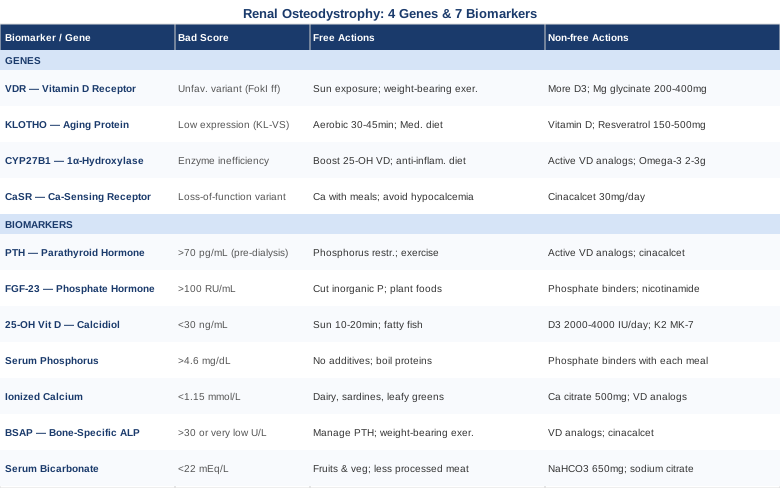
7 Biomarkers to Track in Renal Osteodystrophy
Tracking the right numbers at the right intervals is not about accumulating data for its own sake. In renal osteodystrophy, each biomarker tells a different part of a connected story — about how the kidneys are processing minerals, how bones are responding, and whether a current treatment is working or needs adjustment. The seven markers below were chosen for their combination of clinical relevance, measurability, and actionability. Together they build a picture far more complete than a routine metabolic panel.
1. Parathyroid Hormone (PTH): The Central Driver
Why it matters
PTH is the primary hormone governing bone turnover in CKD. As kidney function declines, phosphorus retention stimulates the parathyroid glands to produce more PTH, which then mobilizes calcium from bone. Sustained high PTH leads to osteitis fibrosa cystica, the most common high-turnover bone disease in CKD. Conversely, over-suppression of PTH — from excessive vitamin D analogs or calcium supplementation — leads to adynamic bone disease, where bone rebuilding becomes inadequate and fracture risk rises through a different mechanism. PTH is the master dial. Every other management decision orbits around it.
How to measure it
PTH is measured via a blood test — specifically intact PTH (iPTH) using an immunoradiometric or chemiluminescent assay. Cost range: $30–$80, often covered by insurance in documented CKD. KDIGO guidelines recommend checking PTH every 6–12 months in CKD stage 3, and every 3–6 months in stages 4–5. The target in CKD not yet on dialysis is generally 35–70 pg/mL. For dialysis patients, KDIGO recommends 2–9 times the upper limit of the normal range (approximately 130–600 pg/mL), acknowledging that mildly elevated PTH may be necessary to maintain bone turnover when active vitamin D synthesis is impaired.
If PTH is elevated — the plan without supplements
The first intervention is dietary phosphorus restriction, prioritizing elimination of inorganic phosphate additives found in processed foods, fast food, and cola beverages. Increasing fruit and vegetable intake alkalinizes the body and reduces the acid-driven stimulation of PTH — this connection is explored further under Biomarker 7. Regular weight-bearing physical activity (even modest walking or resistance training within medical tolerance) signals bone to maintain mass and can slow the PTH escalation curve. Strict avoidance of calcium supplementation at the wrong times is also relevant: calcium taken outside of meals suppresses PTH acutely but can backfire by reducing bone-building signals chronically.
If PTH is elevated — the plan with supplements or medication
Active vitamin D analogs — calcitriol, paricalcitol, or doxercalciferol — suppress PTH synthesis directly and are first-line pharmacological tools. Paricalcitol carries a lower risk of hypercalcemia than calcitriol and is commonly preferred for this reason. When vitamin D analogs alone are insufficient, cinacalcet (a calcimimetic) acts at the calcium-sensing receptor on the parathyroid gland, reducing PTH without raising calcium or phosphorus. For severe refractory hyperparathyroidism unresponsive to medical management, surgical parathyroidectomy becomes necessary. These medications should not be started or stopped without nephrology oversight, as rebound effects — hypercalcemia or paradoxically accelerated bone loss — can occur.
2. FGF-23 (Fibroblast Growth Factor 23): The Earliest Warning Signal
Why it matters
FGF-23 is a hormone produced by osteocytes in response to phosphate loading. It signals the kidney to excrete more phosphorus and suppress vitamin D activation. In CKD, FGF-23 levels rise dramatically — sometimes one-hundred-fold — before phosphorus itself becomes visibly abnormal on standard blood panels. This makes FGF-23 one of the earliest detectable markers of CKD-mineral bone disorder, often rising in CKD stage 2–3 when phosphorus, calcium, and PTH all appear completely normal. Elevated FGF-23 is independently associated with left ventricular hypertrophy, cardiovascular mortality, and accelerated CKD progression — giving it prognostic importance that rivals PTH itself.
How to measure it
FGF-23 is measured with a specialized blood test — either the intact FGF-23 (Kainos assay) or the C-terminal FGF-23 (Immutopics assay). Results are not interchangeable between assay types. Cost range: $150–$350, often not covered by standard insurance without clinical justification. Normal FGF-23 is approximately below 100 RU/mL on the intact assay. In CKD stages 4–5, values routinely exceed 1,000 pg/mL or higher. This test is not yet in routine KDIGO monitoring protocols but is increasingly ordered by precision-oriented nephrologists as an early-stage surveillance tool. It deserves to become standard.
If FGF-23 is elevated — the plan without supplements
Aggressive dietary phosphorus restriction is the most evidence-supported non-pharmacological response. Unlike calcium or vitamin D, you cannot supplement your way out of high FGF-23 — the phosphate burden driving it must be reduced at the source. Processed meats, cola drinks, processed cheese, and packaged baked goods are the worst offenders because they contain inorganic phosphate additives absorbed at near-100% efficiency. Plant-based phosphorus, bound to phytate, is absorbed at only 40–50%, making a whole-food diet a structural phosphate reducer. Modest resistance exercise has also shown preliminary evidence of reducing FGF-23 through improved skeletal signaling.
If FGF-23 is elevated — the plan with supplements or medication
Oral phosphate binders taken with meals reduce phosphorus absorption and, over time, lower FGF-23. Sevelamer carbonate is often preferred as it is calcium-free (avoiding hypercalcemia risk) and carries additional anti-inflammatory effects. Nicotinamide — a form of vitamin B3 — has shown FGF-23-lowering effects in small human trials by inhibiting intestinal sodium-phosphate co-transporters; typical doses of 250–500 mg/day have been studied, though gastrointestinal side effects limit tolerability for some. Iron supplementation in iron-deficient CKD patients can also reduce FGF-23: iron is required for enzymatic cleavage of FGF-23, and deficiency allows intact, biologically active FGF-23 to accumulate. These interventions should be coordinated with a nephrologist given the tight mineral balance involved.
3. 25-OH Vitamin D (Calcidiol): The Storage Tank
Why it matters
In CKD, vitamin D is impaired at two levels: the kidneys convert storage vitamin D (25-OH D3) to the active hormone (1,25-OH D3, calcitriol) less efficiently as GFR falls, and many CKD patients are also profoundly deficient in the storage form itself — due to limited sun exposure, poor diet, or obesity-related fat sequestration. Low storage vitamin D amplifies PTH elevation and accelerates bone loss. Critically, it is also one of the most correctable factors in the entire CKD-MBD picture — yet it is routinely under-monitored in standard nephrology practice.
How to measure it
Standard 25-OH vitamin D blood testing is available at virtually every laboratory. Cost range: $30–$80, widely covered. In CKD, most nephrologists recommend a 25-OH vitamin D level of at least 30 ng/mL, with precision medicine clinicians such as Peter Attia targeting 40–60 ng/mL for optimal bone, immune, and muscle health. Testing every 6 months — or more frequently during supplementation — is appropriate for monitoring response.
If 25-OH vitamin D is low — the plan without supplements
Sensible sun exposure — 10–20 minutes of midday sun on arms and legs, 3–5 times per week — can meaningfully raise levels in lighter-skinned individuals. For darker-skinned individuals, significantly more exposure is required to achieve the same synthesis. Fatty fish (salmon, sardines, mackerel), egg yolks, and fortified foods contribute modestly. Weight loss in overweight patients reduces fat sequestration and can raise circulating levels without any supplementation.
If 25-OH vitamin D is low — the plan with supplements
Plain cholecalciferol (vitamin D3) supplementation is appropriate for CKD stages 1–3 to restore storage levels. Typical doses: 2,000–4,000 IU/day under medical supervision, adjusted by lab response. In CKD stages 4–5, active vitamin D analogs may be added or substituted, since enzymatic conversion is impaired — but this should not replace correcting the underlying storage deficit. Vitamin K2 (MK-7 form, 100–200 mcg/day) is increasingly recommended alongside vitamin D to direct calcium toward bone rather than soft tissue and vasculature — particularly relevant in CKD where vascular calcification is a major concurrent risk. Calcium and phosphorus should be checked every 1–3 months when adjusting vitamin D therapy.
4. Serum Phosphorus: The Mineral That Drives the Cascade
Why it matters
Phosphorus is the linchpin of CKD-MBD. As GFR declines, phosphorus excretion falls, serum levels rise, and the entire compensatory cascade is triggered: higher FGF-23, higher PTH, lower active vitamin D, and progressive secondary hyperparathyroidism. Hyperphosphatemia drives vascular calcification, increases cardiovascular mortality, and accelerates bone disease. It is also a lagging marker: by the time phosphorus is visibly elevated on a standard panel, FGF-23 has often been elevated for years. This is why FGF-23 monitoring begins first, but serum phosphorus monitoring must remain continuous.
How to measure it
Serum phosphorus is included in standard metabolic panels. Cost: typically included in BMP/CMP panels, $10–$30 or covered by insurance. In CKD not on dialysis: target 2.7–4.6 mg/dL. In dialysis patients, KDIGO recommends targeting the normal population range (2.5–4.5 mg/dL). Monitoring every 3–6 months in CKD stage 3+, and monthly in stage 5.
If phosphorus is elevated — the plan without supplements
Dietary phosphorus restriction is the cornerstone — but with nuance. The goal is to limit inorganic phosphate additives (found in processed foods, fast food, and soft drinks) while preserving adequate protein intake. Boiling proteins and discarding the cooking water reduces phosphorus content measurably. Eating whole, unprocessed plant foods provides phytate-bound phosphorus with limited bioavailability. Timing protein evenly across meals — rather than concentrating it in one large sitting — reduces postprandial phosphorus spikes.
If phosphorus is elevated — the plan with supplements or medication
Phosphate binders taken with each meal are the pharmacological standard. Sevelamer carbonate is preferred for dialysis patients due to additional lipid-lowering and anti-inflammatory effects beyond phosphate binding. Calcium-based binders (calcium carbonate, calcium acetate) are lower cost but risk hypercalcemia and vascular calcification at high doses. Lanthanum carbonate and sucroferric oxyhydroxide are highly effective calcium-free alternatives for resistant cases. These medications must be taken with every meal, not once daily — the timing is mechanistically critical and one of the most common adherence errors seen in practice.
5. Ionized Calcium: The Precision Measurement
Why it matters
Standard total calcium on a metabolic panel is significantly influenced by albumin levels, which are commonly low in CKD patients. This means total calcium can appear normal while ionized (free, biologically active) calcium is abnormal. Both hypercalcemia and hypocalcemia carry consequences in CKD: low calcium drives further PTH elevation; high calcium from excessive supplementation suppresses PTH too aggressively, promotes adynamic bone disease, and accelerates vascular calcification. Knowing actual calcium status requires the right measurement, not the most convenient one.
How to measure it
Ionized calcium is measured from a blood gas sample or a specialized serum test. Cost range: $20–$60. Alternatively, albumin-corrected total calcium offers a reasonable approximation: add 0.8 mg/dL to total calcium for every 1 g/dL that albumin falls below 4.0 g/dL. Target ionized calcium: 1.15–1.30 mmol/L. The calcium-phosphorus product (Ca × P should remain below 55 mg²/dL²) provides an additional safety metric for vascular calcification risk.
If ionized calcium is low — the plan without supplements
Focus on dietary calcium from whole foods — dairy if tolerated and phosphorus load is manageable, sardines with bones, bok choy, fortified plant milks. Reducing dietary phosphate burden eases the mineral imbalance. Correcting vitamin D deficiency is prerequisite, since adequate 25-OH vitamin D is required for intestinal calcium absorption to function efficiently.
If ionized calcium is low — the plan with supplements
Calcium supplementation in CKD is complicated: modest doses can beneficially suppress PTH, but higher doses contribute to vascular calcification and adynamic bone disease. When supplementation is needed, calcium citrate (500 mg elemental calcium per dose, taken with meals) is preferred over calcium carbonate in CKD: it does not require stomach acid for absorption and adds an alkalizing citrate effect. Vitamin D must be co-corrected. If hypocalcemia is severe, active vitamin D analogs are more effective than calcium supplementation alone and carry lower risk of phosphorus loading.
6. Bone-Specific Alkaline Phosphatase (BSAP): The Bone Turnover Signal
Why it matters
Total alkaline phosphatase is a crude marker; liver disease confounds it significantly. Bone-specific alkaline phosphatase is produced exclusively by osteoblasts and provides a clean signal of bone formation activity. In high-turnover renal osteodystrophy driven by elevated PTH, BSAP is elevated. In adynamic bone disease, BSAP is low — and this distinction matters enormously because both conditions increase fracture risk, but require opposite management strategies. BSAP is the marker that helps distinguish between them when PTH alone is ambiguous, and it is gaining traction as a more precise bone status indicator in nephrology.
How to measure it
BSAP is measured from a blood sample. Cost range: $80–$200, less commonly covered without documented clinical indication. Normal BSAP: approximately 11–30 U/L (ranges vary by laboratory and sex). A value above the upper normal limit alongside elevated PTH suggests high-turnover disease. A low or normal BSAP alongside suppressed PTH confirms adynamic disease. Some nephrologists also track osteocalcin and C-terminal telopeptide (CTX) as complementary bone resorption markers.
If BSAP is elevated — the plan without supplements
Managing PTH (Biomarker 1) is the most direct way to reduce excessive bone turnover, since BSAP elevation in CKD is almost invariably driven by PTH excess. Weight-bearing exercise within medical tolerance — walking, gentle resistance work — provides mechanical stimulation that modulates osteoblast-osteoclast balance. Correcting metabolic acidosis (Biomarker 7) measurably reduces bone resorption markers independently of PTH management.
If BSAP is elevated — the plan with supplements or equipment
Active vitamin D analogs and cinacalcet reduce PTH and, consequently, normalize bone turnover over weeks to months. Bisphosphonates — commonly used for osteoporosis — are generally avoided in CKD stage 4–5 due to renal clearance impairment and risk of inducing or worsening adynamic bone disease: an important distinction from osteoporosis management in the general population. Whole-body vibration therapy has shown preliminary evidence in small trials for improving bone density markers in CKD patients who cannot perform high-impact exercise. Duration: 10–20 minutes per session, 3 times per week, using a platform vibrating at 30–40 Hz.
7. Serum Bicarbonate: The Acid-Base Connection
Why it matters
Metabolic acidosis is nearly universal in advanced CKD, and it directly damages bone through a mechanism most patients are never told about. When blood pH falls even mildly, the body buffers the acid by dissolving calcium phosphate from bone — accelerating bone loss as a survival mechanism to defend systemic pH. Studies have shown that correcting metabolic acidosis measurably reduces PTH levels, lowers bone resorption markers, and in some randomized trials improves bone mineral density. Serum bicarbonate below 22 mEq/L is a treatable, often overlooked driver of bone disease progression — and it is measured on every standard metabolic panel, making it one of the most accessible actionable values available.
How to measure it
Serum bicarbonate (total CO2) is part of a standard comprehensive metabolic panel. Cost: included in routine CMP, typically $0–$30 with insurance. KDIGO guidelines recommend maintaining serum bicarbonate at 22 mEq/L or above. Many clinicians targeting bone protection set their goal at 24–26 mEq/L for maximum benefit. Monthly monitoring is appropriate when actively treating acidosis; every 3–6 months once stabilized.
If bicarbonate is low — the plan without supplements
A diet rich in fruits and vegetables — producing net base in the body — alkalinizes blood and raises bicarbonate without any medication. Research by Goraya et al. published in Journal of the American Society of Nephrology demonstrated that a fruit and vegetable-rich diet reduced markers of CKD progression comparably to oral bicarbonate supplementation in CKD stage 3 patients (Goraya et al., 2012, PMID 22854643). Reducing dietary acid load — primarily by limiting processed meats and increasing vegetables — is both effective and safe across CKD stages.
If bicarbonate is low — the plan with supplements
Oral sodium bicarbonate (typically 650 mg tablets, 2–3 times daily with meals) is first-line pharmacological therapy. Starting dose: approximately 0.5–1 mEq/kg/day, adjusted to achieve target bicarbonate. Sodium citrate (Bicitra solution) is an alternative for patients with gastric sensitivity to bicarbonate tablets. Key side effect to monitor: additional sodium load from bicarbonate supplementation can worsen blood pressure and fluid retention — this requires active monitoring, particularly in later CKD stages. Potassium citrate provides alkalinization with potassium rather than sodium, which may suit some patients better, but requires close potassium monitoring given CKD's tendency toward hyperkalemia.
The Genetic Layer: 4 Variants That Shape Your Risk and Treatment Response
Biomarker tracking tells you what is happening right now. Genetics tells you why your body responds the way it does — and why identical protocols produce very different outcomes in different patients. In renal osteodystrophy, four genes stand out for their clinical relevance: they influence vitamin D metabolism, calcium sensing, phosphate regulation, and bone aging at a fundamental level. This is not genetic determinism. An unfavorable variant does not guarantee severe disease. But it can explain treatment resistance, guide supplementation choices, and justify closer monitoring in specific areas.
Genetic testing through clinical panels or consumer platforms like 23andMe can reveal these variants, though interpretation benefits from clinical context. Increasingly, practitioners familiar with nutrigenomics and precision nephrology integrate genetic data into CKD-MBD management decisions.
Gene 1: VDR (Vitamin D Receptor) — The Sensitivity Gate
What it does in renal osteodystrophy
The vitamin D receptor is the protein through which calcitriol (active vitamin D) exerts its effects — including suppressing PTH gene transcription, promoting intestinal calcium absorption, and regulating immune function. VDR polymorphisms — particularly BsmI, ApaI, TaqI, and FokI — affect receptor sensitivity and downstream response to any given level of vitamin D. In CKD patients, VDR polymorphisms have been associated with differences in secondary hyperparathyroidism severity and response to vitamin D analog therapy. The FokI ff genotype is associated with a shorter, less active receptor protein and is linked to higher PTH levels and worse bone disease outcomes in multiple CKD cohort studies.
If the gene variant is unfavorable — the plan without supplements
Since unfavorable VDR variants reduce receptor sensitivity, maximizing the upstream vitamin D signal becomes even more important. Optimizing 25-OH vitamin D levels aggressively (the substrate for conversion) gives the less-sensitive receptor its best chance. Sensible sun exposure is more strategically important for these individuals than for average patients. High-quality weight-bearing exercise (within medical constraints) upregulates VDR expression in bone and muscle tissue — one of the most important non-pharmacological compensators available. Maintaining a healthy body weight matters too, since obesity independently downregulates VDR.
If the gene variant is unfavorable — the plan with supplements
Patients with unfavorable VDR variants may require higher doses of vitamin D3 to achieve the same PTH suppression as patients with favorable variants — a clinically important insight that justifies targeting the upper end of the 40–60 ng/mL 25-OH vitamin D range. Magnesium is required both for VDR function and for the enzyme steps that convert vitamin D to its storage and active forms. Magnesium glycinate or citrate: 200–400 mg/day is generally well-tolerated in early-to-moderate CKD. In CKD stages 4–5, magnesium supplementation requires nephrology approval and monitoring given impaired renal excretion. The combination of optimized 25-OH vitamin D plus magnesium repletion may partially compensate for VDR inefficiency in ways that vitamin D alone cannot achieve.
Gene 2: KLOTHO — The Aging Protein That Controls FGF-23 Signaling
What it does in renal osteodystrophy
Alpha-KLOTHO is a protein produced primarily by the kidney that serves as a co-receptor for FGF-23 (it is required for FGF-23 to signal properly to kidney tubules) and independently functions as one of the most studied aging-protective proteins in biology. As CKD advances, KLOTHO expression in the kidney falls precipitously — often before serum KLOTHO becomes measurably low. This decline disrupts FGF-23 signaling, contributes to phosphate retention, and accelerates vascular aging and bone loss. Genetic variants in the KLOTHO gene — particularly the KL-VS haplotype and rs9536314 SNP — have been associated with altered circulating KLOTHO levels and modified risk of aging-related diseases including CKD progression and bone loss.
If the gene variant is unfavorable — the plan without supplements
Exercise is the most potent evidence-based KLOTHO upregulator available without a prescription. Aerobic exercise — 30–45 minutes of moderate intensity, 4–5 times per week — consistently raises circulating KLOTHO in multiple human trials across different populations, including CKD patients. This is one of the most compelling reasons why exercise prescription is not optional in CKD-MBD management. Mediterranean-style dietary patterns — high in vegetables, legumes, olive oil, and fish — are also associated with higher KLOTHO expression. Reducing oxidative stress through whole-food eating and limiting alcohol and processed food supports KLOTHO, as chronic oxidative stress is a primary suppressor of its expression.
If the gene variant is unfavorable — the plan with supplements
No approved KLOTHO pharmaceutical therapy yet exists, though research is actively advancing. Vitamin D — both D3 and active analogs — upregulates renal KLOTHO expression, adding a mechanistic reason to optimize vitamin D beyond mineral balance alone. Resveratrol (150–500 mg/day) has shown KLOTHO-upregulating effects in preclinical and limited human studies; evidence specifically in CKD is preliminary, and discussion with a nephrologist is advisable before adding it in advanced stages. Magnesium supports KLOTHO expression through overlapping pathways. The KLOTHO story is one of the few areas in CKD-MBD where lifestyle modification (specifically aerobic exercise) may rival pharmacological interventions in terms of effect size — a genuinely important finding that deserves more clinical emphasis.
Gene 3: CYP27B1 (1-Alpha Hydroxylase) — The Vitamin D Activation Enzyme
What it does in renal osteodystrophy
CYP27B1 encodes 1-alpha hydroxylase, the enzyme that converts storage vitamin D (25-OH D3) into the active hormone calcitriol (1,25-OH D3) in the kidney. As CKD advances, reduced kidney mass means less enzyme available — a structural problem that worsens with GFR decline. But CYP27B1 genetic polymorphisms can compound this problem: certain variants reduce enzyme efficiency even before kidney disease reaches advanced stages, explaining why some CKD patients develop severe secondary hyperparathyroidism earlier than their GFR alone would predict. These patients take adequate vitamin D but convert it inefficiently — and may be significantly undertreated on standard protocols.
If the gene variant is unfavorable — the plan without supplements
Since CYP27B1 handles conversion, maximizing the input substrate (25-OH vitamin D) is the first leverage point. Every improvement in storage vitamin D translates to as much active vitamin D as the impaired enzyme can produce. Reducing systemic inflammation is also mechanistically important: inflammatory cytokines — particularly TNF-alpha — suppress CYP27B1 expression directly. An anti-inflammatory dietary pattern — rich in omega-3 fatty acids from fatty fish and in polyphenols from berries, leafy greens, and herbs — can partially preserve enzyme function through this pathway. Weight control matters as well: adipose tissue sequesters storage vitamin D and adds to the conversion burden.
If the gene variant is unfavorable — the plan with supplements
This is the scenario where active vitamin D analogs bypass the impaired enzyme entirely. Calcitriol and its analogs (paricalcitol, doxercalciferol) do not require CYP27B1 conversion — they arrive in the active form. Patients who supplement with vitamin D3 but show persistently elevated PTH despite achieving adequate 25-OH vitamin D levels may have CYP27B1 inefficiency as a contributing factor, and would respond better to direct active analog prescriptions. This is a genuinely useful diagnostic insight. Omega-3 fatty acids (EPA/DHA, 2–3 g/day) have shown CYP27B1-supportive effects by reducing inflammatory suppression of the enzyme — and carry a favorable safety profile in early-to-moderate CKD. In advanced CKD, omega-3 dosing requires physician guidance given antiplatelet and other systemic effects.
Gene 4: CaSR (Calcium-Sensing Receptor) — The Parathyroid Gland's Thermostat
What it does in renal osteodystrophy
The calcium-sensing receptor on parathyroid cells detects blood calcium levels and adjusts PTH secretion accordingly. When calcium is high, CaSR activation suppresses PTH; when calcium falls, PTH rises. CaSR gain-of-function variants cause the gland to sense calcium as "high" at lower actual concentrations — suppressing PTH more readily. Loss-of-function variants do the opposite: they allow PTH to rise inappropriately even at normal calcium levels, contributing to more aggressive secondary hyperparathyroidism in CKD. Because cinacalcet works precisely at the CaSR — acting as a calcimimetic — CaSR genotype may predict responsiveness to this drug, making it one of the most clinically actionable genetic variants in CKD-MBD management.
If the gene variant is unfavorable — the plan without supplements
If a CaSR loss-of-function variant is reducing receptor sensitivity to calcium, ensuring optimal dietary calcium timing becomes even more important. Calcium consumed with meals activates CaSR transiently during the postprandial period, providing the suppressive signal the gland relies on. Avoiding hypocalcemia is critical — even brief dips in ionized calcium can trigger disproportionate PTH surges in patients with CaSR insufficiency. More frequent PTH monitoring (every 3 months rather than every 6) is appropriate for patients with known unfavorable CaSR variants and already-elevated PTH, allowing earlier treatment adjustment before escalation.
If the gene variant is unfavorable — the plan with supplements or medication
Cinacalcet works as a calcimimetic — it sensitizes the CaSR to suppress PTH even when actual calcium levels are not high. This makes cinacalcet particularly well-matched to CaSR loss-of-function variants, where the gland is genuinely less responsive to calcium. Knowing your CaSR genotype can meaningfully influence your nephrologist's choice between cinacalcet and active vitamin D analogs when both options are under consideration. Starting dose: typically 30 mg once daily, with gradual upward titration based on PTH response. Key side effects include nausea (often improved by taking with food) and hypocalcemia — frequent calcium monitoring is essential during the first months of cinacalcet therapy.
What Outlive by Peter Attia Teaches About Bone and Mineral Health
Peter Attia's Outlive: The Science and Art of Longevity (2023) is not a kidney disease book. But it is arguably the most rigorous mainstream synthesis of biomarker-driven preventive medicine available in popular form — and several of its frameworks map directly onto renal osteodystrophy management in ways that challenge common clinical assumptions. The following ten insights represent the most relevant lessons for anyone navigating CKD-related bone disease.
1. Biomarkers Are Decisions, Not Data Points
Attia's framework centers on the principle that tracking a biomarker has no value unless it changes what you do. In renal osteodystrophy, this means not merely ordering PTH and phosphorus, but understanding what each value implies for the type and intensity of treatment. A PTH of 300 pg/mL in a pre-dialysis patient with adynamic bone disease has completely different implications than the same number in a dialysis patient with high-turnover disease. Numbers without context mislead.
2. The Earliest Window Matters Most
Attia's "Medicine 3.0" framework is built around early detection — intervening years before disease becomes clinically apparent. FGF-23 is the CKD-MBD embodiment of this principle. It rises before phosphorus, calcium, or PTH become visibly abnormal, offering an intervention window that closes if you wait for standard markers to turn. Measuring FGF-23 in CKD stage 2–3 is the equivalent of measuring coronary artery calcium in someone who appears metabolically normal.
3. Bone Is Not Passive — It Is a Metabolic Organ
One of Attia's recurring themes is that bone is a metabolically active tissue responding continuously to mechanical loading and hormonal signals. Osteocalcin — released by osteoblasts during bone formation — acts as a circulating hormone affecting insulin sensitivity and muscle function. This bidirectional relationship means bone health improvements feed back into systemic metabolic health — and metabolic decline accelerates bone loss. CKD-MBD is not just a bone problem.
4. Muscle and Bone Are Inseparable Partners
Attia emphasizes the muscle-bone axis: muscle contractions generate the mechanical forces that drive bone remodeling. Sarcopenia — loss of muscle mass — is extremely common in CKD and represents a direct driver of bone fragility independent of any hormonal mechanism. Preserving muscle mass through adequate protein intake and resistance training is not a parallel priority to bone health. It is bone health, approached from a different angle.
5. Zone 2 Training Has Specific Biological Effects
Attia advocates for Zone 2 aerobic exercise — the intensity at which you can maintain a conversation — as a metabolic foundation. For CKD patients, Zone 2 is also the intensity range most compatible with reduced cardiovascular reserve and is specifically the level associated with KLOTHO upregulation in published human studies. Three to four sessions of 30–45 minutes per week at this intensity appears to be the threshold where meaningful benefits begin.
6. Protein Is the Most Under-Prescribed Intervention in Chronic Disease
Attia argues that adequate protein intake is chronically under-emphasized in mainstream medicine. In CKD, protein restriction has historically been recommended to slow GFR decline — but this has been significantly revisited. Current evidence suggests that sarcopenia's harms are severe enough that moderate protein intake (0.6–0.8 g/kg/day in pre-dialysis CKD; 1.2 g/kg/day in dialysis patients) is appropriate for most patients, with higher intakes potentially warranted for those with significant muscle wasting. This is an area where standard clinical guidelines and precision medicine recommendations increasingly diverge.
7. Fasting Protocols Require Significant Modification in CKD
Attia discusses time-restricted eating and intermittent fasting as tools for metabolic optimization. However, prolonged fasting in CKD can trigger intracellular release of phosphorus and potassium during refeeding shifts, creating acute mineral imbalances. The aggressive fasting protocols popular in longevity medicine are not safely transferable to advanced CKD without careful medical supervision and modification. This is one area where CKD changes the calculus significantly.
8. Vitamin D Is a Steroid Hormone, Not a Supplement
Attia frames vitamin D as a systemic steroid hormone with effects across immune function, gene expression, cardiovascular health, and metabolic regulation — not simply a bone-focused supplement. In CKD, this matters because the distinction between 25-OH D3 (storage) and 1,25-OH D3 (active hormone) is not academic — it determines what you supplement with and why. Taking vitamin D3 when active analog therapy is needed does not solve the problem. Understanding the pathway clarifies the intervention.
9. DEXA Underestimates the Real Picture in CKD
Attia recommends DEXA scans as a baseline and monitoring tool for bone mineral density. In CKD, however, DEXA measures bone density but not bone quality or turnover type — it cannot distinguish high-turnover from adynamic bone disease, both of which increase fracture risk. Bone histomorphometry (biopsy) remains the gold standard but is rarely performed. This reinforces the argument for biomarker-driven monitoring as the practical alternative.
10. Small Consistent Improvements Compound Over Years
Attia's central longevity argument is that consistent modest improvements across multiple domains — exercise, nutrition, sleep, biomarker optimization — compound significantly over decades. In renal osteodystrophy, this translates to a clear message: consistently maintaining phosphorus in range, correcting acidosis, optimizing vitamin D, and exercising regularly may not produce dramatic single-marker improvements, but together they may meaningfully slow disease progression over years. This is not a glamorous conclusion, but it is a well-supported one.
Evidence-Supported Complementary Approaches
The modalities below do not replace nephrology care. They represent approaches with meaningful — if sometimes limited — human clinical evidence specifically relevant to CKD-related bone disease: fall prevention, quality of life, inflammation reduction, and metabolic support. They work best as structured additions to, not substitutes for, medical treatment.
Tai Chi: Fall Prevention for Fragile Bones
Renal osteodystrophy increases fracture risk through both reduced bone mineral density and progressive muscle weakness, making falls disproportionately dangerous for CKD patients. Tai chi is a slow, deliberate movement practice with well-established benefits for balance, proprioception, lower-body strength, and fall prevention across multiple at-risk populations. In CKD patients, physical deconditioning is common and progressive, making tai chi's low-impact, low-cardiovascular-demand format particularly valuable as an accessible entry point into exercise.
A randomized controlled trial in hemodialysis patients demonstrated significant improvements in balance, lower extremity muscle strength, and health-related quality of life following a 12-week tai chi program compared to a sedentary control group. A Cochrane review on fall prevention in older adults with chronic conditions confirmed tai chi's efficacy as a fall-reducing intervention, with effect sizes comparable to more intensive exercise programs. Evidence specific to renal osteodystrophy bone density outcomes remains limited, but the fall prevention mechanism is highly relevant and the safety profile is excellent.
For practical application, begin with a guided class — in person or video-based — designed for seniors or chronic illness populations. Sessions of 20–30 minutes, 3 times per week match the protocols used in positive trials. Focus initially on weight transfer movements and lower-body stability. Avoid poses requiring extended single-leg balance until baseline stability improves. Tai chi is compatible with most CKD stages, including dialysis patients on non-dialysis days. Consult your nephrologist before starting if you have severe cardiovascular disease or uncontrolled blood pressure.
Mindfulness Meditation / MBSR: Reducing the Biological Cost of Chronic Stress
Living with CKD-related bone disease creates a persistent psychological burden: fear of fractures, dietary restriction fatigue, treatment complexity, and the progressive nature of the disease itself. This burden is not only emotional — it is biological. Chronic psychological stress elevates cortisol, which directly suppresses osteoblast activity and increases bone resorption. Managing the stress response has a genuine bone-protective dimension that is almost never addressed in nephrology appointments.
Mindfulness-Based Stress Reduction (MBSR) is an 8-week structured program combining meditation, body scan practice, and gentle movement that has been studied specifically in CKD populations. A randomized trial in hemodialysis patients found that a simplified mindfulness program significantly reduced depression scores, fatigue, and perceived pain compared to usual care. Reviews in nephrology-focused journals have identified mindfulness-based interventions as among the strongest evidence-supported psychological interventions for CKD quality of life.
The standard MBSR program involves 2.5-hour weekly group sessions plus 45-minute daily home practice for 8 weeks. Scaled-down versions — 10–15 minutes of daily guided meditation using apps such as Insight Timer — show meaningful benefits for stress and sleep in multiple trials. For CKD-MBD specifically, the cortisol-lowering effect is the most relevant biological mechanism. Starting with 10 minutes of breath-focused meditation per day is a realistic, low-barrier entry point that requires no equipment and carries no side effects.
Microbiome-Directed Therapies: The Gut-Kidney-Bone Axis
An emerging and increasingly compelling research area identifies the gut microbiome as a meaningful modulator of CKD-MBD. The gut microbiome influences phosphate absorption efficiency, generates uremic toxins — particularly indoxyl sulfate and p-cresol — that worsen inflammation and bone turnover, and produces short-chain fatty acids (SCFAs) that support intestinal barrier integrity and calcium absorption. CKD patients have significantly disrupted microbiome composition — reduced diversity, loss of SCFA-producing species — and this dysbiosis may independently worsen bone disease through multiple pathways simultaneously.
A randomized trial demonstrated that dietary prebiotic supplementation (resistant starch, inulin-type fructans) in CKD patients significantly reduced uremic toxin production, improved gut barrier markers, and reduced systemic inflammation. Reviews in Nutrients and Journal of Renal Nutrition have summarized evidence for synbiotic (probiotic plus prebiotic combined) interventions in CKD, noting improvements in PTH, inflammatory markers, and oxidative stress in several randomized trials. Evidence linking microbiome therapy directly to bone density improvement in CKD remains preliminary, but mechanistic support for this connection is accumulating.
Practically, a diverse whole-plant-food diet is the primary prebiotic intervention — achievable without supplements and with meaningful phosphorus control. Fermented foods (yogurt, kefir, sauerkraut) may be added cautiously in CKD stages 1–3 with attention to potassium and phosphorus content. Probiotic supplementation — focusing on Lactobacillus acidophilus and Bifidobacterium species studied in CKD — should be discussed with your nephrologist before starting. Introduce prebiotic-rich foods gradually to minimize gas and bloating. Monitor phosphorus, potassium, and bowel function actively during any dietary microbiome intervention in advanced CKD.
Breathing-Based Therapies: A Non-Pharmacological Approach to Metabolic Acidosis
Metabolic acidosis in CKD is primarily managed with bicarbonate supplementation, but breathing-based therapies offer a complementary physiological influence. Controlled slow, deep breathing produces transient respiratory alkalinization — raising blood pH slightly — which can partially offset the metabolic acid burden and reduce the moment-to-moment demand on bone mineral buffering. This mechanism is modest, but physiologically real and additive to dietary and pharmacological management. The secondary benefits — cortisol reduction, improved heart rate variability, and blood pressure lowering — compound the bone-protective relevance.
Slow diaphragmatic breathing at approximately 6 breaths per minute — the physiological resonance frequency — for 15–20 minutes activates the parasympathetic nervous system and produces measurable effects on blood pH and heart rate variability. Clinical studies in CKD patients using structured slow breathing training have reported improvements in blood pressure and subjective wellbeing, though direct bone marker data are absent. The combination of pH support and cortisol reduction provides a rationale for inclusion as a low-risk adjunct.
For practical use, practice diaphragmatic breathing for 15 minutes twice daily — morning and evening work well. Apps such as Breathwrk or Insight Timer can guide pacing. Box breathing (4 counts inhale, 4 hold, 4 exhale, 4 hold) or the 4–7–8 method are accessible entry points. Practice in a seated or supine position. Breathing exercises are safe across virtually all CKD stages and can be performed during dialysis sessions. They should not replace prescribed bicarbonate supplementation, but serve as a genuine complement that also addresses the autonomic dimension of chronic disease.
Conclusion
Renal osteodystrophy is manageable — but only if it is managed with precision. The difference between a patient whose bone disease stabilizes and one whose fractures arrive ahead of schedule often comes down to how carefully the right markers are tracked, how early FGF-23 and PTH trends are recognized, and whether treatment is calibrated to individual biology rather than applied as a uniform protocol. The genetic layer adds a further dimension: understanding whether a VDR variant blunts vitamin D therapy, or whether a CaSR variant predicts cinacalcet responsiveness, can change clinical decisions in meaningful ways.
None of this requires abandoning standard care. It requires expanding it — asking for the markers that matter, considering whether genetic panel testing is relevant when treatment response seems unusually poor, and adding evidence-supported complementary practices that compound benefit over time. The next step is concrete: bring a list of the seven biomarkers in this article to your next nephrology appointment, ask which are currently being tracked and how often, and begin a conversation about what is missing from your current monitoring picture. That one conversation may open more doors than any single supplement or protocol ever could.
Cardiovascular Endocrine & Metabolic
Musculoskeletal: Bone Conditions Muscle Conditions
Urological: Kidney Conditions