This article contains AI generated content.
· UpdatedRheumatoid Nodulosis: 5 Genes And 7 Biomarkers To Track
Introduction
Rheumatoid nodulosis is one of those presentations that standard rheumatology guidelines handle poorly. You can follow every recommended treatment step for rheumatoid arthritis and still watch firm lumps continue forming under your skin near your elbows, fingers, or heels — sometimes because of the treatment itself. Methotrexate, one of the most commonly prescribed disease-modifying drugs for RA, is documented to paradoxically accelerate nodule formation in a subset of patients. If that has happened to you, the gap between generic advice and what you actually need becomes very clear.
The frustration with rheumatoid nodulosis often lies in its specificity. Unlike joint-dominant RA, nodulosis tends to signal a particular immunological phenotype: high seropositivity, specific genetic risk factors, and a granulomatous inflammatory process driven by macrophages rather than the synovial proliferation typical of classic RA. Treating it as ordinary arthritis misses those distinctions entirely, and patients often end up managing symptoms without ever understanding why their disease looks the way it does.
This article is built around measurement and targeted understanding. Knowing which biomarkers to track — and what your specific levels actually mean — turns a vague condition into something more concrete and actionable. Similarly, understanding which genetic variants are associated with rheumatoid nodulosis shifts the question from "why is this happening to me" toward "what upstream factors am I working with, and what can I do about them." Both lines of inquiry are covered here in depth, with practical plans for each.
The article covers two core analytical frameworks: seven biomarkers to track and five gene variants to understand. Beyond those, it includes a summary of a nutrition-first protocol that challenges conventional autoimmune care, followed by four complementary therapies with meaningful clinical evidence for RA. None of these replace your rheumatologist. All of them can make your conversations with that rheumatologist more productive and your own decisions better informed.
Summary
This article covers 7 biomarkers directly relevant to rheumatoid nodulosis — anti-CCP antibodies, rheumatoid factor, hsCRP, IL-6, MMP-3, vitamin D, and ESR — explaining why each matters, how to test it (with cost estimates), what you can do without supplements, and what targeted supplementation or equipment may add. Some markers here, like MMP-3 and IL-6, are rarely discussed outside specialist settings but offer critical information about nodule biology that generic inflammation panels miss.
5 genetic variants — HLA-DRB1, PTPN22, PADI4, STAT4, and TNF-α — are examined with concrete action plans for each, whether you prefer a lifestyle-first approach or want to know which supplements map to specific genetic vulnerabilities.
Beyond lab work, the article summarizes the Wahls Protocol — a nutrition-based strategy that challenges standard autoimmune care with clinical trial support — and reviews four complementary therapies with the best evidence for this condition, including the Autoimmune Protocol from Sarah Ballantyne. If you have been managing RA without tracking these specific markers, there is a high probability that you are missing actionable information about your own disease biology.
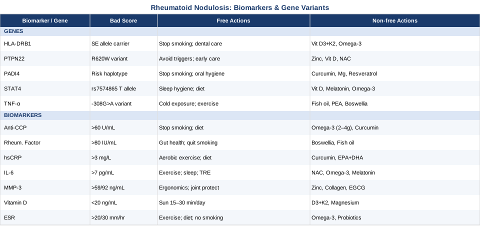
7 Biomarkers to Track for Rheumatoid Nodulosis
Tracking the right biomarkers in rheumatoid nodulosis does more than confirm your diagnosis. It lets you monitor disease activity independently of symptom fluctuation (which can be unreliable), measure the actual biological impact of interventions, and identify specific drivers of nodule formation that may respond to targeted approaches. The seven markers below cover different layers of the disease process — from autoantibody production to tissue destruction to immune regulation — and together form a more complete picture than standard panels alone.
Anti-CCP Antibodies (Anti-Cyclic Citrullinated Peptide)
Why it matters: Anti-cyclic citrullinated peptide antibodies are the most specific serological marker for rheumatoid arthritis, with specificity exceeding 95% using second-generation assays. In the context of rheumatoid nodulosis specifically, anti-CCP seropositivity is found in 70–80% of confirmed cases and correlates with disease severity, nodule burden, and faster radiographic progression. Anti-CCP can appear in blood years before clinical symptoms — indicating that the underlying citrullination-driven immune response precedes and likely causes tissue damage, including nodule formation. The higher the titer, the more aggressive the disease phenotype tends to be. A substantial research base on anti-CCP utility in RA is available through PubMed's anti-CCP literature.
How to measure it: Standard blood draw, ELISA-based assay. Positive threshold is typically 20 units/mL, with high-positive values above 60 units/mL carrying distinct prognostic weight. Cost: $50–150. Most rheumatology panels include it at diagnosis. Annual retesting is reasonable for stable disease; more frequent if disease course is changing or treatment is being adjusted.
If the score is bad, the plan without supplements: Smoking is one of the strongest environmental drivers of anti-CCP production — the association is dose-dependent and replicated across dozens of cohort studies. Stopping smoking reduces RA risk and disease severity in seropositive individuals to a degree no supplement can match. An anti-inflammatory dietary pattern (low in refined carbohydrates and ultra-processed foods, rich in polyphenols, vegetables, and omega-3 fats) reduces the systemic oxidative stress that amplifies the citrullination process. Quality sleep (7–9 hours) and chronic stress reduction lower cortisol-driven immune activation that sustains anti-CCP production. Periodontal health deserves specific attention: Porphyromonas gingivalis, a common gum pathogen, has its own PAD enzyme and can initiate citrullination cascades in susceptible individuals.
If the score is bad, the plan with supplements or equipment: Omega-3 fatty acids (EPA+DHA, 2–4g per day) have a well-documented anti-inflammatory effect in RA, with multiple RCTs demonstrating reductions in disease activity and inflammatory markers. Use fish oil or algal-derived EPA+DHA; start at 2g/day and titrate upward over 4 weeks. Minimum commitment before reassessing: 3 months. Bioavailable curcumin (500–1000mg/day, phospholipid-bound or piperine-enhanced form) reduces NF-κB activation, which drives citrullination gene expression. Side effects at these doses are minimal; watch for mild GI symptoms and interaction with anticoagulants at higher doses. Vitamin D optimization (detailed in its own section below) is also directly relevant to anti-CCP modulation.
Rheumatoid Factor (RF)
Why it matters: Rheumatoid factor is an immunoglobulin — usually IgM — directed against the Fc portion of IgG. While less specific for RA than anti-CCP, RF is a particularly strong marker of extra-articular disease, and rheumatoid nodulosis is one of the most characteristic extra-articular RA manifestations. Studies consistently report RF positivity in over 90% of rheumatoid nodulosis patients, compared to roughly 70–80% of RA patients overall. High-titer RF combined with anti-CCP seropositivity (double seropositivity) represents the immunological profile most strongly associated with aggressive nodule formation. RF reflects immune complex deposition in tissues, which is part of the histological picture in nodule development at the macrophage level.
How to measure it: Blood test, reported in IU/mL. Positive is typically above 20 IU/mL; high-positive above 80 IU/mL carries additional prognostic weight. Cost: $20–80. Routinely ordered at RA diagnosis. Retest annually or when disease course changes significantly.
If the score is bad, the plan without supplements: Smoking increases RF production — another direct argument for cessation. Excess alcohol raises systemic immune complex formation. Gut health matters directly: dysbiosis-associated intestinal permeability increases systemic antigen exposure, which sustains RF production by providing a continuous stream of foreign antigens for the immune system to respond to. Removing ultra-processed food, refined sugar, and excessive grain consumption while increasing fermented foods and dietary fiber directly targets the gut-immune axis relevant to RF elevation.
If the score is bad, the plan with supplements or equipment: Boswellia serrata (100–400mg of standardized extract per day, containing 65%+ boswellic acids) has documented anti-inflammatory effects in autoimmune arthritis, with several RCTs showing reductions in joint pain, stiffness, and inflammatory markers. Take with food; GI symptoms are the main reported side effect. Fish oil at foundational doses (2–4g EPA+DHA/day) remains relevant across all RF-elevated presentations. Cold shower exposure — 2–3 minutes of cold water at the end of each shower, 3–5 days per week — activates norepinephrine-mediated anti-inflammatory signaling and the vagal anti-inflammatory reflex. This mechanism is documented in Wim Hof protocol research and cold immersion studies in inflammatory conditions. All interventions reviewed at 3-month intervals.
High-Sensitivity C-Reactive Protein (hsCRP)
Why it matters: CRP is synthesized by the liver in direct response to pro-inflammatory cytokines, particularly IL-6. In rheumatoid nodulosis, elevated hsCRP reflects the systemic inflammatory burden sustaining both joint disease and nodule growth. High values (above 3 mg/L) are associated with faster nodule expansion, higher DAS28 activity scores, and dramatically increased cardiovascular risk — a concern that is systematically underappreciated in RA patients, whose cardiovascular mortality is roughly 50% higher than the general population. On the practical side, hsCRP is one of the most responsive biomarkers available: it can change meaningfully within weeks of dietary or pharmacological interventions, making it a valuable real-time monitoring tool for tracking whether interventions are actually working.
How to measure it: Blood test using a high-sensitivity assay — specify hsCRP, not standard CRP. Below 1 mg/L is optimal; 1–3 mg/L indicates moderate cardiovascular and inflammatory risk; above 3 mg/L is elevated. Active infection can spike it to 100+ mg/L, invalidating interpretation. Cost: $15–50. Useful at every rheumatology visit, roughly every 3–6 months.
If the score is bad, the plan without supplements: Regular moderate aerobic exercise (30 minutes, 5 days per week) consistently lowers CRP over 8–12 weeks of sustained practice — this is one of the most robustly replicated findings in lifestyle-inflammation research. Mediterranean-style dietary pattern or elimination of refined carbohydrates and fried foods produces significant CRP reductions within 4–6 weeks. Sleep optimization — particularly identifying and treating sleep apnea, since intermittent hypoxia is a potent CRP driver — can produce dramatic improvement. Chronic stress maintains elevated CRP through sustained cortisol and catecholamine activation; structured stress-reduction practices (discussed in the complementary section below) are not peripheral considerations for this marker.
If the score is bad, the plan with supplements or equipment: Bioavailable curcumin (500–1000mg/day) has one of the strongest evidence bases for CRP reduction among supplements, with multiple RCTs demonstrating significant effects in inflammatory conditions. EPA+DHA (2–4g/day) reduces hepatic CRP synthesis. Green tea EGCG extract (400mg/day) and resveratrol (150–500mg/day) have modest but documented anti-inflammatory effects. HIIT exercise (2–3 sessions per week, 20–30 minutes per session) shows both acute post-exercise and chronic CRP reduction in RA intervention trials. All at 3-month review cycles.
Interleukin-6 (IL-6)
Why it matters: IL-6 is not just a generic inflammation marker — it is mechanistically upstream of nodule formation. At the cellular level, IL-6 drives the differentiation of monocytes into the macrophage phenotype that forms the palisading granuloma architecture that defines rheumatoid nodules histologically. Elevated serum IL-6 correlates with nodule number and size, and IL-6 is the primary upstream stimulus for hepatic CRP synthesis, meaning it precedes and predicts the CRP signal. The clinical validation of this pathway is now conclusive: IL-6 receptor inhibitors such as tocilizumab and sarilumab are among the most effective biologics for RA, confirming that IL-6 is not a bystander but an active driver of the disease process. Measuring IL-6 directly provides information that CRP alone cannot — it can be elevated even when CRP is temporarily within range. Relevant literature is available through PubMed on IL-6 and RA pathology.
How to measure it: Blood test by ELISA, reported in pg/mL. Normal is typically below 7 pg/mL, though lab reference ranges vary. This is not routinely ordered in primary care — you may need to specifically request it from a specialist or use a direct-to-consumer lab. Cost: $100–300. Useful as a baseline when beginning treatment, and as a deep-dive assessment 3–6 months into any significant protocol change.
If the score is bad, the plan without supplements: Regular exercise raises IL-6 acutely (from contracting muscle tissue, functioning as a myokine signal) but chronically reduces resting IL-6 through downregulation of the broader inflammatory cytokine network. The net effect of consistent moderate exercise is lower baseline IL-6. Sleep deprivation is one of the fastest and most reliable ways to elevate IL-6 — even partial sleep restriction produces measurable cytokine rises within 2–3 nights. Time-restricted eating (eating within an 8–10 hour window, typically skipping late-night calories) has been shown in several intervention studies to reduce fasting pro-inflammatory cytokine profiles including IL-6.
If the score is bad, the plan with supplements or equipment: NAC (N-acetyl cysteine, 600–1200mg/day) reduces oxidative stress-driven IL-6 production and supports glutathione synthesis relevant to macrophage activation. EPA+DHA (2–4g/day) directly inhibits IL-6 gene transcription in macrophages through competitive arachidonic acid displacement. Melatonin at physiological doses (0.5–3mg, 30 minutes before sleep) has documented immunomodulatory properties including IL-6 suppression. Sauna use (3–4 sessions per week, 15–20 minutes at 80–90°C) activates heat shock proteins and has been associated with reductions in systemic inflammatory cytokines in Finnish cohort data. Begin sauna use cautiously if any cardiovascular history is present. 3-month cycle review.
Matrix Metalloproteinase-3 (MMP-3)
Why it matters: MMP-3 (stromelysin-1) is a tissue-remodeling enzyme secreted by activated synoviocytes and macrophages that degrades extracellular matrix components including fibronectin, laminin, and cartilage proteoglycans. In rheumatoid arthritis, it is a key mediator of structural joint damage. In rheumatoid nodulosis specifically, elevated MMP-3 reflects the tissue destruction occurring both in joints and within the remodeling dynamics of nodule architecture. Unlike CRP and ESR, which measure general inflammatory signal strength, MMP-3 measures whether that inflammation is actively destroying tissue — an important distinction for nodulosis patients. Elevated MMP-3 has been proposed as an independent predictor of radiographic damage progression in RA and tracks with nodule burden in research cohorts.
How to measure it: Blood test by ELISA. Reference ranges are approximately 17–59 ng/mL for women and 17–92 ng/mL for men. Cost: $150–400. Not a standard primary care test — specialist order or specialty lab access is required. Worth establishing as a baseline at diagnosis and retesting 3–6 months into any significant protocol change. Advanced inflammatory panels recommended by specialists including Peter Attia's framework for longevity medicine increasingly incorporate MMP-3 as a marker of active tissue remodeling processes.
If the score is bad, the plan without supplements: Mechanical loading of affected joints directly stimulates MMP-3 secretion — ergonomic adjustments (cushioning over nodule-prone pressure points, activity modification during active flares) have a measurable effect on this pathway. Reducing systemic IL-1β and TNF-α levels through anti-inflammatory diet and the other interventions described in this article will lower MMP-3 indirectly, since these cytokines are its primary upstream transcriptional activators. Joint protection techniques taught by occupational therapists in rheumatology settings are specifically relevant to MMP-3 reduction through mechanical means.
If the score is bad, the plan with supplements or equipment: Zinc (15–30mg/day with food) is a cofactor for metalloproteinase regulation; deficiency has been associated with imbalanced MMP activity in connective tissue studies. Collagen peptides (10–15g/day in divided doses, hydrolyzed type I and III) support connective tissue integrity and have shown cartilage health benefit in small clinical trials. EGCG from green tea (400–600mg/day) inhibits MMP-3 activity in cellular models with some in vivo support. Important: monitor zinc-copper balance with sustained supplementation — zinc competes with copper absorption. Consider 1–2mg copper as a cofactor if using zinc long-term. 3-month review cycle with MMP-3 retesting.
Vitamin D (25-Hydroxyvitamin D)
Why it matters: Vitamin D is a steroid hormone with pervasive immunomodulatory effects that extend far beyond its role in calcium metabolism. Its nuclear receptor (VDR) is expressed on every immune cell type central to rheumatoid nodulosis: T lymphocytes, B lymphocytes, dendritic cells, and macrophages. Mechanistically, adequate vitamin D suppresses Th17 cell differentiation — a major pro-inflammatory arm of adaptive immunity in RA — while simultaneously promoting regulatory T cell (Treg) activity that dampens autoimmune overactivation. It also inhibits the NF-κB pathway that drives multiple inflammatory cytokine genes. Vitamin D deficiency is both extremely common in RA patients and independently associated with higher disease activity, elevated anti-CCP titers, and worse long-term outcomes across multiple systematic reviews. Reference: PubMed meta-analyses on vitamin D and RA.
How to measure it: Blood test for 25-hydroxyvitamin D (25-OH D). Conventional deficiency threshold is below 20 ng/mL (50 nmol/L); functional medicine specialists frequently target 50–70 ng/mL for autoimmune conditions — substantially higher than the bone health threshold. Cost: $30–80. Test in late winter or early spring to capture seasonal nadir. Retest 8–12 weeks after starting supplementation to dial in dose accurately.
If the score is bad, the plan without supplements: Midday sun exposure on large skin surface areas — arms, legs, and face without sunscreen — for 15–30 minutes, 3–5 days per week, can produce meaningful vitamin D3 synthesis depending on latitude, skin tone, and season. Dietary sources (wild-caught fatty fish, egg yolks, beef liver) contribute modestly but rarely enough to correct significant deficiency. For most RA patients in temperate climates, supplementation is practically necessary to reach therapeutic immune-supportive levels.
If the score is bad, the plan with supplements or equipment: Vitamin D3 (cholecalciferol, 2,000–5,000 IU/day — adjust based on blood level targets) should always be paired with vitamin K2 (90–180mcg/day of the MK-7 form) to direct calcium appropriately toward bone rather than arterial tissue. Magnesium (300–400mg/day of glycinate or malate form) is enzymatically required for vitamin D conversion to its active form and is frequently deficient in RA patients — without adequate magnesium, supplemental vitamin D may not be fully utilized. Test and retest is non-negotiable: the goal is 50–70 ng/mL, not just clearing the deficiency threshold. At recommended doses, side effects are rare; toxicity requires very high doses (typically above 10,000 IU/day) maintained for extended periods.
Erythrocyte Sedimentation Rate (ESR)
Why it matters: ESR is the simplest and most accessible measure of systemic inflammation in this list. Red blood cells settle faster in an elevated inflammatory state as pro-inflammatory proteins — fibrinogen, immunoglobulins, and acute-phase reactants — increase plasma viscosity. In rheumatoid arthritis, ESR is a component of the DAS28 disease activity score and reflects the overall inflammatory burden in a way that is complementary to CRP rather than redundant with it: ESR is more influenced by long-lived proteins and immunoglobulins, while CRP responds faster to acute changes. Tracking both together provides a more complete inflammatory picture. In rheumatoid nodulosis, ESR elevation typically corresponds to periods of active nodule growth and systemic disease activity, making it a reliable trend marker over time.
How to measure it: Standard blood test. Normal is below 20 mm/hr for men under 50 and below 30 mm/hr for women under 50, with values increasing modestly with age. Cost: $10–40 — the most affordable marker on this list. Accessible through any primary care physician. Track every 3–6 months as a treatment response signal.
If the score is bad, the plan without supplements: Consistent aerobic exercise, anti-inflammatory diet, smoking cessation, and alcohol reduction all lower ESR meaningfully over sustained practice. Excess adiposity is a substantial source of pro-inflammatory adipokines that sustain ESR elevation; weight management has a documented effect for patients with metabolic comorbidity. Intermittent fasting and time-restricted eating reduce systemic inflammation markers including ESR in several well-designed intervention studies.
If the score is bad, the plan with supplements or equipment: The foundational protocol — omega-3 EPA+DHA (2–4g/day), bioavailable curcumin (500–1000mg/day), and optimized vitamin D3+K2 — applies across all inflammation markers including ESR. Probiotic supplementation (multi-strain, 10–50 billion CFU/day) targeting the gut-immune axis is an emerging but increasingly supported approach: gut dysbiosis is now well-documented in RA and correlates with systemic inflammatory marker elevation including ESR. HIIT exercise (2–3 sessions per week, 20–30 minutes each) shows consistent acute and chronic anti-inflammatory effects in RA intervention trials. All supplements reviewed at 3-month intervals with repeat ESR testing.
The Genetic Architecture of Rheumatoid Nodulosis
Biomarker data tells you what is happening biologically right now — your current inflammatory state, autoantibody levels, and tissue destruction rate. Genetic information adds a different layer: why you are specifically predisposed to this phenotype, and which biological pathways carry structural vulnerabilities that shaped your disease from the beginning. You cannot change your genes, but knowing which variants you carry allows you to address the downstream effects of those variants with precision rather than guesswork.
The five variants below are among the most consistently replicated genetic risk factors for rheumatoid arthritis with documented relevance to extra-articular manifestations including nodulosis. Some are testable through consumer genetic services (23andMe, Ancestry); more complete variant analysis is available through whole exome panels ordered by rheumatology specialists. For each gene, a practical action plan is provided — lifestyle-first, then targeted supplementation or equipment for those who want to go further.
HLA-DRB1: The Shared Epitope
HLA-DRB1 encodes a cell-surface protein on antigen-presenting cells that presents peptide fragments to T cells. The shared epitope (SE) refers to a specific amino acid sequence — QKRAA or related variants — in the DRB1 hypervariable region, encoded by alleles including *04:01, *04:04, and *01:01. This sequence changes how citrullinated peptides are presented to T cells, making the immune system substantially more likely to mount an autoimmune response against citrullinated self-proteins. HLA-DRB1 SE alleles represent the single strongest genetic risk factor for seropositive RA and are specifically associated with anti-CCP positivity, nodule formation, and extra-articular disease severity. Carrying two SE alleles (homozygous) confers significantly higher risk than carrying one. Research available through PubMed on HLA-DRB1 and RA phenotype.
If the gene is bad, the plan without supplements: The shared epitope's risk is dramatically amplified by smoking — individuals who carry SE alleles and smoke have RA risk that is many times higher than non-smoking non-carriers due to the convergence of citrullination-promoting oxidative stress and the SE's efficient presentation of citrullinated antigens. Smoking cessation is the single most impactful non-pharmacological intervention available for SE carriers. Periodontal disease management is equally direct: P. gingivalis-driven citrullination in gum tissue creates the antigenic peptides that SE alleles present most efficiently to T cells, making regular dental care a genuine disease-modifying behavior. Anti-inflammatory diet reduces the overall inflammatory amplification of SE-mediated autoimmunity.
If the gene is bad, the plan with supplements or equipment: Vitamin D3+K2 at optimized doses (targeting 50–70 ng/mL) directly downregulates inflammatory gene expression downstream of HLA-DRB1 antigen presentation — it reduces Th17 differentiation that follows SE-mediated T cell activation. Omega-3 EPA+DHA (2–4g/day) blunts macrophage and T cell activation that SE alleles facilitate through enhanced antigen presentation. Probiotic supplementation targeting both oral and gut microbiome may reduce the antigenic load from microbial-driven citrullination that SE alleles amplify most potently. Ongoing foundational supplementation; reassess with anti-CCP and RF every 6 months.
PTPN22: The R620W Variant
PTPN22 encodes protein tyrosine phosphatase non-receptor type 22, an enzyme that regulates the signaling threshold for T and B cell activation. The R620W variant (rs2476601) is a well-characterized polymorphism that paradoxically lowers the activation threshold of autoreactive immune cells, making them more likely to respond to self-antigens at concentrations that would be ignored in a non-carrier. This variant is associated with multiple autoimmune diseases — RA, type 1 diabetes, lupus, Graves' disease — reflecting a shared autoimmunity mechanism. In RA, PTPN22 R620W carriers tend toward earlier disease onset, higher autoantibody titers, and the aggressive seropositive phenotype consistent with nodular disease. Reference: PubMed on PTPN22 and autoimmune arthritis.
If the gene is bad, the plan without supplements: PTPN22 risk essentially raises the probability that any inflammatory or antigenic trigger will produce a disproportionate autoimmune response. This makes environmental trigger avoidance particularly high-value for R620W carriers. Smoking cessation removes the largest modifiable trigger. Minimizing untreated chronic infections (dental, sinus, urinary) reduces repeated immune activation events that, in R620W carriers, are more likely to generate autoimmune memory. Early rheumatological engagement — rather than waiting for symptoms to worsen — is especially warranted for known R620W carriers with a family history of RA.
If the gene is bad, the plan with supplements or equipment: Zinc (15–25mg/day with food) supports regulatory T cell function and modulates overactivated immune responses. Vitamin D remains the most evidence-backed immune regulator across autoimmune genetics — it directly promotes Treg differentiation that counterbalances the lowered activation threshold imposed by the R620W variant. NAC (600–1200mg/day) reduces oxidative stress that sustains the inflammatory environment in which autoreactive cells are most likely to activate. Ongoing supplementation; monitor with anti-CCP and RF every 6–12 months.
PADI4: The Citrullination Enzyme Gene
PADI4 encodes peptidyl arginine deiminase 4, the enzyme directly responsible for converting arginine residues to citrulline in tissue proteins. Citrullinated proteins are the primary antigens recognized by anti-CCP antibodies, and when presented by HLA-DRB1 SE alleles to T cells, they trigger the adaptive immune cascade central to RA pathogenesis. Functional PADI4 polymorphisms (including rs2240340 and related haplotypes) are associated with increased PADI4 expression and enzyme activity — meaning more citrullination, more anti-CCP production, and higher autoimmune activation. Higher PADI4 activity is a plausible direct contributor to the nodulosis phenotype, since granuloma formation requires activated macrophages processing citrullinated antigens in soft tissue. Reference: PubMed on PADI4 and RA.
If the gene is bad, the plan without supplements: Since PADI4 drives citrullination, reducing both the substrate and the environmental conditions that upregulate PADI4 expression is the first priority. Smoking generates reactive oxygen species that massively upregulate citrullination in lung tissue — cessation is again non-negotiable. Periodontal treatment reduces the oral bacterial citrullination source independently of PADI4 gene status but is amplified in PADI4 risk carriers. PADI4 is calcium-dependent enzymatically, so interventions supporting calcium homeostasis — adequate vitamin D, magnesium, and vitamin K2 — may have direct mechanistic relevance to PADI4 activity levels.
If the gene is bad, the plan with supplements or equipment: Curcumin (bioavailable form, 500–1000mg/day) inhibits NF-κB, reducing PADI4 transcriptional activation, and several in vitro studies demonstrate direct PADI4 inhibitory effects. Resveratrol (150–500mg/day) similarly reduces NF-κB-driven PADI4 expression. Ensuring adequate magnesium (300–400mg/day of glycinate or malate form) and vitamin D supports the calcium regulation that modulates PADI4 enzymatic activity at the molecular level. Cycle at 3-month intervals with anti-CCP retesting to assess impact.
STAT4: Cytokine Signal Amplifier
STAT4 — signal transducer and activator of transcription 4 — mediates intracellular signaling downstream of IL-12 and IL-23 receptors. These cytokines drive Th1 and Th17 lymphocyte differentiation, the cellular arms of the adaptive immune response that are central to RA pathology and granuloma formation. The rs7574865 T allele of STAT4 is a well-replicated RA susceptibility variant that enhances IL-12 and IL-23 signal transduction, increasing the probability of Th1/Th17 polarization in response to inflammatory stimuli. In practical terms, STAT4 risk allele carriers may mount stronger and more sustained pro-inflammatory responses to the citrullinated antigen-driven triggers of RA. Reference: PubMed on STAT4 and RA.
If the gene is bad, the plan without supplements: STAT4 signaling has a documented circadian dimension: IL-12 production and STAT4 responsiveness vary with the day-night cycle. Protecting sleep quality and circadian rhythm consistency — stable sleep and wake times, dark sleep environment, avoiding blue light in the two hours before bed — reduces the nocturnal IL-12/STAT4 spike that is a recognized driver of early-morning stiffness in RA. Anti-inflammatory dietary patterns that reduce processed foods and dietary AGEs (advanced glycation end-products produced in high-temperature cooking) lower the cytokine environment that activates STAT4 signaling throughout the day.
If the gene is bad, the plan with supplements or equipment: Vitamin D is the most evidence-backed direct downregulator of STAT4 pathway activity — it reduces IL-12 production from dendritic cells and promotes STAT4-suppressing Treg differentiation. Melatonin (0.5–3mg before sleep) modulates IL-12 circadian dynamics and has broader immunomodulatory effects documented in autoimmune research. Omega-3 EPA+DHA reduces Th1/Th17 polarization by altering arachidonic acid metabolism in immune cells. 3-month cycles with IL-6 and hsCRP retesting to assess cytokine environment changes.
TNF-α Promoter Polymorphisms
TNFA promoter polymorphisms — notably -308G>A (rs1800629) — increase the transcriptional activity of the TNF-α gene, resulting in higher baseline and stimulus-triggered TNF-α production. TNF-α (tumor necrosis factor alpha) is the quintessential granuloma-forming cytokine: it drives macrophage activation, sustains macrophage survival within granulomatous tissue, and maintains the inflammatory architecture of rheumatoid nodules at the cellular level. The clinical success of TNF inhibitors (etanercept, adalimumab, infliximab, certolizumab) as first-line biologics for RA with nodulosis is the strongest possible clinical validation that TNF-α is a central driver — and TNFA promoter variants partially explain why some patients have particularly active nodular phenotypes. Reference: PubMed on TNF promoter variants and RA.
If the gene is bad, the plan without supplements: Cold exposure — cold showers or cold water immersion — activates norepinephrine release that directly suppresses TNF-α production through adrenergic receptor signaling. This mechanism has been documented in Wim Hof protocol research and general cold exposure physiology studies. Regular aerobic exercise reduces resting TNF-α through multiple pathways including IL-6-induced IL-10 upregulation. Dietary patterns low in saturated fat and refined carbohydrates reduce TLR4-mediated TNF-α activation from the gut. Quality sleep is essential: even moderate sleep deprivation dramatically upregulates TNF-α production in a dose-dependent fashion documented across multiple sleep restriction studies.
If the gene is bad, the plan with supplements or equipment: Fish oil (EPA+DHA, 2–4g/day) directly reduces TNF-α gene transcription by competing with arachidonic acid in macrophage membrane phospholipids. Palmitoylethanolamide (PEA, 400–1200mg/day) is an endocannabinoid-related compound with documented anti-inflammatory and TNF-α-modulating effects in multiple cell types and clinical studies, and an excellent tolerability profile. Boswellia serrata (100–400mg/day standardized) reduces TNF-α production in macrophages through a distinct mechanism. Combine with cold exposure for potential synergistic effects. 3-month review cycles.
The Wahls Protocol: A Nutrition-First Framework That Challenges Standard Autoimmune Care
Beyond individual markers and variants, a few coherent frameworks have tried to address autoimmune disease holistically rather than one biomarker at a time. The Wahls Protocol is among the most clinically credible of these — because it emerged from documented personal reversal of severe autoimmune disability and has since been subject to formal clinical trial evaluation.
Dr. Terry Wahls is a clinical professor of medicine at the University of Iowa who was diagnosed with secondary progressive multiple sclerosis in 2000 and became wheelchair-dependent by 2007. Using a self-designed nutritional protocol derived from the functional medicine framework and mitochondrial biology research, she regained full neurological function within a year. The protocol she developed has since undergone published clinical trials and has been increasingly applied to a broad spectrum of autoimmune conditions including rheumatoid arthritis. What makes it relevant beyond MS is its mechanistic targeting: it addresses mitochondrial function, gut barrier integrity, and micronutrient sufficiency — three pathways that are demonstrably compromised in RA and rheumatoid nodulosis. It is explicitly not a supplement strategy; it is a food-first, micronutrient-density-driven intervention that challenges the symptom-suppression model of conventional management.
The 10 most impactful insights from the Wahls Protocol for rheumatoid nodulosis follow below.
1. Mitochondrial Dysfunction Is Central to Autoimmune Disease
Wahls identified mitochondrial dysfunction as a common pathway across autoimmune conditions, not a downstream symptom. Mitochondria require specific micronutrients — B vitamins, CoQ10, carnitine, and sulfur compounds — to produce ATP efficiently. When cells are micronutrient-depleted, ATP production drops and inflammatory signaling escalates as a compensatory stress response. In RA synoviocytes and macrophages, this mitochondrial stress drives the inflammatory cytokine production that sustains both joint disease and nodule formation. Correcting mitochondrial nutrition through whole food density — not supplements patching a deficient diet — addresses the energy deficit that perpetuates the inflammatory cycle.
2. Nine Cups of Plants Per Day Is the Non-Negotiable Foundation
The core dietary prescription is 9 cups of plants per day, structured in three distinct categories: 3 cups of leafy greens (kale, collards, chard — dense in vitamins K, B9, B6, and trace minerals); 3 cups of sulfur-rich vegetables (cabbage family, onions, garlic, mushrooms — supporting glutathione synthesis and mitochondrial electron transport); and 3 cups of deeply colored produce (beets, berries, carrots, red cabbage — providing antioxidant polyphenols and anthocyanins that reduce inflammatory NF-κB signaling). This level substantially exceeds standard dietary guidelines and is designed to produce micronutrient density that supplementation cannot replicate because it comes embedded in fiber, phytonutrient cofactors, and bioavailable food matrices.
3. Organ Meats Are the Original Multivitamin
Wahls specifically recommends eating organ meats — liver, kidney, heart — at least weekly as nutritionally irreplaceable foods. Beef liver contains retinol (preformed vitamin A), the complete B-vitamin complex including B12, CoQ10, choline, and dense mineral profiles that no multivitamin replicates structurally. These nutrients are required for the methylation cycle, immune cell differentiation, and mitochondrial energy production. For autoimmune patients, B12 and folate adequacy is particularly important because their deficiency amplifies homocysteine-driven vascular inflammation — a real concern in RA patients already at elevated cardiovascular risk from systemic inflammation.
4. Gluten and Dairy Must Be Completely Removed
Wahls considers gluten and dairy removal foundational for autoimmune conditions, not optional or experimental. The mechanistic case is now reasonably well-supported: gluten proteins activate zonulin-mediated gut barrier disruption even in non-celiac individuals, increasing intestinal permeability and systemic antigen exposure. Dairy proteins, particularly casein A1, have cross-reactive potential with certain autoimmune antigens. Complete elimination for a minimum of 3 months is required before assessing clinical impact — partial compliance does not achieve gut barrier restoration. This is one of the clearest points of divergence from conventional dietary guidelines in this protocol.
5. Your Gut Microbiome Writes Your Inflammatory Phenotype
The gut microbiome in RA patients is documentably different from healthy controls, with specific dysbiosis patterns — notably elevated Prevotella copri and reduced overall diversity — identified in several published cohort studies. Wahls' dietary framework systematically shifts microbiome composition toward an anti-inflammatory configuration through high-diversity plant fiber, fermented foods, and elimination of gut-permeability-promoting substances. This is not a peripheral consideration for RA: gut bacteria metabolites (short-chain fatty acids, bile acid derivatives, postbiotics) directly regulate immune cell differentiation — including the Treg/Th17 balance that is central to RA pathology and nodule formation.
6. Omega-6 Reduction Matters as Much as Omega-3 Addition
The Western diet has a pathological omega-6/omega-3 ratio — 20:1 or worse in many individuals — driven by ubiquitous use of refined vegetable seed oils (soybean, corn, canola) rich in linoleic acid. Wahls emphasizes that reducing omega-6 consumption is as important as increasing omega-3 intake, because eicosanoid pathways are competitive: both use the same desaturase and elongase enzymes, and a high omega-6 background neutralizes the benefit of added omega-3. Eliminating seed oils and processed foods containing them — while emphasizing wild-caught fatty fish, pasture-raised meat, walnuts, and flaxseed — shifts eicosanoid production toward pro-resolving lipid mediators (resolvins, protectins) that actively resolve inflammation.
7. Vitamin D Is a Hormone, Not a Supplement
The Wahls Protocol considers vitamin D optimization non-negotiable for autoimmune patients — and frames it as hormonal repletion rather than supplementation. As established in the biomarker section of this article, vitamin D acts as a master regulator of immune tolerance through VDR activation on immune cells. Wahls emphasizes sun exposure as the biological source and recommends blood testing to ensure levels reach the functional immune range (50–70 ng/mL), not merely the deficiency threshold set for bone health. This is a consistent and important point of divergence from conventional guidelines.
8. Sleep Is When Your Immune System Resets
Wahls frames sleep as a therapeutic intervention with direct autoimmune relevance, not a passive recovery period. Glymphatic clearance during deep sleep clears inflammatory protein aggregates from the central nervous system. Circadian cytokine rhythms — including IL-6, TNF-α, and IL-12 — are profoundly disrupted by insufficient or poorly-timed sleep. In RA patients, sleep disturbance is extremely common and creates a self-sustaining cycle: pain disrupts sleep, disrupted sleep worsens inflammatory cytokine profiles, worsened inflammation increases pain. The protocol addresses sleep proactively through consistent sleep timing, morning light exposure, magnesium before bed, and avoidance of late eating that disrupts melatonin onset.
9. Exercise Is a Pro-Resolving Therapy
Exercise is treated in the Wahls Protocol as a biological intervention with measurable immune effects — not optional fitness activity. Contracting muscle tissue releases myokines including IL-6 (acutely, as a metabolic signal) and irisin (chronically), which shift macrophage polarization toward the anti-inflammatory M2 phenotype — the same cellular direction targeted pharmacologically by TNF inhibitors. Walking, yoga, swimming, and resistance training all have documented anti-inflammatory benefits in RA trials. Wahls' emphasis is on consistent low-to-moderate intensity activity as the baseline, escalated gradually according to disease activity and tolerance.
10. Stress Activates the Same Inflammatory Pathways as Poor Diet
Chronic psychological stress activates the HPA axis, sustaining elevated cortisol that dysregulates the immune system, increases intestinal permeability, and amplifies TNF-α and IL-6 production through direct neuroimmune pathways. Wahls treats stress management — mindfulness practice, social connection, purposeful activity — as a physiological intervention with real anti-inflammatory effects, not a lifestyle preference. For autoimmune patients, the gut-brain-immune axis means that psychological interventions produce measurable changes in biomarkers. This connects directly to the MBSR evidence reviewed in the next section, which demonstrates significant reductions in inflammatory markers in chronically stressed autoimmune patients.
Complementary Approaches With Clinical Evidence for RA
The approaches below are selected for their specific relevance to rheumatoid nodulosis and the strength of their supporting evidence. They are not alternatives to rheumatological care — they function as adjunct strategies that address mechanisms and dimensions of the condition that pharmacological treatment alone does not cover.
The Autoimmune Protocol (AIP) by Sarah Ballantyne
The Autoimmune Protocol was developed by Dr. Sarah Ballantyne and is detailed in her book The Paleo Approach. It is an elimination-based dietary and lifestyle framework specifically designed for autoimmune conditions, including inflammatory arthritis. The elimination phase removes grains, legumes, dairy, eggs, nightshade vegetables, nuts, seeds, alcohol, and all processed foods — categories associated with gut barrier disruption, molecular mimicry, or immune activation in predisposed individuals. This is followed by a systematic reintroduction phase to identify individual-specific dietary triggers. The lifestyle component of the protocol addresses sleep, stress, movement, and social connection with equal rigor to the dietary component. For rheumatoid nodulosis — an autoimmune granulomatous condition — AIP directly targets gut permeability, microbiome dysbiosis, and food-driven immune activation, all of which are now associated with RA onset and severity in published research.
A pilot RCT by Konijeti et al. (2017), published in Inflammatory Bowel Diseases, demonstrated significant reductions in endoscopic disease activity and inflammatory biomarkers in IBD patients following a 6-week AIP, providing proof-of-concept for the dietary protocol's anti-inflammatory mechanisms. A 2019 pilot study by Abbott et al. demonstrated significant improvements in quality of life and inflammatory markers in Hashimoto's thyroiditis patients after 10 weeks on AIP. For RA specifically, practitioner-reported outcomes and emerging case data suggest benefit; large RCTs are lacking but ongoing. Research on AIP and autoimmune inflammation is accessible through PubMed on autoimmune elimination diets.
For rheumatoid nodulosis, the standard AIP elimination phase should be maintained for 30–90 days before beginning structured food reintroductions. Reintroduce one food at a time, allowing 3–5 days between introductions to detect reactions. Work with a dietitian familiar with AIP to prevent nutritional deficiencies during the elimination phase, particularly calcium (dairy excluded), vitamin D, and diverse fiber (grains excluded). The lifestyle component — 8+ hours of sleep, daily movement, and stress management — is not optional; Ballantyne's framework treats it as biologically equivalent to the dietary component. Do not apply this protocol without medical supervision if you are on immunosuppressive medications, as significant dietary changes can affect drug absorption and immune regulation.
Mindfulness-Based Stress Reduction (MBSR)
MBSR was developed by Jon Kabat-Zinn at the University of Massachusetts and consists of an 8-week structured program of mindfulness meditation, body scan practice, and gentle yoga. For rheumatoid arthritis and nodulosis, its relevance is both psychological and measurably physiological. Chronic psychological stress sustains elevated cortisol, disrupts the Treg/Th17 immune balance, worsens gut barrier function, and drives IL-6 and TNF-α production — mechanisms directly relevant to nodule formation and systemic disease activity. MBSR targets these pathways through a reproducible, evidence-based protocol. Beyond subjective stress reduction, there is now documented evidence that consistent MBSR practice produces measurable changes in inflammatory biomarkers including CRP and IL-6 in chronically ill populations.
A study by Zangi et al. published in Annals of the Rheumatic Diseases demonstrated significant improvements in psychological distress, fatigue, and general well-being in inflammatory arthritis patients following an MBSR-based intervention compared to controls. A 2013 systematic review in Annals of Behavioral Medicine covering mind-body interventions in RA found consistent evidence for pain reduction, psychological well-being, and quality of life improvements across multiple study designs. Research is available through PubMed on MBSR and RA.
The standard MBSR program runs for 8 weeks with a 2.5-hour weekly group session and daily home practice of 20–45 minutes. For rheumatoid nodulosis patients, daily practice consistency matters more than session length. Even 10–15 minutes of focused body-scan or breath-awareness meditation produces measurable cortisol and inflammatory marker reductions in consistent practitioners. Digital programs (Insight Timer, structured online MBSR courses from universities) make the protocol accessible without in-person attendance. Begin with 10 minutes daily for the first 2 weeks and build gradually. Guided audio practice lying down is appropriate during flares if position-holding causes discomfort. Combining MBSR with yoga (below) produces synergistic benefits for RA patients in several intervention studies.
Yoga for Inflammatory Arthritis
Yoga for inflammatory arthritis is increasingly supported by randomized trial data. For rheumatoid nodulosis, it offers several specific and complementary benefits: gentle joint mobilization that preserves range of motion, strengthening that reduces mechanical load on inflamed joints, parasympathetic nervous system activation that lowers cortisol-driven inflammation, and the mindfulness component that overlaps with MBSR effects on cytokine profiles. It is important to distinguish therapeutic yoga for RA from general yoga: adapted sessions avoid compressive loading on affected joints and accommodate nodule-prone areas (elbows, small finger joints, heels) through props and modified positioning. An experienced yoga therapist with knowledge of inflammatory arthritis makes a significant practical difference.
A randomized controlled trial by Evans et al. published in Musculoskeletal Care found that RA participants completing an 8-week yoga program showed significant improvements in disease activity, pain, mood, and functional ability compared to a waitlist control group — effects that were sustained at follow-up. A systematic review in Current Rheumatology Reviews summarizing yoga interventions in RA concluded that yoga produced consistent improvements in pain, DAS28 disease activity, and psychological outcomes with no significant adverse events across the reviewed trials. Research is accessible through PubMed on yoga and RA.
For rheumatoid nodulosis, gentle or restorative yoga styles are recommended — hatha, yin, or therapeutic yoga rather than power, hot, or vinyasa practices. Two to three sessions per week of 45–60 minutes represents the evidenced protocol. Avoid sessions during active flares; resume when inflammation is managed. Inform the yoga teacher about your specific condition and which joints are affected — props (blocks, bolsters, chairs, wrist supports) allow full participation without compromising vulnerable tissue. The Yoga for Arthritis Foundation provides freely accessible resources specifically adapted for inflammatory arthritis. Consistent practice over 8–12 weeks is needed before evaluating impact on disease activity scores.
Low-Level Laser Therapy (Photobiomodulation)
Low-level laser therapy (LLLT), also called photobiomodulation, applies near-infrared or red-wavelength light at specific power densities to tissue without generating thermal damage. In rheumatoid arthritis, LLLT has been studied for its effects on joint inflammation, pain, and functional limitation. The mechanism involves photon absorption by cytochrome c oxidase in the mitochondrial respiratory chain, increasing ATP production, reducing reactive oxygen species, and modulating the local inflammatory microenvironment. For rheumatoid nodulosis specifically, LLLT is relevant both to affected joints and to the soft tissue inflammation associated with nodules themselves, since photobiomodulation reduces the macrophage-driven inflammatory activation that is central to nodule pathology.
A Cochrane systematic review by Brosseau et al. on LLLT for RA included 7 randomized controlled trials and found that LLLT significantly reduced pain — by approximately 70% relative to placebo — and morning stiffness, with moderate improvements in range of motion. Effects were greatest with doses of 4 J/cm² applied to the hand joints. A 2019 meta-analysis in Lasers in Medical Science confirmed pain reduction and functional improvements in RA patients treated with LLLT across multiple device parameters. Research is accessible through PubMed on LLLT and RA.
For home use, photobiomodulation devices emitting red (660nm) and near-infrared (850nm) light at 10–100mW power are commercially available and widely used. Apply to affected joints (elbows, small hand joints, ankles) for 5–15 minutes per area, 3–5 sessions per week. Home devices cost $100–600 depending on power output and treatment area size. Professional LLLT delivered by physiotherapists typically costs $50–150 per session and uses higher-powered clinical devices with more precise dosing. Contraindicated directly over the eyes and in the presence of active malignancy. Avoid applying over open skin lesions or infected nodules. Allow 4–6 weeks of consistent treatment before evaluating impact on pain, stiffness, and joint function. Side effects at proper dosing are minimal.
Conclusion
Rheumatoid nodulosis is a specific immunological phenotype that responds best to targeted understanding rather than broad-spectrum symptom management. The seven biomarkers covered here — anti-CCP, RF, hsCRP, IL-6, MMP-3, vitamin D, and ESR — provide a measurable biological picture of what is driving your disease activity and allow you to assess whether interventions are producing real change. The five genetic variants give structural context for why your immune system behaves the way it does, and which specific pathways deserve the most attention.
The practical first step is to review which of these markers have been tested and which gaps exist in your current monitoring. Requesting a more complete panel — including MMP-3, IL-6, and 25-OH vitamin D alongside standard CRP and RF — gives meaningfully more information about the biological mechanisms at work in your case specifically. If you have access to genetic testing, the HLA-DRB1 shared epitope, PTPN22, PADI4, STAT4, and TNF-α promoter variants are all clinically relevant to understanding your baseline risk profile.
Bring this framework to a conversation with your rheumatologist or a functional medicine physician. Better data leads to better decisions — and that shift in specificity is where real progress in managing rheumatoid nodulosis tends to begin.
Musculoskeletal: Joint Conditions
Autoimmune: Inflammatory Conditions Connective Tissue Conditions