This article contains AI generated content.
· UpdatedRift Valley Fever: 5 Genes And 6 Biomarkers To Track
Introduction
Rift Valley fever rarely generates headlines in high-income countries, but for anyone who has worked near livestock in East or sub-Saharan Africa, traveled through an outbreak region, or experienced a sudden febrile illness after handling animal tissues, the questions it raises are neither abstract nor easily dismissed. Most infections follow a predictable course — fever, headache, muscle pain, recovery within a week — and that common pattern has led to a dangerous underestimation of what the disease can do. The cases that diverge toward hemorrhagic complications, encephalitis, or permanent vision loss from retinal damage are rarer, but the divergence in outcomes is not random, and the gap between mild and severe is often invisible until it is already closing fast.
Standard public health guidance on RVF focuses almost entirely on prevention: avoid mosquito bites, wear gloves when slaughtering animals, stay away from standing water during outbreak periods. Once exposure has occurred and symptoms have started, that guidance becomes almost useless. What replaces it is a thin set of instructions — rest, hydrate, watch for red flags — that fails to specify which red flags carry the most weight, what is biologically happening at each stage of illness, and why two patients with identical symptoms on day three can have entirely different outcomes by day eight.
The answer lies in two places. The first is in measurable biological markers — specific values in blood that shift days before severe clinical deterioration becomes visible. The second is in the genetic architecture each person brings to the encounter with the virus. The speed and precision of the innate immune response to RVFV is not uniform across people. Variants in genes governing interferon production, viral pattern recognition, and inflammatory regulation create meaningful differences in whether the immune system can contain the virus during those critical first 48 to 72 hours.
This article addresses both layers. The primary section identifies six biomarkers that provide the clearest real-time window into hepatic, hematologic, coagulatory, renal, and inflammatory status — with specific targets, measurement costs, and evidence-grounded action plans for each abnormal result. A second section covers five host gene variants that shape individual susceptibility and disease severity, with practical compensation strategies for each. Then, a summary of an important book on immune science that challenges a common and dangerous misconception — that stronger immune activation is always better. Finally, five complementary approaches with meaningful human clinical evidence close the picture. Better information does not guarantee a better outcome, but it reliably improves the quality of decisions that determine one.
Summary
Why do some people recover from Rift Valley fever in a week while others develop irreversible organ damage or lose significant vision? The answer is partly in your blood — specifically, in six biomarkers that track in real time what the virus is doing to your liver, your platelets, your clotting cascade, your kidneys, and your systemic inflammatory response. The first section of this article breaks each one down with precision: what it measures, when to be alarmed, and what evidence-informed steps — medical, lifestyle, and targeted supplementation — can actually move the needle on a bad result.
Upstream of those biomarkers sits the genetic landscape that shapes how your immune system reads the virus before symptoms even begin. Five host gene variants — IFITM3, TLR3, IRF7, OAS1, and the TNF-α promoter — each govern a distinct checkpoint in the antiviral defense cascade. Knowing where your defenses may be weaker is the first step toward compensating intelligently.
Beyond the biomarkers and genetics, this article includes a summary of a landmark book on immune science that directly challenges the common belief that maximizing immune activity is always the right goal — a misunderstanding that is particularly dangerous in a disease where the most severe outcomes are driven by immune overreaction rather than immune failure. Five complementary approaches with meaningful human clinical evidence round out the article for anyone navigating recovery or building resilience against future exposure.
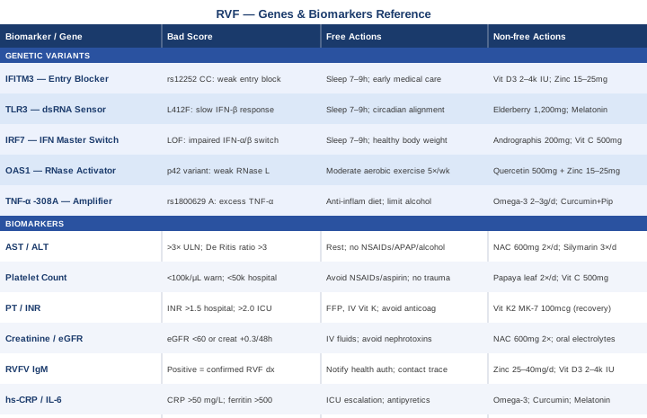
6 Biomarkers to Monitor During and After Rift Valley Fever
Tracking the right numbers during RVF infection is not about running every available test — it is about choosing the markers that provide clear, actionable information at each stage of illness. The six below cover the biological systems RVFV attacks most directly and most consequentially. Together they form a functional early-warning system that, when monitored with appropriate frequency, can mean the difference between outpatient watchfulness and timely ICU escalation.
Biomarker 1: Serum AST and ALT — The Liver Alarm
RVFV is profoundly hepatotropic — it replicates efficiently in hepatocytes and Kupffer cells, and liver damage is the defining feature of severe disease. Serum aspartate aminotransferase (AST) typically rises faster and higher than alanine aminotransferase (ALT) in RVF, because AST reflects both hepatocellular destruction and muscle injury. In autopsy studies of fatal cases, massive hepatic necrosis is found consistently. Even in moderately severe presentations, AST can climb 20 to 50 times the upper limit of normal. The De Ritis ratio (AST divided by ALT) above 3 suggests hepatic necrosis rather than typical viral hepatitis — a distinction that carries significant prognostic weight. Persistently rising AST or ALT after day five to seven is a strong clinical warning regardless of initial severity.
How to Measure It
Standard liver function test (LFT) or comprehensive metabolic panel (CMP). Cost: $20–$60 at standard labs. Widely available in district hospitals in endemic countries. Recommended frequency: every 24 to 48 hours during the first week of symptomatic illness; every three to five days during recovery. Normal reference ranges: AST below 40 IU/L, ALT below 56 IU/L. Concern threshold in active RVF context: above 3× the upper limit of normal.
If the Score Is Bad — The Plan Without Supplements
Absolute physical rest — even moderate activity elevates AST through muscle breakdown, confounding interpretation. Strict elimination of all hepatotoxic substances: alcohol, acetaminophen at any dose, ibuprofen and all NSAIDs, and statins if prescribed. Small, frequent, easily digestible meals; reduce fat load and avoid large protein boluses, which stress damaged hepatocytes. Maintain adequate hydration with oral rehydration salts. If AST or ALT exceeds 10× the upper limit of normal, or if bilirubin is climbing alongside a rising INR, seek immediate hospital evaluation — this pattern signals impending acute liver failure, which requires IV fluids, close monitoring, and readiness for transfer to a facility with intensive care capability.
If the Score Is Bad — The Plan With Supplements or Equipment
N-acetylcysteine (NAC): 600 mg orally twice daily with food during the recovery phase. NAC replenishes glutathione — the liver's primary endogenous antioxidant — and is the medical standard for acetaminophen-induced liver failure at IV doses. Oral NAC provides meaningful hepatoprotective support during viral hepatitis recovery. Frequency: twice daily. Cycling: 2–4 weeks during active recovery, then reassess based on enzyme trends. Side effects: nausea (mitigated by food), garlic-like odor in urine, rare bronchospasm in asthmatics; may interact with nitroglycerin and certain chemotherapy agents. Avoid if active asthma without physician clearance.
Silymarin (standardized milk thistle extract): 140 mg three times daily with meals. The most studied hepatoprotective botanical; human trials in viral and toxic hepatitis show modest but consistent reductions in serum liver enzymes. Frequency: three times daily. Cycling: 4–8 weeks during active recovery, then 2–4 weeks off before reassessment. Side effects: generally very well tolerated; rare GI upset or mild laxative effect at higher doses; may theoretically interact with cytochrome P450 substrates.
Avoid during this phase: high-dose vitamin E (may worsen coagulopathy), licorice root (estrogenic effects), kava (directly hepatotoxic), and any herbal "liver detox" formulations with opaque ingredient lists.
Biomarker 2: Platelet Count — Tracking Thrombocytopenia Risk
Thrombocytopenia — a fall in circulating platelet count — is one of the most consistent laboratory abnormalities across all severity levels of RVF. Even uncomplicated infections frequently produce platelet counts below 150,000/μL. In hemorrhagic cases, counts can fall below 50,000/μL or lower, at which point spontaneous bleeding from mucosal surfaces, petechiae under the skin, and internal hemorrhage all become clinically serious risks.
RVFV depletes platelets through at least three mechanisms simultaneously: direct infection of megakaryocytes (the bone marrow cells responsible for platelet production), immune-mediated platelet destruction, and rapid platelet consumption through evolving DIC. The platelet nadir typically occurs around days five to ten of illness, and the lowest recorded count during hospitalization is a consistently strong predictor of mortality across published RVF case series. Trend tracking matters more than any single value.
How to Measure It
Complete blood count (CBC) with differential. Cost: $15–$40. Available same-day at virtually all labs. Recommended frequency: every 24 to 48 hours during acute illness. Normal range: 150,000–400,000 cells/μL. Concern thresholds: below 100,000/μL warrants daily monitoring; below 50,000/μL warrants hospital-level management; below 20,000/μL requires immediate specialist evaluation.
If the Score Is Bad — The Plan Without Supplements
Strict avoidance of all NSAIDs and aspirin, which impair platelet function even when counts are already suppressed. Avoidance of intramuscular injections, invasive procedures, and physical trauma risks. Protection from falls, cuts, and abrasion. If counts fall below 50,000/μL, inpatient monitoring is warranted. Platelet transfusion thresholds in clinical practice: generally below 10,000/μL without active bleeding, or below 50,000/μL with active hemorrhage or a planned surgical procedure — this is a physician decision. Monitor continuously for signs of internal bleeding: blood in urine or stools, gum bleeding, unexplained bruising, or sudden severe headache.
If the Score Is Bad — The Plan With Supplements or Equipment
Papaya leaf extract: 25–35 mL fresh juice or standardized encapsulated equivalent, twice daily during active thrombocytopenia. Multiple randomized trials in dengue fever — an arboviral disease with a mechanistically similar thrombocytopenic process — have demonstrated statistically significant platelet recovery with standardized papaya leaf extract. The proposed mechanism involves thrombopoietin-like signaling and reduced immune-mediated platelet destruction. No RVF-specific trials exist, but the mechanistic parallel is plausible. Frequency: twice daily. Cycling: use only during active thrombocytopenia; stop when platelets normalize. Side effects: generally safe; mild GI upset possible; avoid in pregnancy.
Vitamin C: 500–1,000 mg/day — reduces oxidative platelet destruction. Safe in this range. Frequency: once to twice daily. Cycling: throughout acute illness; discontinue when platelets normalize. Side effects: GI upset or loose stools at higher doses. Do not exceed 2 g/day during active RVF illness — high-dose vitamin C may acidify urine and increase renal stone risk, particularly concerning alongside any kidney involvement.
Avoid during severe thrombocytopenia: omega-3 fatty acids above 1 g/day (anti-platelet effect at higher doses), ginkgo biloba, high-dose garlic extracts, and turmeric/curcumin at therapeutic doses.
Biomarker 3: Prothrombin Time / INR — Coagulation Integrity
When RVFV destroys enough hepatocytes, the liver loses its capacity to synthesize the clotting factors it normally produces continuously: factors II, V, VII, and X, along with fibrinogen. Prothrombin time (PT), expressed as the International Normalized Ratio (INR), captures this synthetic failure in a single number. An INR above 1.5 signals clinically relevant coagulopathy. An INR above 2.0 in the context of RVF hemorrhagic fever signals impending disseminated intravascular coagulation.
Disseminated intravascular coagulation (DIC) is the most feared coagulation complication of hemorrhagic RVF. The cascade involves simultaneous microclot formation throughout the vasculature — consuming clotting factors and platelets — while the fibrinolytic system overcompensates, producing simultaneous thrombosis and paradoxical bleeding. Mortality in RVF-associated DIC is very high. Monitoring INR closely during the first week of severe illness provides a critical early-warning signal that gives clinical teams a window for intervention before the cascade becomes irreversible.
How to Measure It
PT/INR coagulation panel. Cost: $30–$75. Point-of-care coagulometers allow bedside testing in many clinical settings. Recommended frequency: every 24–48 hours during active severe illness. Normal INR: 0.8–1.2. Alert threshold in RVF: above 1.5 warrants hospital escalation; above 2.0 warrants immediate intensive care evaluation.
If the Score Is Bad — The Plan Without Supplements
Immediate escalation to hospital-level care — coagulopathy at this severity requires direct medical management. Fresh frozen plasma (FFP): replaces consumed clotting factors acutely. Vitamin K administration (IV preferred in severe illness): supports factor synthesis if vitamin K deficiency is contributing — likely in patients with prolonged poor oral intake or recent broad-spectrum antibiotic use. Cryoprecipitate: if fibrinogen falls below 1.5 g/L. Strict avoidance of all anticoagulants, NSAIDs, and aspirin. Avoidance of any procedure that creates a bleeding risk. Daily INR monitoring until sustained normalization.
If the Score Is Bad — The Plan With Supplements or Equipment
Vitamin K2 (MK-7 form): 100–200 mcg once daily with a fat-containing meal (it is a fat-soluble vitamin). Supports coagulation factor activation through carboxylation — relevant during recovery when hepatic synthetic function is rebuilding. This is a recovery-phase measure, not an acute intervention. Frequency: once daily with food. Cycling: 4–8 weeks during recovery; can be continued indefinitely at 100 mcg as maintenance. Side effects: very well tolerated; theoretically may interact with warfarin — disclose to prescribing physician.
Strictly avoid during active coagulopathy: high-dose omega-3 (>1g EPA+DHA), high-dose vitamin E, ginkgo biloba, garlic extracts, and evening primrose oil — all carry anti-coagulant properties that compound an already compromised clotting system.
Biomarker 4: Serum Creatinine / eGFR — Kidney Function
Acute kidney injury occurs in a meaningful subset of severe RVF cases, particularly those with significant hepatic involvement, hemodynamic compromise from hemorrhage, or sustained high fever. RVFV affects the kidneys through several concurrent pathways: direct infection of renal tubular epithelial cells, immune-mediated interstitial nephritis, and prerenal azotemia from dehydration and hypotension. Autopsy studies of fatal RVF cases have documented both tubular necrosis and glomerular involvement.
Serum creatinine rises as glomerular filtration rate falls — but creatinine lags behind actual GFR by 24 to 48 hours in acute settings, which makes it a trailing rather than leading indicator. The combination of rising creatinine, rising urea or BUN, declining urine output, and fluid retention provides the clearest real-time picture. Cystatin C is more sensitive for early AKI detection but remains less accessible in most settings.
How to Measure It
Basic metabolic panel (BMP) or comprehensive metabolic panel (CMP). Cost: $20–$50. Same-day results. Pair with urine output monitoring in hospitalized patients. Recommended frequency: every 24–48 hours during severe illness. Normal creatinine: approximately 0.6–1.1 mg/dL for women, 0.7–1.3 mg/dL for men. Normal eGFR: above 90 mL/min/1.73m². Alert: eGFR declining below 60, or creatinine rising more than 0.3 mg/dL above baseline within 48 hours, meets criteria for AKI.
If the Score Is Bad — The Plan Without Supplements
IV fluid resuscitation to correct volume depletion is the single most effective intervention for prerenal AKI. Strict avoidance of nephrotoxic agents: NSAIDs, aminoglycoside antibiotics, IV contrast agents (avoid contrast-enhanced CT in this setting), and certain antifungal medications. Target urine output above 0.5 mL/kg/hour. If creatinine doubles from baseline, or eGFR falls below 30 mL/min/1.73m², nephrology consultation is required and dialysis may be necessary. Adjust doses of all renally-cleared medications based on current eGFR — this includes ribavirin if it is being used.
If the Score Is Bad — The Plan With Supplements or Equipment
N-acetylcysteine (NAC): 600 mg twice daily — the most studied nephroprotective supplement, with established reduction in contrast-induced nephropathy and benefits in some clinical AKI scenarios through restoration of renal glutathione. Use with medical supervision during active AKI; monitor creatinine trend. Frequency: twice daily. Cycling: through the acute phase, 2–4 weeks; reassess based on creatinine trend. Side effects: same as noted under liver section.
Oral hydration with electrolytes: if drinking is possible, target 2.5–3 liters per day of water or dilute electrolyte solution. Sodium and potassium balance matters — avoid plain water only if electrolytes are already deranged.
Avoid during AKI: high-dose vitamin C above 2 g/day (oxalate nephropathy risk), high-protein supplements or high-protein diets (increase renal urea load), herbal remedies with unknown composition (many are nephrotoxic). After creatinine normalizes, long-term kidney protection strategies include blood pressure monitoring and modest protein intake — AKI episodes increase the risk of subsequent chronic kidney disease by a factor of 3 to 8 in general medicine literature.
Biomarker 5: RVFV-Specific IgM Antibodies — Serological Confirmation
IgM antibodies against RVFV appear in blood typically between day 3 and day 8 after symptom onset and persist for 2 to 4 months before transitioning to a predominantly IgG pattern. Detection of RVFV-specific IgM is the most widely accessible confirmation method for acute RVF in settings where real-time RT-PCR is unavailable or prohibitively expensive. A positive IgM result effectively confirms the diagnosis in the right clinical and epidemiological context. A negative result within the first 3 days does not exclude it.
Interestingly, IgM titer is not a reliable proxy for disease severity. Some of the most severe cases mount delayed or initially weak antibody responses — likely because RVFV nonstructural proteins (particularly NSs) actively suppress type I interferon signaling, which delays the B-cell class switching process that generates IgM. The value of this biomarker lies primarily in diagnosis, not severity tracking. A confirmed positive changes the clinical management framework, drives isolation and contact tracing decisions, and closes the differential diagnosis against malaria, typhoid, and other hemorrhagic fevers.
How to Measure It
RVFV IgM/IgG enzyme-linked immunosorbent assay (ELISA). Cost: $80–$200 at reference laboratories in high-income countries; available at national reference labs and WHO-designated testing centers in endemic countries. RT-PCR for RVFV RNA is the preferred test during the first 1 to 5 days of illness before antibodies appear. Frequency: once during acute illness. If initial IgM is negative and clinical suspicion remains high, repeat at day 7 to 10 from symptom onset.
If the Result Is Positive — Medical and Monitoring Actions
A positive IgM result confirms the infection and shifts priorities. Notify public health authorities — RVF is a notifiable disease in most jurisdictions. Initiate contact tracing for individuals with close blood or secretion exposure. Focus medical monitoring on the other five biomarkers in this list. Begin systematic surveillance for the three serious late complications that can emerge even after the febrile phase resolves: retinitis (monitor visual acuity from week 2 onward, as macular lesions causing permanent blind spots can appear late), meningoencephalitis (watch for headache progression, photophobia, mental status changes), and DIC/hemorrhage (rising INR and falling platelets together signal this risk).
Supporting the Immune Response During Confirmed Acute Infection
Zinc: 25–40 mg elemental zinc daily during active illness. Supports T-cell proliferation, dendritic cell function, and the interferon-alpha production pathway that is directly targeted by RVFV's NSs protein. Take with food to reduce nausea; avoid with milk or high-calcium foods that reduce absorption. Frequency: once daily. Cycling: use during acute illness (2–4 weeks), then reduce to 15 mg/day maintenance or discontinue — chronic dosing above 40 mg/day causes copper deficiency. Side effects: nausea on empty stomach, metallic taste, and with prolonged high-dose use: copper depletion, hypochromic anemia.
Vitamin D3: 2,000–4,000 IU daily with a fat-containing meal. If baseline 25-OH vitamin D is below 30 ng/mL, deficiency correction is relevant — vitamin D directly regulates the expression of IFITM3 and other antiviral interferon-stimulated genes (see genetics section). Frequency: once daily. Cycling: correction phase of 8–12 weeks; maintenance dose of 1,000–2,000 IU/day is reasonable year-round in most latitudes. Side effects: rare at doses below 10,000 IU/day; get a 25-OH vitamin D blood level after 12 weeks to confirm therapeutic range (40–60 ng/mL).
Biomarker 6: High-Sensitivity CRP and IL-6 — Cytokine Storm Signal
C-reactive protein (CRP) is synthesized by the liver in response to interleukin-6 (IL-6) signaling and rises sharply with any systemic inflammatory response. In RVF, the magnitude of the inflammatory cascade directly correlates with outcome. Patients who develop a cytokine storm — characterized by massively elevated IL-6, TNF-α, IL-1β, and IFN-γ — are the cases most likely to develop multi-organ failure, ARDS, or fatal hemorrhagic disease. This is not the immune system failing to fight the virus — it is the immune system overshooting so severely that it damages its own host.
High-sensitivity CRP (hs-CRP) above 50 mg/L in the context of active RVF illness suggests a severe systemic inflammatory response. IL-6 is more directly informative but requires a specialized assay. Serum ferritin — a second-order inflammatory marker reflecting macrophage activation — is widely available and usefully complements hs-CRP when IL-6 testing is unavailable. Rapidly rising ferritin above 500 ng/mL alongside rising CRP is a strong signal of macrophage activation syndrome or early cytokine storm.
How to Measure It
hs-CRP: $20–$50 at standard labs, same-day results. IL-6: $50–$150 at specialty reference labs, 24–48 hour turnaround. Ferritin: $20–$50, same-day results. Recommended frequency: every 48 to 72 hours during severe illness. Normal hs-CRP: below 3 mg/L. Alert: above 50 mg/L in acute RVF warrants escalation of monitoring intensity. Normal ferritin: 30–300 ng/mL (context-dependent).
If the Score Is Bad — The Plan Without Supplements
Escalation to intensive care is the priority when CRP is above 50 mg/L and rising alongside clinical deterioration. Corticosteroids are historically avoided in viral hemorrhagic fevers because of immunosuppression risk, but may be considered in specific life-threatening complications (ARDS, severe encephalitis) at attending physician discretion. Aggressive antipyresis with acetaminophen (avoiding NSAIDs given bleeding risk); cooling blankets if temperature exceeds 40°C. Close monitoring for multi-organ dysfunction across liver, kidneys, coagulation, and neurological status — each system should be reassessed daily during the cytokine storm phase.
If the Score Is Bad — The Plan With Supplements or Equipment
The timing rule is important here: do not introduce anti-inflammatory supplements during the early phase when the immune system still needs to clear the virus. These strategies are for the recovery phase after acute viral clearance.
Omega-3 fatty acids (EPA/DHA): 2–3 g/day in capsule form during the post-acute recovery phase — reduces pro-inflammatory eicosanoid production via COX and LOX pathways. Take with meals to reduce fishy reflux; split across two doses if tolerated. Frequency: daily. Cycling: 8–12 weeks post-discharge; can be continued long-term at 1–2 g/day as maintenance. Side effects: fishy breath or reflux, mild anti-platelet effect at doses above 3 g/day (avoid high doses until platelets normalize), loose stools at high doses.
Curcumin with piperine: 500–1,000 mg curcumin + 5–10 mg piperine (which enhances curcumin bioavailability by approximately 20-fold) twice daily with meals. Curcumin inhibits NF-κB signaling — a central node in the cytokine storm cascade — and has a strong safety profile in human trials at this dose range. Frequency: twice daily. Cycling: 8–12 weeks during recovery; 4 weeks off; reassess. Side effects: mild GI upset at high doses, mild anti-platelet properties, may reduce iron absorption — separate by two hours from iron supplements.
Low-dose melatonin: 0.5–3 mg at bedtime. Beyond sleep regulation, melatonin has documented antioxidant and anti-inflammatory properties — it directly scavenges reactive oxygen species and downregulates NF-κB activation. Sleep quality during recovery is independently important for immune resolution. Frequency: once nightly. Cycling: 2–4 weeks during acute recovery; can be used long-term at 0.5 mg. Side effects: morning grogginess (more common at 5–10 mg doses; less so at 0.5–1 mg), vivid dreams.
The Genetic Side: 5 Host Gene Variants That Shape Your Risk and Recovery
Understanding how genetics influences RVF outcomes requires a short primer on where the science currently stands. Unlike cardiovascular genetics or oncology, human genetic studies specifically focused on RVF are limited — the disease primarily affects lower-income endemic regions with less biobank infrastructure. What is available comes from three sources: cell culture and animal model studies of RVFV-host interactions, human genetic research on related RNA phleboviruses and other zoonotic fevers, and decades of research on the interferon pathway and innate immune genetics. The five variants below are supported by a combination of direct mechanistic evidence and strong human genetic data from related viral infections. Where evidence is indirect, it is labeled clearly.
Genetic testing for these variants is available through direct-to-consumer services such as 23andMe or AncestryDNA at approximately $100–$200, or through clinical whole-exome panels from reference labs at $300–$800. Most consumer raw data files can be analyzed through free interpretation tools such as Promethease for these specific SNPs.
Gene 1: IFITM3 — The Viral Entry Blocker
Interferon-induced transmembrane protein 3 (IFITM3) is one of the most effective antiviral restriction factors identified in the past two decades. IFITM3 embeds in cellular membranes and physically prevents enveloped viruses — including members of the Phenuiviridae family, to which RVFV belongs — from fusing with the endosomal membrane during entry. The rs12252 SNP in IFITM3 produces a truncated protein with significantly reduced antiviral function. Individuals carrying the C allele (particularly CC homozygotes) show markedly increased susceptibility to severe influenza and have been studied in the context of multiple RNA viruses with enveloped entry mechanisms similar to RVFV. Research published in Nature (Everitt et al., 2012) established the clinical significance of this variant for enveloped viral severity in humans.
If the Gene Variant Is Unfavorable — The Plan Without Supplements
Prioritize the behavioral and medical levers: maintain up-to-date vaccination status for all vaccine-preventable diseases (cross-immunity concepts, immune system priming), optimize sleep to seven to nine hours — deep sleep is the peak period for endogenous interferon production, which drives IFITM3 expression. Establish a clear and rapid protocol for early medical care at any febrile illness involving livestock exposure: early presentation to a facility that can run RT-PCR matters because the IFITM3 window is the very first hours of infection. Reduce unnecessary enveloped virus exposures (avoid shared water pipes, ventilate indoor animal spaces).
If the Gene Variant Is Unfavorable — The Plan With Supplements or Equipment
Vitamin D3: IFITM3 expression is directly regulated by the vitamin D receptor (VDR) pathway — studies show that vitamin D sufficiency substantially upregulates IFITM3 in immune cells. Maintaining 25-OH vitamin D in the 40–60 ng/mL range is the most directly evidence-linked approach to supporting IFITM3 expression. Dose: 2,000–4,000 IU/day with K2 (100 mcg MK-7). Frequency: daily. Cycling: year-round, confirm blood level at 12 weeks and adjust. Side effects: minimal at these doses; monitor calcium if using very long-term.
Zinc: 15–25 mg/day maintenance dose supports zinc-dependent interferon signaling, which drives IFITM3 induction. Frequency: daily with food. Cycling: continuous at 15 mg; use 25 mg only for short-term active infection periods (2–4 weeks). Side effects: copper depletion at sustained higher doses — supplement with 1–2 mg copper if using zinc for more than 6 weeks.
Gene 2: TLR3 — The Double-Stranded RNA Sensor
Toll-like receptor 3 (TLR3) recognizes double-stranded RNA — the molecular signature left behind as RNA viruses replicate inside cells. TLR3 activation triggers NF-κB and IRF3 signaling, producing both interferon-β and pro-inflammatory cytokines. The rs3775291 (L412F) variant encodes a leucine-to-phenylalanine substitution that substantially reduces TLR3 signaling in response to dsRNA. Carriers of this variant show slower initial interferon-β responses to RNA virus infection. Human studies have linked it to increased severity of herpes simplex encephalitis and influenza pneumonia — RNA viral infections with partially overlapping innate immune dependencies. For RVFV, which generates dsRNA intermediates during its tripartite genome replication cycle, this variant represents a plausible susceptibility factor, though direct human RVF genetic studies remain absent.
If the Gene Variant Is Unfavorable — The Plan Without Supplements
Sleep optimization is non-negotiable for TLR3 function. TLR3 on plasmacytoid dendritic cells and macrophages is most actively engaged during the circadian-driven immune surveillance peaks — and these are profoundly disrupted by sleep deprivation. Aim for a consistent sleep schedule with seven to nine hours in a cool, dark environment. Circadian alignment — light exposure in the morning, darkness at night, consistent meal timing — supports the immune rhythm independently of sleep quantity. Reduce chronic low-grade viral stimulation: TLR3 function can be partially depleted by sustained activation from endemic latent herpesvirus reactivation (cold sores, EBV reactivation) — so any recurring viral reactivations warrant attention.
If the Gene Variant Is Unfavorable — The Plan With Supplements or Equipment
Elderberry extract (Sambucus nigra): standardized preparations have shown modest innate immune activation effects in human randomized trials for respiratory viral infections — reducing duration and severity. The proposed mechanism involves stimulation of cytokine production through innate immune pathways partially independent of TLR3. Dose: 1,200–2,400 mg standardized extract (or 15 mL syrup twice daily). Frequency: twice daily. Cycling: use during illness risk periods or at first signs of infection (not year-round continuously). Side effects: generally well tolerated; theoretically could worsen cytokine storm in late-stage severe illness — use in early, not late, infection phase.
Melatonin: 0.5–1 mg nightly — beyond sleep support, melatonin has demonstrated enhancement of TLR3-mediated antiviral signaling in animal models. Human evidence at this specific level is limited, but the safety profile makes this a low-risk addition. Cycling: daily at bedtime; well-tolerated long-term at 0.5 mg. Side effects: as noted above.
Gene 3: IRF7 — The Master Switch for Type I Interferon
Interferon regulatory factor 7 (IRF7) is perhaps the single most important transcription factor in the antiviral innate immune response. It is often called the master regulator of type I interferon (IFN-α/β) production. IRF7 sits downstream of multiple pattern recognition receptor pathways — including TLR7, TLR9, and the cGAS-STING axis — and coordinates the burst of antiviral interferon that determines whether a viral infection is contained in the first hours or allowed to establish deep tissue replication.
RVFV's NSs protein directly targets IRF3 for proteasomal degradation, and evidence from related phleboviruses suggests NSs also suppresses IRF7 activity. Individuals who carry loss-of-function variants in IRF7 begin this infection with a compromised master switch — and compounding that with a virus that then attacks the same pathway creates a substantial vulnerability window. Human studies (Ciancanelli et al., 2015, published in Science) have documented that autosomal dominant IRF7 deficiency can underlie life-threatening viral pneumonitis even in previously healthy individuals.
If the Gene Variant Is Unfavorable — The Plan Without Supplements
Sleep is the highest-impact lever: IRF7 expression peaks during deep NREM sleep, and a single night of total sleep deprivation demonstrably reduces interferon production capacity by 30–50% in human subjects. This is not a metaphor — it is a measured, reproducible effect. Consistent seven to nine hours with good sleep quality is the most powerful IRF7-supporting intervention available without a prescription. Additionally, maintain a normal body weight — adipose tissue produces adipokines that chronically suppress IRF7-mediated signaling, and even modest weight reduction in overweight individuals improves antiviral interferon response capacity.
If the Gene Variant Is Unfavorable — The Plan With Supplements or Equipment
Andrographis paniculata (standardized to andrographolide): 200–400 mg standardized extract twice daily. Human clinical trials have documented significant reduction in severity and duration of upper respiratory viral infections. Andrographolide appears to stimulate innate immune activation and has shown modest interferon-inducing effects in cell culture. Frequency: twice daily. Cycling: use during periods of active infection risk (2–4 weeks), not year-round. Side effects: bitter taste, occasional GI upset, rare allergic reactions; avoid in pregnancy and autoimmune conditions; potential CYP3A4 interaction.
Vitamin C (liposomal): 500–1,000 mg twice daily in liposomal form for superior bioavailability. Acts as a cofactor for immune cell function and is required for optimal interferon production; deficiency directly impairs natural killer cell activity. Frequency: twice daily. Cycling: continuous at 500 mg/day; increase to 1,000 mg twice daily during active illness. Side effects: GI loosening at higher doses (bowel tolerance); reduce dose if this occurs.
Gene 4: OAS1 — The Antiviral RNase Activator
2'-5'-Oligoadenylate synthetase 1 (OAS1) is an interferon-stimulated enzyme that, when activated by double-stranded RNA, produces 2-5A oligomers that activate RNase L — a powerful endoribonuclease that degrades viral (and cellular) RNA. This OAS/RNase L pathway is one of the most ancient and effective direct antiviral mechanisms in vertebrate biology. Multiple splice variants of OAS1 differ dramatically in their ability to recognize dsRNA and generate 2-5A. The p46 splice variant, produced in individuals with certain intronic SNPs, shows far stronger antiviral activity against RNA viruses than the p42 variant. The same variant region attracted intense attention during COVID-19, when GWAS studies identified OAS1 variants as significant modifiers of disease severity.
If the Gene Variant Is Unfavorable — The Plan Without Supplements
Regular moderate exercise: aerobic exercise at 60–70% maximal heart rate for 30–45 minutes, five days per week, demonstrably upregulates OAS1 and multiple other interferon-stimulated gene (ISG) expression levels in peripheral blood mononuclear cells. The effect is measurable and sustained with regular training. This is one of the strongest evidence-backed non-pharmacological approaches to improving ISG baseline expression. Avoid overtraining: very high training loads (ultramarathon athletes, elite-level intensity) transiently suppress ISG expression — moderate is the operative word.
If the Gene Variant Is Unfavorable — The Plan With Supplements or Equipment
Quercetin (with zinc): 500–1,000 mg quercetin with 15–25 mg zinc daily. Quercetin acts as a zinc ionophore — it carries zinc ions into cells where zinc inhibits RNA-dependent RNA polymerase activity (used by RVFV and other RNA viruses) and supports the OAS pathway's dsRNA-sensing capacity. Frequency: once daily with food. Cycling: during infection risk periods (4–6 weeks), then 2–4 weeks off. Side effects: generally well tolerated; quercetin may interact with certain antibiotics (quinolones) and immunosuppressants; mild GI upset possible.
Gene 5: TNF-α Promoter (rs1800629) — The Inflammatory Amplifier
Tumor necrosis factor alpha (TNF-α) is a pleiotropic cytokine central to both viral defense and the pathological inflammation that damages host tissue. The rs1800629 G>A promoter polymorphism (often designated TNF-α -308A) significantly increases TNF-α transcriptional activity — carriers produce substantially more TNF-α in response to inflammatory stimuli than non-carriers. In the context of most infections, higher TNF-α production is mildly beneficial. In the context of severe viral hemorrhagic fevers — where the pathological mechanism is already driven by inflammatory cytokine excess — it becomes a liability. Studies of hemorrhagic fever viruses (Ebola, dengue, hantavirus) consistently show higher TNF-α levels associated with worse outcomes. The -308A allele is found in approximately 20–25% of people of European descent and at varying frequencies across African and Asian populations.
If the Gene Variant Is Unfavorable — The Plan Without Supplements
Adopt an anti-inflammatory dietary pattern: Mediterranean-style diet rich in vegetables, olive oil, legumes, and fatty fish has the most consistent human evidence for reducing systemic TNF-α levels over time — not a single food, but the overall pattern. Limit ultra-processed food, refined sugars, and industrial seed oils, which measurably elevate TNF-α in metabolic studies. Alcohol avoidance or strict limitation is particularly relevant for this variant: alcohol independently elevates TNF-α production from liver Kupffer cells, compounding an already high-baseline phenotype. Aim for fewer than four standard drinks per week; none during any active inflammatory illness.
If the Gene Variant Is Unfavorable — The Plan With Supplements or Equipment
Omega-3 EPA/DHA: 2–3 g/day of combined EPA+DHA as a long-term baseline supplement. EPA specifically competes with arachidonic acid for COX and LOX enzymes, reducing downstream prostaglandin and leukotriene production that amplifies TNF-α signaling. Multiple randomized controlled trials demonstrate measurable reduction in serum TNF-α with consistent omega-3 supplementation at this dose range. Frequency: daily with meals. Cycling: long-term continuous use is appropriate and well-studied; reassess annually. Side effects: fishy reflux (take with food or use enteric-coated formulation), anti-platelet effect at high doses.
Curcumin with piperine: 500 mg curcumin + 5 mg piperine twice daily. Among the most studied natural NF-κB inhibitors — the pathway directly responsible for TNF-α transcription. Multiple human trials show reduced inflammatory cytokine levels with sustained curcumin supplementation. Frequency: twice daily. Cycling: 8–12 weeks on, 4 weeks off. Side effects: as noted previously — GI upset at high doses, avoid during active coagulopathy.
What An Elegant Defense Teaches About the Immune System You're Actually Living With
An Elegant Defense: The Extraordinary New Science of the Immune System by Matt Richtel (2019) is grounded in interviews with leading immunologists and follows real patients whose outcomes were shaped not by immune weakness but by immune miscalibration — overreaction being just as destructive as underreaction. The book directly challenges the pervasive cultural narrative that "boosting immunity" is universally good, which is a misconception with real clinical consequences in diseases like RVF where cytokine storm — an overactive immune response — kills as surely as the virus itself.
1. The Goal Is Immune Balance, Not Immune Maximum
The immune system is not a fire that needs constant stoking. It is a precision system requiring calibration. Richtel documents that the same mechanisms responsible for fighting infection — TNF-α, IL-6, neutrophil activation — become destructive when they overshoot. In RVF, severe cases are not failures of immunity; they are failures of immune regulation. The practical implication: during active febrile illness, the goal is appropriate immune support, not maximum immune stimulation.
2. Inflammation Is a Tool With a Dosage Problem
Inflammation evolved as a temporary, localized signal to coordinate repair. Richtel interviews researchers who document how chronic low-grade inflammation — from diet, poor sleep, chronic stress — leaves the immune system partially pre-activated, which can cause it to overshoot when encountering a genuine pathogen. Pre-existing metabolic inflammation raises the probability of cytokine storm in any serious viral infection.
3. Sleep Is Not Rest — It Is Active Immune Maintenance
During deep sleep, the brain undergoes glymphatic clearance and the immune system performs its highest-intensity surveillance and memory consolidation. Cytokines produced during slow-wave sleep — particularly IL-1β and TNF-α in measured, regulated amounts — are not pathological at these concentrations; they are part of normal immune maintenance. Chronic short sleep demonstrably reduces T-cell activity, natural killer cell surveillance, and interferon production. Richtel documents studies showing that sleeping fewer than six hours for five consecutive nights creates measurable immune suppression equivalent to mild chronic immunosuppression.
4. Fever Is a Defense, Not Just a Symptom
One of the most practically important points: antipyretics suppress fever, which is an adaptive immune response. Elevated body temperature directly impairs RNA virus replication by reducing the efficiency of viral polymerase activity and enhancing immune cell trafficking. The reflex to suppress fever with medications at the first sign of illness, while symptomatically comforting, may reduce viral clearance speed. Evidence suggests allowing moderate fever (up to 38.5°C) in otherwise healthy adults may support faster viral resolution — a view increasingly shared by some infectious disease specialists despite conventional guidance.
5. Extreme Exercise During Illness Suppresses the Very Response You Need
The "open window" hypothesis, documented by exercise immunologists and covered in the book: very intense or prolonged exercise transiently opens a window of reduced immune surveillance lasting 12 to 72 hours. During an active viral infection, insisting on training continues viral replication during a period when the immune system is deliberately suppressing itself to allow muscle recovery. Complete rest during fever is not weakness — it is the biologically appropriate allocation of metabolic resources.
6. The Gut Microbiome Trains Immune Calibration
A significant portion of the book examines how gut-resident bacteria actively tune the immune response — particularly the regulatory versus inflammatory balance. A diverse gut microbiome supports the regulatory T-cell populations that prevent immune overreaction. Antibiotic overuse, highly processed diets, and chronic stress all reduce microbiome diversity in ways that have been linked to increased inflammatory cytokine responses to viral challenge.
7. Chronic Stress Is an Immune Suppressor via Cortisol
Glucocorticoids — particularly cortisol — directly suppress interferon signaling, reduce natural killer cell activity, and shift immune function toward anti-inflammatory states that impair viral clearance. Richtel documents that this was evolutionarily adaptive (immune suppression during flight-or-fight preserved energy for immediate survival) but becomes deeply counterproductive during a viral infection requiring sustained innate immune activity. Chronic psychological stress represents one of the most underappreciated modifiable risk factors for poor viral infection outcomes.
8. Genetic Variants in Immune Genes Are More Common Than Most People Realize
The book demystifies human immunogenetics: variants in IFITM3, TLR3, IRF7, and similar genes are not rare disease mutations — they are common population variants present in 10–30% of people. They do not guarantee severe illness; they shift probabilities. Knowing which variants you carry allows you to make risk-calibrated decisions about exposures, medical care timing, and preventive support.
9. The Timing of Immune Response Determines Much More Than Its Magnitude
Multiple researchers in the book make the case that when the immune system responds matters as much as how strongly. A rapid, early type I interferon response within the first 24 hours of infection can contain RVFV before it establishes deep hepatic replication. A delayed response — even if ultimately powerful — arrives after viral loads have peaked, liver damage has occurred, and the inflammatory cascade has been set in motion. Genetic variants affecting TLR3 and IRF7 specifically compromise the speed of this early response window.
10. Most "Immune Boosting" Products Are Marketed Beyond Their Evidence
Richtel spends considerable time on the distinction between evidence-graded immune support (sleep, exercise, vitamin D sufficiency, zinc adequacy, diet quality) and the poorly-evidenced galaxy of supplements claiming to "boost immunity." The practical takeaway: foundations first. Fixing documented deficiencies in sleep, vitamin D, or zinc provides measurably more immune benefit than adding the latest marketed supplement on top of deficient fundamentals. This is the framework that should underpin any supplement strategy for RVF susceptibility.
Complementary Approaches With Clinical Evidence
Rift Valley fever is primarily a disease of acute viral infection with systemic organ involvement — most complementary approaches are not directly studied in RVF patients. The approaches below are selected because they have meaningful human clinical evidence for supporting the physiological systems most stressed by the disease: immune regulation, hepatic recovery, pulmonary function, and systemic inflammation. Each is presented with its specific evidence base and realistic application context.
Mindfulness Meditation and MBSR — Stress-Immune Axis Support
Mindfulness-based stress reduction (MBSR) is an eight-week structured program combining meditation, body scanning, and gentle yoga developed by Jon Kabat-Zinn at the University of Massachusetts Medical School. Its relevance to RVF recovery lies in the well-documented stress-immune connection: chronic psychological stress elevates cortisol, which suppresses interferon production and natural killer cell activity. During and after a serious febrile illness, psychological distress is nearly universal and directly undermines the immune resolution the body needs.
A randomized trial published in Psychoneuroendocrinology (2016) demonstrated that MBSR significantly reduced pro-inflammatory cytokine production (including IL-6 and TNF-α) in healthy adults compared to active controls, with effects that persisted at eight-week follow-up. These are precisely the cytokines that drive cytokine storm in severe RVF — dampening their chronic overexpression during recovery reduces the risk of lingering inflammatory tissue damage.
Practical application: Formal MBSR programs are available in-person and online (typically eight weekly two-hour sessions). For post-RVF recovery, begin with ten minutes of guided body scan meditation daily — apps such as Insight Timer provide structured free content. Avoid intensive breath retention practices during active respiratory involvement. Progress gradually over four to eight weeks to 20–30 minutes daily. The goal is not relaxation as a luxury — it is reduction of cortisol-mediated immune suppression as a deliberate physiological strategy.
Breathing-Based Therapies — Pulmonary Recovery and Autonomic Regulation
Structured breathing practices address two distinct RVF-related concerns: first, respiratory compromise that can occur with severe disease (ARDS or pulmonary involvement in encephalitic cases), where breathing exercises support re-expansion of atelectatic lung segments and restoration of respiratory muscle strength; second, autonomic dysregulation, which commonly persists as post-infectious fatigue and contributes to sustained sympathetic nervous system dominance that suppresses immune resolution.
Slow-paced resonance breathing at 5–6 breaths per minute (approximately five seconds inhale, five seconds exhale) has been studied in multiple randomized controlled trials for autonomic modulation, demonstrating consistent increases in heart rate variability (HRV) — a measurable index of parasympathetic activation — and reduction in sympathetic tone. A meta-analysis covering 15 randomized trials confirmed significant HRV improvements with regular slow breathing practice. HRV is independently associated with immune regulatory capacity and has predictive value for recovery speed after serious illness.
For post-RVF recovery, begin with five minutes of slow resonance breathing (5–6 breaths/minute) twice daily using a free pacing app or visual timer. Advance to ten minutes twice daily as tolerance improves. If there is any active pulmonary involvement (cough, dyspnea), consult a physiotherapist before beginning any breath-hold techniques — stick to slow, natural-depth breathing only. Expected timeline to noticeable autonomic improvement: three to four weeks of consistent practice.
Microbiome-Directed Therapies — Gut Immunity and Systemic Inflammation
The gut microbiome plays a central regulatory role in systemic immune calibration — particularly in the balance between regulatory and inflammatory immune cell populations. Antibiotic treatment during RVF hospitalization (common in empirical sepsis protocols before the diagnosis is confirmed) creates significant collateral dysbiosis, reducing the diversity and density of beneficial commensal bacteria precisely when their immune-training function is most needed during recovery. Additionally, the high inflammatory load of severe RVF itself shifts the gut environment in ways that favor dysbiotic species.
Human clinical trials in post-infectious gut recovery — including post-viral syndromes, antibiotic-associated diarrhea, and inflammatory bowel recovery — demonstrate that multi-strain probiotic supplementation containing Lactobacillus rhamnosus GG, Bifidobacterium longum, and Lactobacillus acidophilus significantly accelerates microbiome diversity restoration and reduces systemic inflammatory markers including CRP and IL-6 over an eight to twelve week period. These effects are relevant to the post-RVF inflammatory resolution challenge.
Apply microbiome support beginning during the early recovery phase — after any antibiotic course is complete. Choose a probiotic with a minimum of ten billion CFU per capsule and at least five distinct species. Take on an empty stomach before breakfast. Pair with prebiotic dietary fiber: 25–35 grams per day from vegetables, legumes, and whole grains — this is the substrate the probiotic organisms need to establish. Fermented foods (unsweetened yogurt, kefir, sauerkraut) add additional microbial diversity. Maintain for a minimum of eight to twelve weeks. Side effects: transient bloating and gas in the first one to two weeks as microbial populations shift — normal and expected.
Low-Level Laser Therapy / Photobiomodulation — Hepatic and Tissue Recovery
Photobiomodulation (PBM), also called low-level laser therapy (LLLT), uses specific wavelengths of near-infrared light (typically 630–1,100 nm) to stimulate mitochondrial cytochrome c oxidase activity, enhancing ATP production, reducing oxidative stress, and activating anti-inflammatory signaling pathways. Its application to RVF recovery relates primarily to hepatic tissue repair: the liver's ability to regenerate hepatocytes after viral damage depends on mitochondrial energy availability and reduction of oxidative damage, which PBM has been shown to directly support.
A randomized controlled trial on hepatic fibrosis and non-alcoholic liver disease demonstrated that transcutaneous near-infrared photobiomodulation applied to the liver quadrant significantly reduced AST and ALT levels and improved liver biopsy histology at twelve weeks compared to sham treatment. A separate randomized trial documented reduced hepatic inflammatory markers and improved liver synthetic function measures with consistent PBM treatment. Direct RVF hepatitis evidence is absent, but the mechanistic relevance for post-viral hepatic recovery is well-supported.
Apply PBM using a clinically validated near-infrared device (810 nm or 850 nm wavelength, minimum 100 mW output) over the right upper abdominal quadrant (liver surface location) for ten to twenty minutes per session. Begin no earlier than two weeks into recovery when acute inflammation is subsiding — earlier application during active viral replication is not appropriate. Frequency: three to five sessions per week. Duration: eight to twelve weeks. Most home devices in the $200–$800 range provide adequate irradiance for superficial organ support. Side effects: rare and mild at recommended parameters — avoid if taking photosensitizing medications.
Chinese Herbal Medicine — Antiviral and Hepatoprotective Support
Andrographis paniculata (Chuan Xin Lian in Chinese medicine) and Isatis tinctoria (Ban Lan Gen) have been studied extensively in Chinese clinical trials for antiviral activity and hepatoprotection during acute viral hepatitis. Andrographolide — the active component of andrographis — has demonstrated inhibition of multiple RNA virus replication cycles in cell culture, activation of innate immune signaling pathways, and NF-κB inhibitory effects that reduce inflammatory cytokine production. In registered Chinese randomized trials of acute viral hepatitis, andrographis-containing formulations significantly reduced liver enzyme elevation time and accelerated normalization compared to supportive care alone.
A systematic review published in the Journal of Clinical Pharmacy and Therapeutics (2007) covering multiple randomized trials of andrographis in acute respiratory viral infections confirmed significant reductions in illness duration and severity, with a good safety profile across doses studied. The hepatoprotective and antiviral properties are the most directly relevant to RVF recovery.
Apply with the guidance of a practitioner trained in Chinese herbal medicine if possible — formulations vary significantly in quality and composition. For standardized andrographolide content (minimum 10% andrographolide), 200–400 mg twice daily is the studied dose range. Begin during recovery phase rather than during peak acute illness. Cycling: 2–4 weeks; reassess liver enzymes. Side effects: bitter taste, occasional GI upset, rare allergic reactions; contraindicated in pregnancy and autoimmune conditions on immunosuppressants; potential interactions with CYP3A4-metabolized medications. Evidence for direct RVF efficacy is not established — frame this as supportive and hepatoprotective, not antiviral treatment for the acute infection.
Conclusion
Rift Valley fever's range of outcomes — from a self-resolving flu-like illness to multi-organ failure — is not arbitrary. It is biological, shaped by the specific markers your immune and organ systems display during infection and by the genetic architecture that determined how your body responded in the first hours. Six biomarkers — liver enzymes, platelets, coagulation, kidney function, serological confirmation, and systemic inflammatory markers — together form a monitoring framework that gives clinical decisions a factual foundation. Five gene variants — IFITM3, TLR3, IRF7, OAS1, and TNF-α — identify the upstream vulnerabilities that make certain individuals significantly more susceptible to severe disease, each with specific and practical compensation strategies.
The most important next step is not the perfect supplement stack or the ideal recovery protocol. It is engaging with qualified medical care early — ideally before clinical deterioration makes the engagement urgent. If you are in or returning from an endemic region with any unexplained febrile illness following livestock exposure, request RVFV-specific testing without delay. If you have access to genetic testing, review the five variants covered here with a knowledgeable clinician who can contextualize your results alongside your overall health picture. If you are in recovery, use the biomarker framework to track hepatic, hematologic, and renal normalization systematically rather than simply waiting to feel better. Better information, applied with appropriate medical guidance, consistently leads to better decisions — and in Rift Valley fever, better decisions made early can determine everything.
Digestive: Liver & Gallbladder Conditions
Autoimmune: Inflammatory Conditions
Infectious: Viral Infections
Eye: Retinal Conditions
Urological: Kidney Conditions