This article contains AI generated content.
Sacroiliac Joint Dysfunction: 5 Genes and 7 Biomarkers to Track
If you've been told your sacroiliac joint is "just inflamed" or "out of alignment" and left with a stretching sheet and a follow-up appointment in six weeks, you're not imagining that something is missing. Sacroiliac joint dysfunction is a real, mechanically definable problem, but it is also a label that gets applied to very different underlying situations: a joint that's simply irritated from asymmetrical loading, a joint destabilized by hormonal changes after pregnancy, and — in a meaningful minority of cases — a joint that is the first visible sign of an underlying inflammatory disease. Generic advice tends to treat all three the same way, which is why it so often plateaus.
The reason lab work and family history matter here is not to give you a scary diagnosis. It's to help you and your clinician sort out which of those three situations you're actually dealing with, because the right next step is different for each one. A course of core stabilization exercises will do very little for a joint that is inflamed by an autoimmune process, and a rheumatology referral is unnecessary overkill for a joint that's unstable because of a collagen variant or a postpartum hormone shift. Precision here isn't a luxury — it changes what you do on Monday morning.
This article takes the more specific route. It walks through the blood-based biomarkers that can tell you whether your sacroiliac joint pain is mechanical, inflammatory, or metabolic in nature, what each one costs to check, and what a realistic improvement plan looks like — with and without supplements — when a number comes back outside the range you'd want. It then covers the genetic and epigenetic side of the picture, including the one gene variant that changes sacroiliac joint management more than any other, a research-backed book that reframes how the joint should be trained rather than just treated, and a short review of complementary approaches with genuine supporting evidence.
None of this replaces an examination by a clinician who can actually move your pelvis and test your joint directly. What it can do is help you show up to that appointment with better questions, and help you understand which of the numbers on your lab report are actually worth acting on.
Summary
Sacroiliac joint dysfunction sits at a crossroads between orthopedics and rheumatology, and the biomarkers below are what help tell those two paths apart. Inflammatory markers like hs-CRP and ESR can flag whether a "mechanical" SI joint problem is actually an early sign of axial spondyloarthritis — a distinction that changes treatment entirely. Vitamin D, magnesium, HbA1c, homocysteine, and relaxin round out a panel that captures bone and connective tissue health, nerve function, tissue glycation, and hormonal joint laxity, each with its own affordable test and its own fix.
On the genetic side, one variant — HLA-B27 — carries outsized weight in sacroiliitis risk, and it's joined by ERAP1, IL23R, COL5A1, and COMT, each nudging inflammation, connective tissue integrity, or pain sensitivity in a different direction. Further down, you'll also find a research-backed reframe of how the joint should actually be trained (drawn from one of the most influential books in spine mechanics), plus a look at which complementary approaches — from spinal manipulation to biofeedback — actually have human evidence behind them for this specific joint.
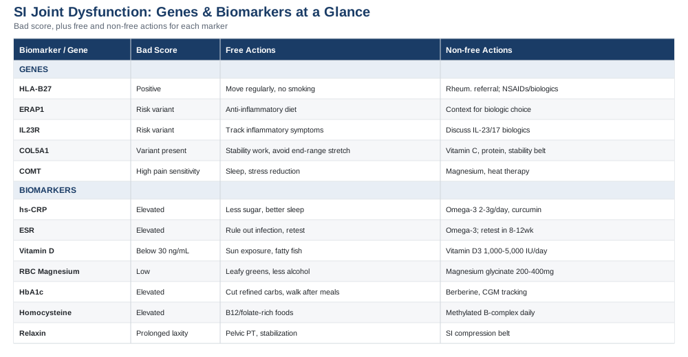
7 Biomarkers Worth Tracking for Sacroiliac Joint Dysfunction
The sacroiliac joint is small, but it sits at the junction of a lot of different systems: load-bearing bone, ligament, hormone-sensitive tissue, and — in some people — the earliest target of a systemic inflammatory disease. That's part of why "SI joint pain" is such a frustrating diagnosis to receive. Two people can have nearly identical symptoms — one-sided low back and buttock pain, worse with prolonged standing or transitional movements — for completely different underlying reasons. Blood work won't replace a physical exam or imaging, but it does something imaging often can't: it tells you whether the joint's problem is purely mechanical or whether it's part of something bigger.
The seven biomarkers below are chosen because each answers a specific question relevant to SI joint dysfunction: is there systemic inflammation, is bone and ligament health compromised, is tissue repair impaired, or is there a hormonal driver of joint laxity. Where the evidence is strongest — inflammatory markers in suspected axial spondyloarthritis — the guidance is more directive. Where it's more exploratory — like relaxin testing — that's stated plainly.
1. High-Sensitivity C-Reactive Protein (hs-CRP)
hs-CRP is a general marker of systemic inflammation produced by the liver in response to inflammatory signaling, primarily interleukin-6. In the context of SI joint pain, it's one of the two most useful screening tools (alongside ESR) for distinguishing a purely mechanical joint problem from sacroiliitis driven by axial spondyloarthritis, a form of inflammatory arthritis that often targets the SI joints first. A prospective community-based study found that baseline elevations in hs-CRP predicted incident ankylosing spondylitis years later, reinforcing that this marker isn't just a snapshot — it can flag risk before a formal diagnosis is made (Muñoz-Ortego et al., PLoS One 2019).
How to Measure It
hs-CRP is a standard blood draw, widely available through primary care or direct-to-consumer lab services, typically costing $10–$30 out of pocket when not covered by insurance. Morning fasting isn't required, but avoid testing during an acute cold, flu, or injury, since any of these will transiently raise it regardless of your SI joint status.If hs-CRP Is Elevated: The Plan Without Supplements
The first move isn't a supplement — it's context. A single elevated reading should be repeated in 4–6 weeks, since transient infections and poor sleep both raise hs-CRP. If it stays elevated alongside inflammatory-pattern symptoms (morning stiffness lasting more than 30 minutes, improvement with movement rather than rest, alternating buttock pain), this is a genuine reason to request a rheumatology referral and HLA-B27 testing rather than pushing harder on stretching or manual therapy. Lifestyle levers that reliably lower hs-CRP include reducing ultra-processed food and added sugar intake, improving sleep consistency, and, if applicable, weight loss — visceral fat is itself a source of inflammatory cytokines.If hs-CRP Is Elevated: The Plan With Supplements or Equipment
Omega-3 fatty acids (EPA/DHA, roughly 2–3g/day combined) have the best-supported anti-inflammatory effect on CRP among over-the-counter options, typically taken with food to reduce GI upset, reassessed at 8–12 weeks. Curcumin (500–1000mg/day of a bioavailability-enhanced formulation) has more modest evidence but a good safety profile; it can interact with blood thinners, so it's worth flagging to a physician if you're on anticoagulants. Neither of these should be used as a substitute for identifying and treating an underlying inflammatory arthritis if that's what's actually driving the number — they're supportive, not corrective.2. Erythrocyte Sedimentation Rate (ESR)
ESR measures how quickly red blood cells settle in a test tube, which happens faster in the presence of inflammatory proteins. It's a blunter, slower-moving marker than hs-CRP, but it's still part of the standard workup for suspected axial spondyloarthritis, and the two are often ordered together because they don't always move in lockstep — some people run a high ESR with a normal CRP, and vice versa.
How to Measure It
ESR is one of the cheapest tests in this entire panel, usually $5–$15, run on the same blood draw as most standard panels. It's widely available and rarely requires special preparation.If ESR Is Elevated: The Plan Without Supplements
As with CRP, the first step is repeating the test and ruling out confounders — anemia, recent infection, and pregnancy all raise ESR independent of joint disease. If it remains elevated with SI-joint-specific symptoms, the practical next step is the same: request imaging aimed specifically at sacroiliitis (MRI is more sensitive than X-ray for early inflammatory change) and HLA-B27 testing, rather than treating it as a purely mechanical issue.If ESR Is Elevated: The Plan With Supplements or Equipment
There's no supplement that meaningfully lowers ESR on its own; it tracks the same inflammatory drivers as CRP, so the omega-3 and anti-inflammatory dietary approach above applies here too. Where ESR is genuinely useful with equipment is longitudinal tracking — if you're diagnosed with an inflammatory cause and started on treatment, retesting ESR every 8–12 weeks is a standard, low-cost way to see whether the treatment plan is actually working.3. 25-Hydroxyvitamin D
Vitamin D does more for the SI joint than most people expect: it plays a role in bone mineralization at the joint surfaces, in modulating inflammatory signaling, and possibly in pain processing itself. A systematic review and meta-analysis of observational studies found a consistent association between low vitamin D status and low back pain, and more recent work suggests inflammatory markers may partly explain that link (Zadro et al., 2017; Xu et al., 2021).
How to Measure It
A 25-hydroxyvitamin D blood test costs roughly $30–$60 out of pocket, or is often bundled into an annual physical. Test in late winter or early spring if you want to see your seasonal low point, since levels drift with sun exposure.If Vitamin D Is Low: The Plan Without Supplements
Fifteen to twenty minutes of midday sun exposure on bare arms and legs several times a week, when climate and skin tone allow, can meaningfully raise levels over 8–12 weeks. Fatty fish (salmon, sardines, mackerel) and fortified dairy or plant milk contribute smaller but real amounts. This route is slower and more variable than supplementation, so it's best paired with a retest rather than assumed to be working.If Vitamin D Is Low: The Plan With Supplements or Equipment
Vitamin D3 (cholecalciferol) at 1,000–2,000 IU/day is a reasonable general maintenance dose; if levels are significantly below the 30 ng/mL threshold most labs flag, a physician-guided higher dose (often 5,000 IU/day for 8–12 weeks) followed by retesting is standard practice. Take it with a meal containing some fat to improve absorption. Cycling isn't typically necessary, but retesting every 3–6 months during correction avoids overshooting — vitamin D toxicity is rare but real at sustained high doses, and can raise blood calcium. A UVB lamp is a niche equipment-based alternative for people with very limited sun exposure, though it carries its own skin-safety tradeoffs and is a distant second choice to supplementation.4. Red Blood Cell (RBC) Magnesium
Magnesium is a cofactor in muscle relaxation, nerve conduction, and connective tissue metabolism, and low intracellular levels are linked to muscle cramping and heightened neuromuscular excitability — relevant for the muscles (glutes, piriformis, multifidus) that stabilize the SI joint. RBC magnesium is a more accurate reflection of tissue stores than standard serum magnesium, which the body tightly regulates even when whole-body magnesium is depleted. A clinical review notes that magnesium deficiency, whether genetic or acquired, produces a "wide range of clinical effects including muscle cramps, fatigue, seizures and arrhythmias" (Blaine & Achinger, 2021).
How to Measure It
RBC magnesium is a specialty test, roughly $40–$70, not always included in standard panels — you may need to specifically request it rather than accept a serum magnesium result, since serum levels can look normal even when tissue stores are low.If RBC Magnesium Is Low: The Plan Without Supplements
Increasing intake of leafy greens, pumpkin seeds, almonds, black beans, and dark chocolate can meaningfully shift dietary magnesium over several weeks. Reducing alcohol intake also helps, since alcohol increases renal magnesium excretion.If RBC Magnesium Is Low: The Plan With Supplements or Equipment
Magnesium glycinate (200–400mg elemental magnesium/day, taken in the evening) is well absorbed and less likely than magnesium oxide to cause the loose stools that limit compliance with cheaper forms. Effects on muscle tension and sleep are often noticeable within 2–4 weeks; retest RBC magnesium at the 8–12 week mark. The main side effect to watch for is GI upset at higher doses — if that happens, splitting the dose morning and evening usually resolves it. People with kidney impairment should not supplement magnesium without medical supervision, since impaired clearance can lead to accumulation.5. HbA1c (Glycated Hemoglobin)
HbA1c reflects average blood glucose over roughly three months, and it matters for the SI joint because chronically elevated glucose accelerates the formation of advanced glycation end-products (AGEs) in collagen — the structural protein in the ligaments that stabilize the joint. Glycated collagen becomes stiffer and less resilient, which can both predispose to and slow recovery from ligamentous strain around the SI joint. This connection is more established in tendon and disc tissue research broadly than in SI-joint-specific studies, so it's worth treating as a contributing-factor marker rather than a diagnostic one.
How to Measure It
HbA1c is inexpensive and widely available, typically $15–$35, requires no fasting, and is often already part of an annual metabolic panel.If HbA1c Is Elevated: The Plan Without Supplements
The most impactful lever is reducing refined carbohydrate and added sugar intake while keeping protein intake adequate to support collagen turnover. Post-meal walks (even 10 minutes) measurably blunt glucose spikes and, over months, contribute to lower HbA1c. Resistance training two to three times a week improves insulin sensitivity independent of weight change.If HbA1c Is Elevated: The Plan With Supplements or Equipment
Berberine (500mg two to three times daily with meals) has glucose-lowering evidence comparable to some pharmaceutical options in trials, but it can cause GI upset and interacts with several medications, so it warrants a conversation with a physician before starting, especially if already on glucose-lowering drugs. A continuous glucose monitor (roughly $50–$100/month) is the most useful piece of equipment here — not to obsess over, but to identify which specific foods and meal patterns spike your glucose the most, so changes can be targeted rather than generic. Retest HbA1c every 3 months while making changes, since that's the natural turnover window of the marker.6. Homocysteine (with B12 and Folate)
Homocysteine is an amino acid that accumulates when B12, folate, or B6 metabolism is impaired. Elevated homocysteine has been linked to impaired collagen cross-linking and reduced connective tissue quality, and B12 deficiency independently causes nerve-related pain and altered sensation that can overlap with or mimic SI joint referral patterns into the buttock and leg. This is a more indirect marker for SI joint dysfunction specifically, but a useful one when symptoms include numbness, tingling, or pain that doesn't track cleanly with mechanical movement testing.
How to Measure It
Homocysteine testing runs about $30–$60; pairing it with serum B12 and folate (another $20–$40 combined) helps pinpoint which nutrient is the bottleneck rather than guessing.If Homocysteine Is Elevated: The Plan Without Supplements
Increasing dietary B12 (meat, eggs, dairy, fortified nutritional yeast) and folate (leafy greens, legumes) is sufficient for many people with mild elevations from dietary insufficiency alone. Reducing alcohol intake also helps, since alcohol impairs folate absorption.If Homocysteine Is Elevated: The Plan With Supplements or Equipment
A B-complex supplement providing methylated B12 (methylcobalamin) and methylfolate, taken once daily with food, is the standard approach, particularly for people with MTHFR variants that impair conversion of synthetic folic acid. Improvement is typically visible on retest at 8–12 weeks. Side effects are minimal at standard doses; very high-dose B6 (above 100mg/day sustained) has been linked to peripheral neuropathy, so more is not better here — stick to standard multivitamin-range dosing unless a specialist directs otherwise.7. Relaxin
Relaxin is a hormone that rises sharply during pregnancy to loosen pelvic ligaments in preparation for childbirth, and it's the most SI-joint-specific biomarker on this list — but also the most specialized, relevant mainly to pregnant and postpartum individuals rather than the general population. A case series on postpartum pelvic and SI joint dysfunction describes how this hormonally-driven laxity can persist well beyond delivery, producing pain that looks mechanical but has a distinct hormonal driver (Roh et al., Cureus 2021).
How to Measure It
Relaxin testing is not routine and is typically only available through specialty or research labs, often $80–$150; most clinicians will diagnose peripartum SI joint laxity clinically rather than via blood test, so this is more of a "know it exists" marker than one to request by default.If Relaxin-Driven Laxity Is Suspected: The Plan Without Supplements
The evidence-backed approach is a pelvic support belt worn during activity, combined with SI-joint-specific stabilization exercises (deep core and glute activation, avoiding asymmetrical single-leg loading early in recovery) under the guidance of a pelvic floor or postpartum physical therapist. Relaxin's effects typically fade over the months following delivery, so patience combined with targeted stabilization work outperforms aggressive early stretching, which can worsen laxity-driven pain.If Relaxin-Driven Laxity Is Suspected: The Plan With Supplements or Equipment
There's no supplement that meaningfully counteracts relaxin's effect on ligament laxity. The most useful "equipment" is a properly fitted SI joint compression belt, worn during upright activity (not continuously, to avoid muscle deconditioning) for as long as symptoms persist, generally weeks to a few months. If pain persists beyond 6 months postpartum, this stops being a "wait it out" situation and becomes one worth revisiting with a specialist, since prolonged instability can lead to compensatory patterns elsewhere in the spine and hips.What Genetics and Epigenetics May Add to the Picture
Blood biomarkers tell you what's happening right now. Genetics tells you about predisposition — the terrain the joint is operating on. Researchers like Ali Torkamani, who has written extensively on translating genomic risk into actionable prevention, and Gary Brecka, who has popularized proactive genetic and biomarker panels for identifying "fixable" risk factors, both emphasize the same principle: a gene variant is rarely destiny, but it does tell you where to look harder and which levers are worth pulling first. That framing applies well to the SI joint, where genetic risk is concentrated in a small number of well-studied variants rather than spread thin across hundreds of unknowns.
HLA-B27
HLA-B27 is, by a wide margin, the single most consequential genetic variant relevant to sacroiliac joint problems. It's a variant of a gene involved in presenting proteins to the immune system, and it's carried by a large majority of people with ankylosing spondylitis and related axial spondyloarthritis — conditions in which the SI joints are frequently the first and most severely affected site. A 2023 review in Nature Reviews Rheumatology describes how HLA-B27 carriage is specifically linked to inflammatory changes concentrated at the peri-fibrocartilaginous regions of the sacroiliac joint (McGonagle et al., 2023), and it's estimated to account for roughly 20% of the overall genetic risk of ankylosing spondylitis (Agrawal et al., Cureus 2024). Importantly, most people who carry HLA-B27 never develop spondyloarthritis — it raises risk, it doesn't guarantee disease.
If the Gene Is Positive: The Plan Without Supplements
A positive HLA-B27 result in someone with genuine inflammatory-pattern SI joint symptoms (morning stiffness over 30 minutes, improvement with activity, night pain that wakes you, alternating buttock pain) warrants a rheumatology referral rather than continued generic physical therapy alone. Regular movement — spondyloarthritis responds better to consistent activity than to rest — and smoking cessation (smoking is one of the few modifiable factors shown to worsen structural progression in axial spondyloarthritis) are the two highest-leverage non-drug actions.If the Gene Is Positive: The Plan With Supplements or Equipment
There is no supplement that meaningfully alters HLA-B27-driven inflammation — this is a case where the honest answer is that pharmaceutical treatment (NSAIDs first-line, biologics for more severe or NSAID-unresponsive cases) managed by a rheumatologist is the evidence-based path, not a supplement stack. Omega-3s and the anti-inflammatory dietary approach described under hs-CRP above are reasonable adjuncts, not substitutes. Retesting isn't really the model here since the gene doesn't change — tracking hs-CRP and ESR periodically as treatment response markers is the more useful ongoing measurement.ERAP1
ERAP1 encodes an enzyme that trims proteins before they're presented by HLA molecules, and specific ERAP1 variants interact with HLA-B27 to modify spondyloarthritis risk — carrying both a high-risk ERAP1 variant and HLA-B27 confers more risk than either alone. A study on ERAP1 polymorphisms found associations with altered inflammatory cytokine profiles in ankylosing spondylitis patients (Mohammadi et al., 2018). Evidence here is solid at the population level but still maturing at the level of individual clinical action.
If the Gene Is Flagged: The Plan Without Supplements
There's no separate lifestyle protocol specific to ERAP1 — its clinical relevance mainly matters in combination with HLA-B27 status, so the practical action is the same inflammatory-symptom vigilance and anti-inflammatory lifestyle pattern described above, with slightly lower urgency if HLA-B27 is negative.If the Gene Is Flagged: The Plan With Supplements or Equipment
Same caveat as HLA-B27: this isn't a supplement-fixable variant. Its main value is contextual — it modestly raises or lowers the confidence you place in an HLA-B27 result when deciding how aggressively to pursue rheumatology follow-up.IL23R
IL23R variants affect the IL-23/IL-17 inflammatory signaling pathway, which is increasingly understood as a central driver of spondyloarthritis and is now the direct target of several modern biologic therapies. Research on IL23R variation in ankylosing spondylitis patients has linked it to altered cytokine generation, including effects on TNF production (Nossent et al., 2018).
If the Gene Is Flagged: The Plan Without Supplements
As with ERAP1, this is a risk-context gene rather than one with its own standalone action plan. It reinforces the value of not dismissing inflammatory-pattern SI symptoms as purely mechanical, and it's worth mentioning to a rheumatologist if biologic therapy targeting the IL-23/IL-17 pathway becomes a treatment consideration, since it can inform which class of medication might be more relevant to discuss.If the Gene Is Flagged: The Plan With Supplements or Equipment
No supplement meaningfully modulates IL-23/IL-17 signaling at a clinically relevant scale. This is squarely in "discuss with a specialist" territory rather than a do-it-yourself category.COL5A1
COL5A1 encodes a component of type V collagen, a minor but structurally important collagen that regulates the assembly of the more abundant type I collagen fibers found in ligaments, including the strong posterior sacroiliac ligaments that are the joint's primary stabilizers. Variants in COL5A1 are well established in classic Ehlers-Danlos syndrome, a connective tissue disorder characterized by joint hypermobility and skin hyperextensibility (Malfait et al., GeneReviews). Most people with SI joint dysfunction don't have full Ehlers-Danlos syndrome, but milder collagen variation along this spectrum may still contribute to joint laxity that predisposes to instability-driven SI pain, particularly in people who also show hypermobility elsewhere (double-jointed fingers, ability to hyperextend elbows or knees).
If the Gene Is Flagged: The Plan Without Supplements
The priority shifts from stretching (which people with lax collagen often already have too much of) to stability and proprioception work: closed-chain strength training, single-leg balance work, and glute/deep core strengthening to actively compensate for what the passive ligaments aren't doing as well. Avoiding end-range stretching of the hips and low back is a genuinely useful adjustment for this population, since it can further destabilize an already lax joint.If the Gene Is Flagged: The Plan With Supplements or Equipment
Vitamin C (as a cofactor for collagen synthesis, 500–1000mg/day) and adequate dietary protein support whatever collagen-building capacity is present, though they can't correct a structural variant. A pelvic stability belt during high-demand activity, similar to the relaxin-related recommendation above, is a reasonable equipment-based support during flare-ups or high-load periods (long travel days, heavy lifting). There's no cycling requirement — this is a standing accommodation rather than a temporary fix, since the underlying collagen variant doesn't change.COMT
COMT (catechol-O-methyltransferase) regulates the breakdown of catecholamines and, through effects on dopamine and endorphin signaling, is one of the better-studied genes associated with individual differences in pain sensitivity. A study on COMT haplotypes in fibromyalgia patients linked specific variants to heightened pain sensitivity (Vargas-Alarcón et al., 2013), and similar variation likely contributes to why two people with structurally similar SI joint findings can report very different pain intensity. This gene doesn't change what's happening in the joint — it changes how loudly the nervous system reports it.
If the Gene Is Flagged (High Pain Sensitivity Variant): The Plan Without Supplements
This is where nervous-system-focused approaches earn their place alongside joint-focused treatment: graded exposure to movement rather than avoidance, sleep prioritization (sleep debt measurably lowers pain thresholds), and stress reduction practices, since COMT-related sensitivity tends to be amplified under psychological stress. This isn't about "it's in your head" — it's about recognizing that a more reactive pain system benefits from calming inputs in addition to structural fixes.If the Gene Is Flagged (High Pain Sensitivity Variant): The Plan With Supplements or Equipment
Magnesium (see above) has a plausible supportive role given its effect on nerve excitability. Heat therapy (heating pad, 15–20 minutes as needed) is a low-risk, equipment-based way to modulate pain signaling locally before or after activity. There's no supplement that changes COMT genotype itself, so the goal here is managing downstream sensitivity, not the gene.The Book That Reframes How the SI Joint Should Actually Be Trained
Most people with SI joint pain are handed a stretching routine. Stuart McGill — a spine biomechanics researcher whose lab-based work has shaped decades of clinical thinking on low back and pelvic pain — argues in his book Back Mechanic that this is frequently the wrong instinct, particularly for joints and discs that are painful because they're unstable or irritated by repeated flexion, not because they're "tight." His broader research career, including foundational work on lumbar spine stability mechanics (Cholewicki & McGill, 1996), underlies the approach. Below are ten of the book's most useful, occasionally counter-intuitive ideas as they apply to SI joint dysfunction.
1. Find Your Specific Pain Triggers Before Choosing Exercises
McGill's central diagnostic tool is testing which specific postures and movements (sitting, standing, bending, twisting) reproduce pain, rather than applying a generic exercise list to everyone labeled with the same diagnosis. For SI joint pain, this often means identifying whether pain is worse in flexion (bending forward), extension (arching back), or rotation, since the corrective exercise differs meaningfully by trigger.2. Stop Stretching a Joint That's Unstable, Not Tight
A joint that feels "tight" because surrounding muscles are guarding against instability will often feel temporarily better after stretching and worse afterward, because stretching removes the very muscular splinting that was protecting it. This is especially relevant for SI joints affected by ligament laxity (postpartum, or collagen-variant-related), where more range of motion is the last thing the joint needs.3. Build a "Big Three" Stability Base Before Adding Load
McGill's well-known core sequence — the curl-up, side plank, and bird-dog — is designed to build spinal and pelvic stability without provoking the specific flexion or rotation patterns that aggravate an irritated disc or joint. For SI-specific work, the bird-dog in particular reinforces the glute and multifidus co-contraction pattern that directly supports the joint.4. Walking Is an Underrated Rehabilitation Tool
Rather than treating walking as a neutral activity, McGill frames it as an active rehabilitation input — its gait pattern rhythmically loads and unloads the SI joint and surrounding musculature in a way that promotes tissue health, provided the pace and posture don't reproduce pain.5. Sitting Posture Matters More Than People Assume, But Not the Way They Think
Rather than a single "correct" posture, McGill emphasizes changing position frequently, since any static posture — even a supposedly good one — becomes provocative over time for an irritated joint. Micro-breaks every 20–30 minutes matter more than achieving a perfect seated position.6. Loaded Movements Should Be Re-Learned From the Ground Up
Deadlift-pattern movements (picking things up from the floor) are often the highest-risk daily movement for an unstable SI joint, and McGill advocates deliberately relearning a hip-hinge pattern that keeps the spine and pelvis stable under load, rather than assuming "good form" is intuitive.7. Pain Location Can Mislead You About the Actual Source
Referred pain patterns mean SI joint dysfunction can mimic hip or disc pathology and vice versa — McGill stresses careful movement-based assessment over assuming the painful spot is the actual problem location, which is part of why self-diagnosis from symptoms alone is unreliable.8. Athletes and Sedentary People Need Opposite Advice
A highly active person with SI pain often needs to temporarily reduce spine-loading volume and address a specific technical flaw; a sedentary person often needs graded reintroduction of movement and load. Applying the same "rest" or the same "push through it" advice to both groups is a common and counterproductive mistake.9. Fear of Movement Can Become Its Own Problem
Once a stable, pain-appropriate movement pattern is confirmed safe, hesitation to use it can itself prolong disability. McGill's approach includes deliberately rebuilding confidence in movement once the destabilizing pattern has been identified and removed.10. Full Recovery Is a Process of Building Capacity, Not Just Removing Pain
The end goal in this framework isn't just a pain-free joint — it's a joint and surrounding musculature that can tolerate the actual physical demands of your life without recurring irritation, which requires progressively rebuilding strength and control, not stopping rehab the moment pain subsides.Complementary Approaches With Real Supporting Evidence
Beyond biomarkers, genetics, and structured rehabilitation, a handful of complementary approaches have meaningful human evidence specifically relevant to SI joint and low back pain — not as replacements for the strategies above, but as reasonable additions.
Spinal Manipulation
Spinal manipulation, delivered by a chiropractor or trained manual therapist, applies controlled, targeted force to the SI joint and surrounding segments to restore normal movement and reduce pain, and it's one of the more directly condition-specific options on this list. A randomized controlled trial found that combining manipulation with exercise therapy outperformed exercise alone for sacroiliac joint dysfunction specifically (Nejati et al., Pain Physician 2019), and a separate study found manipulation beneficial even in patients with concurrent disc herniation and SI joint hypomobility (Shokri et al., 2018). In practice, this means a short course (often 4–8 sessions) combined with — not instead of — active stabilization exercise is a reasonable, evidence-aligned starting point, ideally with a provider experienced specifically in pelvic and SI joint assessment.Massage Therapy
Massage therapy works on the muscular guarding and compensatory tension that often surrounds an irritated SI joint — the glutes, piriformis, and paraspinals frequently tighten protectively, and that secondary tension can become its own pain source. A Cochrane systematic review found massage has the potential to reduce pain and improve function in low back pain, though effects are often short-to-medium term rather than curative on their own (Furlan et al., Cochrane 2015). Realistically, this works best as a symptom-management adjunct during flare-ups or ahead of exercise sessions, roughly weekly during an active flare, tapering as an active rehabilitation plan takes over the main workload.Biofeedback
EMG (electromyographic) biofeedback uses sensors to give real-time visual or auditory feedback on muscle activation, which is particularly useful for SI joint stabilization work because it helps confirm whether you're actually recruiting the deep stabilizing muscles (like the multifidus and transverse abdominis) rather than compensating with more superficial muscles. The BEAT-pain study protocol specifically investigates EMG biofeedback for chronic low back pain, reflecting growing clinical interest in this approach (Chan et al., Digital Health 2023). This is most useful early in a rehabilitation program, for a limited number of sessions (often 6–10), specifically to correct movement patterns rather than as an indefinite standalone treatment.Mindfulness-Based Stress Reduction (MBSR)
MBSR is a structured, typically 8-week program combining meditation, body awareness, and gentle movement, and it's relevant here because chronic pain — including SI joint pain — is modulated significantly by the nervous system's threat response, not just tissue status. A large randomized clinical trial published in JAMA found MBSR produced meaningful improvement in back pain and functional limitation, comparable to cognitive behavioral therapy and superior to usual care (Cherkin et al., JAMA 2016). This fits particularly well for people whose COMT genetics or history suggest heightened pain sensitivity, and works best as a parallel track alongside physical rehabilitation rather than a substitute for it.Yoga
Structured yoga — distinct from unsupervised stretching — combines controlled movement, breath regulation, and body awareness, and has a strong evidence base in chronic low back pain broadly, though caution is warranted for SI-joint-specific hypermobility given some poses' emphasis on end-range flexibility. A randomized noninferiority trial found yoga performed comparably to physical therapy for chronic low back pain, with both outperforming an education-only control (Saper et al., Annals of Internal Medicine 2017). For SI joint dysfunction specifically, a stability-oriented style (rather than a deep-flexibility-focused style) with an instructor briefed on avoiding provocative end-range poses is the more sensible application, once or twice weekly as a complement to targeted stabilization exercise.Bringing It Together
The most useful thing you can take from all of this isn't a single number or a single gene — it's the sorting logic. Inflammatory markers and HLA-B27 status help separate a joint problem that needs a rheumatologist from one that needs a physical therapist. Vitamin D, magnesium, HbA1c, and homocysteine round out the metabolic and connective-tissue picture that generic advice usually skips entirely. Collagen and pain-sensitivity genetics explain why the same exercise plan can work beautifully for one person and do nothing for another. And the training philosophy behind approaches like McGill's work reframes the goal from "make the pain stop" to "build a joint and surrounding system that can actually handle your life."
None of this requires doing everything at once. A sensible next step is choosing two or three biomarkers most relevant to your symptom pattern — inflammatory markers if your pain has an inflammatory rhythm, vitamin D and magnesium if it doesn't — getting them checked, and bringing the results to a clinician who can combine them with a hands-on assessment of the joint itself. Precision, not intensity, is usually what moves a stalled SI joint problem forward.
Disclaimer
This article is for educational purposes and does not replace individualized medical advice. Discuss testing, supplementation, and treatment decisions with a qualified healthcare provider, particularly before starting new supplements or making changes if you are pregnant, postpartum, or managing an existing diagnosis.Women's Health Endocrine & Metabolic Autoimmune
Musculoskeletal: Bone Conditions Tendon & Ligament Conditions
Autoimmune: Inflammatory Conditions