This article contains AI generated content.
· UpdatedSaphenous Nerve Entrapment – 4 Genes and 6 Biomarkers To Track
Introduction
If you have been dealing with a sharp, burning, or tingling sensation along the inner knee, the medial side of the lower leg, or even into the inner ankle, and nobody seems to find anything definitive on imaging, you are probably familiar with the frustration. The saphenous nerve, a purely sensory branch of the femoral nerve that runs the entire length of the inner leg, can get entrapped at several anatomical chokepoints — most commonly within Hunter's canal in the mid-thigh, at the knee joint line near the pes anserinus, or along the infrapatellar branch. The pain is real, the anatomy is specific, and yet diagnosis and management are often vague.
Generic advice — rest, stretch, take an anti-inflammatory — misses something important. Two people with identical imaging findings can have wildly different pain levels, recovery speeds, and responses to the same treatments. That gap is not random. It reflects individual biology: how much systemic inflammation is running in the background, how well your myelin is maintained, how your nervous system processes and amplifies pain signals, and how your genes shape all of those processes.
Understanding the specific biological drivers in your case does not replace good clinical care, but it makes that care far more precise. Rather than hoping that a generic protocol lands, you can track the variables that most directly affect nerve health, nerve recovery, and pain sensitivity — and adjust based on what the data actually shows. Affordable blood tests and a consumer genetic panel can now give you more insight into your situation than most standard appointments will.
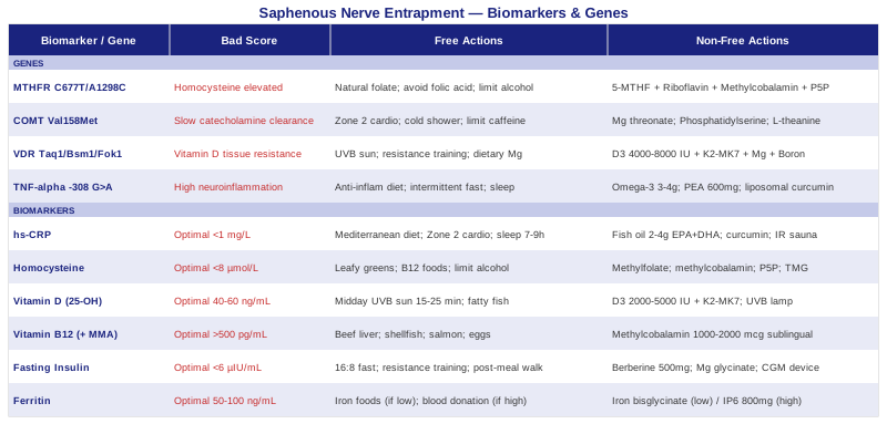
This article covers two complementary approaches. The primary section walks through six biomarkers that can be measured with routine blood work, explaining what each one reveals specifically about nerve health, how to test it, and what concrete steps you can take based on your results. The bonus section covers four genes that shape pain sensitivity, myelin synthesis, and neuroinflammation — with practical plans based on your genetic profile. Two additional sections then cover science-grounded insights from podcast research and complementary modalities with meaningful clinical evidence for nerve pain.
6 Biomarkers to Track for Saphenous Nerve Entrapment
The biomarkers below are not generic "wellness" panels. Each one has a specific mechanistic connection to peripheral nerve health, pain sensitization, or the inflammatory environment around entrapped nerves. They can be ordered as standard blood tests, most are covered by insurance, and the results give you an actionable target — not just a vague indication to "reduce inflammation."
Biomarker 1: High-Sensitivity C-Reactive Protein (hs-CRP)
Why it matters and what it reveals
hs-CRP is the most accessible marker of systemic low-grade inflammation. When the immune system is chronically activated — even at sub-clinical levels — it drives neuroinflammation in and around peripheral nerves. Perineural inflammation thickens fascial tunnels, reduces the nerve's tolerance to mechanical compression, and lowers the threshold for nociceptor activation. In plain terms: if hs-CRP is elevated, the same amount of nerve compression will hurt more, and the nerve will recover more slowly.
Optimal hs-CRP for long-term health is under 1 mg/L. Values between 1 and 3 mg/L indicate moderate risk and active biological noise. Above 3 mg/L suggests high inflammatory load. These thresholds are used by Peter Attia as a minimum standard before assuming any pain or nerve issue is "purely mechanical."
How to measure it
Standard blood test at most laboratories. Cost: $10–30 out of pocket, often covered by insurance. Always order hs-CRP (not standard CRP, which is less sensitive at low levels). Do not test within two weeks of any acute infection, surgery, or heavy exercise — all will falsely elevate the result.
If the score is bad, the plan without supplements
Remove refined carbohydrates, sugar-sweetened beverages, and seed oils (soybean, canola, sunflower, corn) from the diet — these are the highest-impact dietary drivers of chronic hs-CRP elevation. Shift to a Mediterranean-style pattern with daily fatty fish, olive oil, leafy greens, and colorful vegetables. Add 150–200 minutes per week of Zone 2 cardio (a pace where you can hold a conversation) — this is one of the most consistent interventions for lowering hs-CRP in controlled studies. Prioritize 7–9 hours of sleep per night: even two nights of poor sleep significantly elevates hs-CRP. Address visceral adiposity if present — fat stored around the abdomen is itself an inflammatory organ.
If the score is bad, the plan with supplements or equipment
Omega-3 fish oil (2–4g EPA+DHA daily): This is the most consistently evidence-backed supplement for lowering hs-CRP. Take with meals. Can be used continuously. Side effects: mild GI upset, fish burps (use enteric-coated or refrigerate); very high doses may mildly thin blood — consult a physician if on anticoagulants.
Curcumin with piperine (500–1000 mg/day): Curcumin inhibits NF-kB, a master inflammatory transcription factor. Piperine (from black pepper) increases bioavailability by ~20x. Use in 8–12 week cycles with a 4-week break. Side effects: may interact with blood thinners; avoid at high doses in gallbladder disease.
Infrared sauna (15–20 min sessions, 3–4x/week): Consistent thermal stress has been shown in multiple Finnish cohort studies to reduce hs-CRP and systemic inflammatory markers. Equipment cost: $300–1500 for home portable units. The heat-shock protein response it activates has direct anti-inflammatory effects.
Biomarker 2: Homocysteine
Why it matters and what it reveals
Homocysteine is an amino acid produced during methionine metabolism. When methylation pathways are impaired — due to genetic variants like MTHFR, or nutritional deficiencies in B12, B6, or folate — homocysteine accumulates. Elevated homocysteine is directly neurotoxic: it damages the myelin sheath surrounding peripheral nerves, impairs endothelial function in the vasa nervorum (the tiny blood vessels that feed peripheral nerves), and increases oxidative stress within axons.
For the saphenous nerve specifically, high homocysteine creates a context where compression injury is more damaging, recovery is slower, and neuropathic pain is more likely to persist. This is not speculative — the association between elevated homocysteine and peripheral neuropathy is well-documented in the clinical literature.
Optimal homocysteine: under 8 µmol/L. Elevated is generally defined as above 12–15 µmol/L (some neurologists use 10 µmol/L as a clinical threshold). Levels above 15 µmol/L are associated with significantly impaired nerve recovery and increased demyelination risk.
How to measure it
Standard fasting blood test. Cost: $30–80 out of pocket. Often not included in standard panels — you will need to request it specifically. For a more complete methylation picture, add methylmalonic acid (MMA) ($50–100), which reveals intracellular B12 deficiency even when serum B12 appears normal.
If the score is bad, the plan without supplements
Increase dietary natural folate (dark leafy greens, asparagus, legumes, avocado) — not folic acid from fortified foods, which is a synthetic form that can actually impair methylation in MTHFR variants. Increase B12-rich animal proteins: beef liver (the richest food source), shellfish, salmon, eggs. Limit alcohol consumption strictly — alcohol actively depletes B12, folate, and B6. Address gut health: intrinsic factor production can be compromised by chronic PPI use, H. pylori infection, or autoimmune gastritis, all of which impair B12 absorption regardless of dietary intake.
If the score is bad, the plan with supplements or equipment
Methylfolate (400–800 mcg/day): Use the active methylated form (5-MTHF), not folic acid. This bypasses the MTHFR enzyme bottleneck. Take daily, continuously. Side effects: in some people with MTHFR variants, introducing methyl donors causes irritability, anxiety, or insomnia (an "overmethylation" reaction) — if this occurs, reduce dose or add niacin (flush form), which acts as a methyl buffer.
Methylcobalamin B12 (1000–2000 mcg/day sublingually): Sublingual delivery bypasses the intrinsic factor pathway and is superior to oral cyanocobalamin for raising functional B12 levels. Daily, continuous. Side effects: extremely safe; rarely causes mild acne at high doses.
P5P (Pyridoxal-5-Phosphate, active B6) 10–25 mg/day: The active form of B6, which participates directly in homocysteine remethylation. Frequency: daily, continuous. Side effects: doses above 50 mg/day for prolonged periods have been associated with peripheral neuropathy (paradoxically) — stay within the 10–25 mg range.
TMG/Betaine 500–2000 mg/day: Provides an alternative methyl group donor pathway (BHMT pathway), particularly useful when the MTHFR pathway is severely impaired. Take with meals. Can be used continuously. Side effects: GI discomfort at high doses; mild fishy odor in some individuals.
Biomarker 3: 25-OH Vitamin D
Why it matters and what it reveals
Vitamin D receptors (VDR) are found not only in bone and immune cells, but on neurons, Schwann cells (the myelin-producing cells of peripheral nerves), and cells of the dorsal root ganglia — the nerve cell bodies that process sensory signals from the leg and foot. Low vitamin D impairs myelin maintenance, reduces anti-inflammatory gene expression in perineural tissue, and has been consistently associated with increased pain sensitivity and peripheral neuropathy in observational studies. The NIH Office of Dietary Supplements notes the expanding role of vitamin D in neurological function.
Low vitamin D does not cause saphenous nerve entrapment, but it can dramatically worsen the pain experience and slow recovery from it.
Optimal targets: Peter Attia recommends 40–60 ng/mL (100–150 nmol/L). Levels below 30 ng/mL are associated with higher pain sensitivity and slower peripheral nerve regeneration. Most people in Northern latitudes, office workers, and those who cover their skin are chronically below 30 ng/mL without knowing it.
How to measure it
Fasting blood test: 25-hydroxyvitamin D. Cost: $30–60, often covered when ordered with a physician's note. Test twice per year if supplementing — once in late winter (typically the annual low) and once in late summer.
If the score is bad, the plan without supplements
Get direct midday sun exposure (when the sun is high enough for UVB to penetrate) for 15–25 minutes on large skin areas (arms, legs, back) daily — without sunscreen on the targeted areas during this brief window. Eat vitamin D-rich foods: fatty fish (salmon, mackerel, sardines), egg yolks, UV-exposed mushrooms. Recognize that dietary sources alone are insufficient for most people to maintain optimal levels, particularly in winter months or above 35° latitude.
If the score is bad, the plan with supplements or equipment
Vitamin D3 (2000–5000 IU/day for maintenance; up to 10,000 IU/day for correction of documented deficiency): Always pair with Vitamin K2-MK7 (100–200 mcg/day) — K2 directs calcium to bone rather than soft tissue and blood vessels. Take both with a fat-containing meal for optimal absorption. Frequency: daily, continuous. Test 25-OH vitamin D every 3 months when adjusting dose to avoid over-correction. Side effects: vitamin D toxicity (hypercalcemia) is rare but possible at sustained doses above 10,000 IU without monitoring; K2 may interact with warfarin — consult a physician.
UVB lamp (narrowband 311 nm) — $100–300: For winter months or northern latitudes, a medical-grade UVB lamp used 3–4 times per week can stimulate endogenous vitamin D synthesis. More physiologically natural than oral supplementation. Follow manufacturer's exposure guidelines carefully to avoid sunburn.
Biomarker 4: Vitamin B12 (with Methylmalonic Acid)
Why it matters and what it reveals
B12 is arguably the single most critical micronutrient for peripheral nerve integrity. It is required for myelin synthesis via the methylation cycle, and for the regeneration of damaged axons. Without adequate functional B12, myelin around the saphenous nerve degrades progressively, making the nerve more susceptible to entrapment at anatomical narrowings, slower to conduct sensory signals, and more likely to generate spontaneous pain and paresthesias. The NIH Office of Dietary Supplements B12 fact sheet documents the well-established relationship between B12 deficiency and peripheral neuropathy.
The problem with standard serum B12 testing is that it measures B12 in circulation, not B12 actually being used inside cells. Many patients with "normal" serum B12 (200–500 pg/mL) have functional deficiency. This is why methylmalonic acid (MMA) is the more sensitive marker — MMA rises when cells cannot use B12, even if serum levels appear adequate.
Optimal targets: Serum B12 above 500 pg/mL (many neurologists use 600–800 pg/mL as a functional threshold). MMA should be below 0.28 µmol/L. If serum B12 is "normal" but MMA is elevated, functional deficiency is present.
How to measure it
Serum B12: $20–40. Add MMA: $50–100. Both can be ordered fasting. MMA is especially important for vegetarians, vegans, the elderly, and anyone on long-term proton pump inhibitors or metformin (both significantly impair B12 absorption).
If the score is bad, the plan without supplements
Increase consumption of animal proteins that are high in bioavailable B12: beef liver (highest source), shellfish (oysters, clams), salmon, sardines, eggs, and dairy. If following a vegan or vegetarian diet, dietary correction alone is insufficient — supplementation is essentially mandatory. Discontinue or reduce PPI use if clinically appropriate (discuss with your physician). Address any H. pylori infection, which reduces intrinsic factor production.
If the score is bad, the plan with supplements or equipment
Methylcobalamin or adenosylcobalamin (1000–2000 mcg/day, sublingual): Sublingual delivery bypasses the intrinsic factor-dependent absorption pathway in the gut, making it effective even in malabsorption states. Avoid cyanocobalamin — it requires conversion steps and is less bioavailable for neural tissue. Frequency: daily, continuous. Retest B12 and MMA at 3 months. Side effects: extremely safe at these doses; mild acne reported rarely at very high doses.
For confirmed severe functional deficiency with neurological symptoms, intramuscular B12 injections (1000 mcg methylcobalamin, 1x/week for 4–6 weeks, then monthly) can be considered — this route entirely bypasses absorption. Cost: $5–20 per injection at a clinic. Discuss with your physician.
Biomarker 5: Fasting Insulin and HOMA-IR
Why it matters and what it reveals
Insulin resistance does not require a type 2 diabetes diagnosis to affect peripheral nerve health. Even subclinical metabolic dysfunction — what Peter Attia calls "the insulin resistance iceberg" — creates a chronic state of systemic inflammation, impairs microvascular blood flow to peripheral nerves (vasa nervorum dysfunction), and increases oxidative stress within axons. This biochemical environment makes any peripheral nerve more sensitive to mechanical compression and more likely to remain symptomatic after that compression resolves.
The connection is supported by clinical observation: patients with metabolic syndrome have significantly higher rates of peripheral neuropathy and chronic nerve pain, even in the absence of diabetic neuropathy. The saphenous nerve, running through the metabolically active tissues of the thigh, is not exempt from this effect.
Optimal targets: Peter Attia considers fasting insulin above 6 µIU/mL as suboptimal, while most conventional labs flag it only above 25. HOMA-IR above 2.0 is generally considered clinically significant. A value of 1.0 or below is excellent.
How to measure it
Fasting insulin: $15–40. HOMA-IR is calculated: (fasting insulin × fasting glucose) ÷ 405. Both require 12-hour fasting. Critically, fasting insulin is not included in most standard metabolic panels — you must request it explicitly from your physician or order it separately. Many people are insulin resistant for years without this ever being measured.
If the score is bad, the plan without supplements
Time-restricted eating (14:10 or 16:8): Compressing food intake to a 8–10 hour window significantly reduces fasting insulin over 4–8 weeks without caloric restriction. This is one of the highest-leverage, cost-free interventions for insulin resistance.
Resistance training 3x/week: Muscle tissue is the primary disposal site for blood glucose. Building and maintaining muscle is the most durable long-term strategy for insulin sensitivity. Focus on compound movements (squats, deadlifts, rows).
Post-meal walking (10–15 minutes after each meal): Dramatically blunts post-prandial glucose spikes. This reduces the insulin demand placed on the pancreas and, over time, improves overall insulin sensitivity.
Sleep 7–9 hours: One night of 4–5 hours sleep increases insulin resistance to levels comparable to early pre-diabetes. This is well-established in controlled sleep deprivation studies.
If the score is bad, the plan with supplements or equipment
Berberine (500 mg, 2–3x/day with meals): Multiple RCTs have shown berberine comparable to metformin for lowering fasting glucose and fasting insulin, with additional gut microbiome-modulating effects. Use in 3-month cycles with a 1-month break to prevent gut adaptation. Side effects: GI discomfort, especially at higher doses; lowers blood sugar, so caution in anyone on diabetes medications; do not combine with medications sensitive to CYP3A4 metabolism without physician guidance.
Magnesium glycinate or magnesium malate (300–400 mg/day): The majority of insulin-resistant individuals are magnesium deficient. Magnesium is required for insulin receptor signaling. Daily, continuous. Side effects: loose stools at higher doses (switch to glycinate form if this occurs, as it is gentler).
Continuous glucose monitor (CGM) — $70–200/month for sensors: Wearing a CGM for 2–4 weeks as a biofeedback tool gives direct insight into which foods, activities, and sleep patterns drive glucose variability. This data is often transformative for motivation and behavior change around insulin resistance. Devices include the Libre 3 (requires prescription in some countries) and the Stelo (available OTC in the US).
Biomarker 6: Ferritin
Why it matters and what it reveals
Ferritin is the body's primary iron storage protein. For peripheral nerve health, it sits at the intersection of two distinct problems. First, iron is required for myelin synthesis and for mitochondrial energy production within axons — low ferritin (even without frank anemia) impairs nerve repair and can cause or worsen neuropathic symptoms, including restless legs syndrome (which shares neural pathways with the saphenous distribution). Second, elevated ferritin — when not explained by acute infection or inflammation — may reflect iron overload, which generates significant oxidative stress and damages peripheral neurons.
Ferritin is also an acute phase reactant, meaning it rises with systemic inflammation. An elevated result should therefore be interpreted alongside hs-CRP to distinguish true iron excess from inflammatory elevation of a storage protein.
Optimal targets: Approximately 50–100 ng/mL is associated with good nerve and mitochondrial function. Below 30 ng/mL: likely functional iron deficiency. Above 200 ng/mL (women) or above 300 ng/mL (men) without inflammation warrants investigation for iron overload, including hereditary hemochromatosis.
How to measure it
Standard blood test: $15–30, often included in CBCs or iron panels. For a complete iron picture, combine with serum iron, TIBC, and transferrin saturation ($40–80 complete panel). A transferrin saturation consistently above 45% alongside elevated ferritin is a stronger signal for iron overload.
If the score is bad (low ferritin), the plan without supplements
Increase dietary heme iron: red meat (especially beef and lamb), organ meats (beef liver is extremely iron-dense), oysters and clams. Pair iron-rich foods with vitamin C-rich foods (bell peppers, citrus) to enhance non-heme iron absorption. Avoid coffee, tea, or calcium-rich foods within 1 hour of iron-rich meals — these compounds strongly inhibit iron absorption. Address any occult blood loss: heavy menstrual bleeding, GI blood loss, or high-impact running (hemolysis) are common causes.
If the score is bad (low ferritin), the plan with supplements or equipment
Iron bisglycinate 18–25 mg/day, on alternate days: Research by Tomas Ganz and others has shown that alternate-day dosing (rather than daily) maximizes iron absorption by avoiding the hepcidin rebound that blocks GI iron uptake after daily dosing. Bisglycinate form is significantly gentler on the GI tract than ferrous sulfate. Always recheck ferritin at 3 months. Side effects: GI discomfort and constipation (take with food and increase dietary fiber); never supplement iron without laboratory-confirmed deficiency.
If the score is bad (high ferritin), the plan without supplements
Avoid cooking in cast iron and limit red meat to 2–3 servings/week. Stop any iron-containing supplements immediately. Regular blood donation (every 56 days as allowed, or more frequently if approved by a physician) is the most effective way to reduce iron stores — hemochromatosis patients are treated almost exclusively with therapeutic phlebotomy. Get tested for HFE gene variants (C282Y and H63D) if ferritin is consistently elevated without explanation.
If the score is bad (high ferritin), the plan with supplements or equipment
IP6 (Inositol hexaphosphate) 800 mg, twice daily between meals: IP6 is a natural iron chelator found in legumes and grains. Taken away from food on an empty stomach, it can bind excess iron in the GI tract and reduce iron absorption. Frequency: daily during active reduction phase; reassess ferritin every 3 months. Side effects: may also reduce absorption of zinc and calcium — cycle off for 1 month every 3 months if using long-term. Do not use if iron status is already normal or low.
The biomarkers above give you a concrete, testable picture of your nerve's biochemical environment. With a baseline and a follow-up at 3–6 months, you can see whether your interventions are actually changing the biological terrain — not just managing symptoms.
4 Genes That May Shape Your Saphenous Nerve Pain
A consumer genetic panel (23andMe, AncestryDNA) combined with a third-party analysis tool (Genetic Genie, Strategene, or SelfDecode) can reveal the gene variants below. These are not destiny — they are tendencies that environmental and nutritional inputs can substantially modify. The goal is not to worry about your genetics, but to make smarter decisions about where to focus your interventions.
MTHFR (C677T and A1298C)
What it affects
MTHFR encodes the enzyme methylenetetrahydrofolate reductase, which converts dietary folate into its biologically active form needed for the methylation cycle. The NIH MedlinePlus genetics page documents two well-studied variants: C677T and A1298C. The homozygous C677T variant reduces enzyme activity by approximately 70%. Compound heterozygosity (one copy of each variant) reduces it by roughly 50%.
Reduced MTHFR function leads to elevated homocysteine (directly neurotoxic), reduced production of SAM-e (the universal methyl donor needed for myelin synthesis), and impaired DNA repair in neurons. For saphenous nerve entrapment, this matters: the nerve's ability to repair itself after compression or traction injury is substantially dependent on the methylation cycle running efficiently.
If the gene is bad, the plan without supplements
Shift aggressively toward natural folate sources (not fortified grains, which contain synthetic folic acid that can actually block active folate receptors in MTHFR-variant individuals). Prioritize leafy greens, legumes, asparagus, and avocado. Avoid long-term alcohol use and chronic PPI exposure, both of which further impair the already-compromised methylation pathway. Minimize folic acid in multivitamins — check labels explicitly.
If the gene is bad, the plan with supplements or equipment
Methylfolate (5-MTHF) 400–800 mcg/day: The active form that bypasses the impaired enzyme. Start at the lower end to avoid "overmethylation" reactions (anxiety, irritability, insomnia). Riboflavin (B2) 100–400 mg/day: This is the cofactor for the MTHFR enzyme itself — often overlooked, but particularly important for the C677T variant. Studies show riboflavin supplementation meaningfully lowers homocysteine in C677T homozygous individuals. Methylcobalamin B12 1000 mcg/day and P5P (active B6) 10–20 mg/day complete the methylation support stack. Frequency: daily, continuous. Reassess homocysteine every 3–6 months. Side effects: introduce methyl donors slowly; overmethylation symptoms respond to niacin (flush-form, 50–100 mg) as a methyl group buffer.
COMT (Val158Met)
What it affects
COMT encodes catechol-O-methyltransferase, the enzyme responsible for breaking down dopamine, norepinephrine, and epinephrine in the prefrontal cortex and sympathetic nervous system. The NIH MedlinePlus genetics page for COMT describes the Val158Met variant: the Val/Val variant produces a fast enzyme (catecholamines cleared quickly), while the Met/Met variant produces a slow enzyme (catecholamines accumulate).
For nerve pain, the Met/Met variant is the more relevant concern. Under chronic stress conditions, slow COMT leads to excess catecholamine accumulation in the spinal cord dorsal horn, which amplifies incoming pain signals from peripheral nerves. This is part of the mechanism behind central sensitization — a state where the central nervous system over-amplifies pain independent of the peripheral injury. Met/Met individuals are significantly more susceptible to chronic pain after nerve injury and typically rate the same nerve compression as more painful than Val/Val carriers.
If the gene is bad, the plan without supplements
Stress management is the highest-leverage intervention for Met/Met individuals: not as a vague suggestion, but because excess sympathetic tone (driven by slow catecholamine clearance) is a direct amplifier of pain signaling. Aerobic exercise is the most effective natural metabolizer of excess catecholamines. Zone 2 cardio 4–5 days/week substantially lowers sympathetic baseline over time. Deliberate cold exposure (1–3 minutes of cold shower daily) provides an acute catecholamine spike followed by a recovery period that trains the system toward faster clearance. Limit excessive caffeine (raises norepinephrine significantly).
If the gene is bad, the plan with supplements or equipment
Magnesium threonate (300–400 mg/day at night): Crosses the blood-brain barrier more effectively than other magnesium forms; supports GABA activity in the prefrontal cortex, counterbalancing the excess excitatory tone from slow COMT. Phosphatidylserine (100 mg, 2–3x/day): Well-studied for blunting cortisol and stress reactivity; helps reduce the sympathetic hyperactivation that amplifies pain in Met/Met individuals. Use in 3-month cycles. L-theanine (200 mg) combined with morning caffeine can blunt the norepinephrine spike from coffee without eliminating mental clarity. Side effects: all three are generally well-tolerated. Phosphatidylserine at very high doses (> 600 mg/day) may cause GI discomfort.
VDR (Taq1, Bsm1, Fok1 variants)
What it affects
VDR encodes the vitamin D receptor — the protein that vitamin D must bind to in order to activate gene transcription. The NIH MedlinePlus genetics page for VDR outlines multiple well-studied variants. Even if your circulating vitamin D levels are adequate, poor VDR function means that neurons, Schwann cells, and immune cells cannot respond properly to it. This creates a form of "vitamin D resistance" at the tissue level.
VDR is expressed in peripheral neurons, dorsal root ganglia, and the immune cells that patrol perineural tissue. Impaired VDR function reduces myelin maintenance, blunts anti-inflammatory gene expression in nerve tissue, and impairs nerve regeneration after compression injury.
If the gene is bad, the plan without supplements
Maximize UVB-driven vitamin D synthesis (which engages slightly different pathways than oral supplementation). Resistance training upregulates VDR expression in muscle — a finding that transfers partially to other tissues. Ensure adequate dietary magnesium, which is required for VDR activation. Reduce adiposity if present — excess body fat sequesters vitamin D and reduces its availability to target tissues.
If the gene is bad, the plan with supplements or equipment
Poor VDR responders typically need higher circulating vitamin D levels to achieve the same tissue effect: target 50–70 ng/mL rather than 40 ng/mL. This may require 4000–8000 IU of D3/day, always with K2-MK7 (200 mcg/day) and magnesium glycinate (400 mg/day). Monitor 25-OH vitamin D every 3 months. Boron (3–6 mg/day) has preliminary evidence supporting improved VDR sensitivity by extending vitamin D's half-life and influencing steroid hormone receptor activity. Cycling: D3 and K2 are continuous; recheck levels seasonally. Side effects: at doses above 4000 IU, close monitoring of calcium levels is prudent; K2 interacts with warfarin.
TNF-alpha (-308 G>A Polymorphism)
What it affects
The TNF -308 G>A variant (rs1800629) increases the promoter activity of the tumor necrosis factor-alpha gene. Carriers of the A allele — particularly AA homozygotes — produce significantly higher amounts of TNF-alpha in response to inflammatory stimulation. TNF-alpha is one of the key cytokines directly involved in peripheral and central sensitization: it sensitizes nociceptors, promotes demyelination, disrupts the blood-nerve barrier, and maintains neuroinflammation in compressed nerve tissue long after the initial mechanical insult.
In practical terms: high TNF producers with the same degree of saphenous nerve compression will experience more pain, are more likely to develop chronic neuropathic symptoms, and may not respond as well to purely mechanical interventions (physical therapy, bracing) without simultaneously addressing the neuroinflammatory environment.
If the gene is bad, the plan without supplements
An anti-inflammatory dietary pattern is the foundation: Mediterranean or Mediterranean-ketogenic hybrid, eliminating ultra-processed foods, refined carbohydrates, and industrial seed oils. Intermittent fasting (16:8 or alternate-day caloric restriction) consistently reduces TNF-alpha in human studies. Regular moderate exercise reduces circulating TNF-alpha through myokine signaling (muscle contractions during exercise release IL-6 transiently, which subsequently drives an anti-inflammatory cascade). Prioritize sleep above all — chronic sleep restriction dramatically amplifies TNF-alpha production, and for AA carriers, this effect can be disproportionately severe.
If the gene is bad, the plan with supplements or equipment
Omega-3 EPA+DHA (3–4g/day): The most consistently evidence-backed supplement for reducing TNF-alpha, through its conversion to resolvins and protectins that actively resolve neuroinflammation. Continuous use is appropriate. Side effects: high-dose fish oil mildly thins blood — caution with anticoagulants.
Palmitoylethanolamide (PEA) 600 mg, twice daily: PEA is an endogenous lipid mediator with specific relevance to neuropathic pain. It acts partly via PPAR-alpha receptors and the cannabinoid system to downregulate mast cell activation and TNF-alpha signaling specifically within perineural tissue. Multiple human trials support its use in peripheral neuropathy. Use in 3-month cycles. Side effects: generally very well tolerated; no significant drug interactions reported.
Liposomal curcumin (500–1000 mg/day): NF-kB inhibition from curcumin specifically addresses one of TNF-alpha's downstream signaling pathways. Liposomal form has substantially better bioavailability than standard curcumin with piperine. Cycle 8–10 weeks on, 4 weeks off. Side effects: may interact with blood thinners; avoid in known gallbladder disease.
Moving from biology toward practical daily habits, the next section draws on neuroscience research that has meaningfully changed how clinicians think about chronic nerve pain.
The Huberman Lab Lens on Nerve Pain: 10 Things That May Change How You Think About Your Recovery
Andrew Huberman's Huberman Lab podcast has released multiple episodes covering the neuroscience of pain, inflammation, peripheral nerve function, and recovery. Drawing on his interviews with pain neuroscientists, the following insights are grounded in peer-reviewed literature and consistently challenge the passive, rest-and-wait approach that most patients receive. These are not motivational points — they are mechanistic principles with direct relevance to saphenous nerve entrapment.
1. Central Sensitization Is Often a Bigger Driver Than the Compression Itself
The saphenous nerve may be physically released from its entrapment — surgically or via conservative treatment — and yet pain persists. This is central sensitization: the spinal cord and brain have become amplifiers that maintain pain independent of the peripheral signal. Huberman covers this in depth: the nervous system is plastic, and it can learn pain as a default state. Addressing central sensitization requires active intervention — not just rest.
2. Sleep Is the Highest-Leverage Intervention in Chronic Nerve Pain
Huberman repeatedly emphasizes that the glymphatic system — which clears neuroinflammatory waste — operates almost exclusively during deep sleep. Chronic poor sleep (< 7 hours, or fragmented sleep) dramatically elevates hs-CRP, TNF-alpha, and IL-6 while simultaneously increasing pain sensitivity. For nerve pain specifically, there is a bidirectional loop: pain worsens sleep, and poor sleep worsens pain. Breaking this loop through sleep hygiene, melatonin, and NSDR (non-sleep deep rest/yoga nidra) is often the first meaningful intervention.
3. Physiological Sighs Directly Modulate Pain Gating
The gate control theory of pain includes descending modulatory pathways from the brain to the spinal cord that can either amplify or suppress incoming nerve signals. Sympathetic activation (fight-or-flight) opens the gate and worsens pain. Huberman describes the physiological sigh — a double inhale through the nose followed by a full extended exhale — as the fastest way to manually shift from sympathetic to parasympathetic tone. Practicing this during acute pain episodes (3–5 repetitions) can measurably reduce pain intensity within minutes.
4. Chronic Stress Sensitizes the Exact Receptors That Make Nerve Entrapment Worse
Sustained cortisol and norepinephrine exposure sensitizes peripheral nociceptors and primes spinal cord interneurons for amplified response. For COMT Met/Met carriers specifically, this mechanism is even more pronounced. Huberman's work on stress physiology frames this clearly: managing the autonomic nervous system is not "soft" medicine — it directly changes pain receptor sensitivity at a molecular level.
5. Zone 2 Cardio Is the Best Anti-Inflammatory Exercise Dose
Huberman and collaborators consistently identify Zone 2 aerobic exercise (60–70% of maximum heart rate, conversational pace, 45–60 minutes per session) as the exercise modality with the strongest anti-inflammatory evidence. Muscle contractions release myokines that reduce systemic TNF-alpha and IL-6, improve vascular function of the vasa nervorum, and over time reduce the hs-CRP background noise that amplifies nerve sensitization. Target 150–200 minutes/week.
6. Deliberate Cold Exposure Reduces Neuroinflammatory Cytokines
Huberman references multiple studies showing that deliberate cold exposure — 1–3 minutes in cold water (10–15°C) — triggers a norepinephrine release followed by a recovery state that activates descending noradrenergic pain inhibition pathways. Cold exposure also consistently lowers circulating IL-6 and TNF-alpha baseline levels over weeks of practice. For nerve pain, this dual effect (acute pain inhibition + chronic inflammatory reduction) makes it one of the more promising low-cost tools.
7. Morning Light Sets the Circadian Pain Threshold
Huberman's most foundational recommendation — morning sunlight exposure within 30–60 minutes of waking — sets circadian rhythm through retinal photoreceptors. Circadian timing governs cortisol's daily pattern, and correct circadian rhythm is associated with significantly lower pain sensitivity and better descending inhibition of nerve pain signals throughout the day. Disrupted circadian rhythm, common in shift workers and screen-heavy lifestyles, raises pain sensitivity measurably.
8. DHA Is the Structural Foundation of Neuronal Membrane Health
Huberman covers the critical role of docosahexaenoic acid (DHA) in neuronal membrane fluidity and synapse function. DHA is the primary fatty acid in peripheral axon membranes and Schwann cell myelin. It is also the precursor to resolvins and protectins — specialized pro-resolving lipid mediators that actively terminate neuroinflammation. Without adequate DHA (from fatty fish or supplementation), the inflammatory response around an entrapped nerve lacks the molecular tools to fully resolve.
9. Sensorimotor Training Drives Cortical Remapping and Reduces Maladaptive Pain
Huberman discusses how the brain's representation of body parts shifts during prolonged pain — regions associated with the painful area can "smear" or over-represent, contributing to hypersensitivity. Targeted sensorimotor training (gentle, deliberate movements of the affected limb within pain-free range) can reverse this cortical drift over weeks to months. This is one of the neurological mechanisms behind why graded motor imagery and pain-free movement protocols outperform passive rest for most peripheral nerve pain.
10. The Placebo and Nocebo Effect Are Neurobiologically Real — and Modifiable
Huberman dedicates considerable attention to the neuroscience of expectation. In chronic nerve pain, negative expectation (nocebo) physiologically amplifies pain through the same descending pathways that inhibit it. Understanding this is not about dismissing pain as psychological — it is about recognizing that the meaning and expectation context around pain symptoms directly activates or suppresses real neurochemical pain-modulating pathways. Patients who understand their diagnosis, believe recovery is possible, and engage actively in management have better measurable outcomes than those in a state of anxious passivity.
Complementary Approaches with Meaningful Evidence
The four modalities below were selected because they have meaningful human clinical evidence for nerve pain, peripheral neuropathy, or chronic musculoskeletal pain — not for general wellness claims. Each is described practically with appropriate caveats.
Low-Level Laser Therapy (LLLT) / Photobiomodulation
What it is and why it may be relevant
LLLT (also called photobiomodulation) uses specific wavelengths of red and near-infrared light (typically 600–1000 nm) to stimulate mitochondrial activity in target tissue. At therapeutic doses, it increases ATP production, reduces oxidative stress, and promotes local anti-inflammatory signaling in nerve tissue. For peripheral nerve entrapment, the photobiomodulation effect on Schwann cells and axon mitochondria is the mechanistically relevant pathway — it may accelerate nerve repair and reduce local neuroinflammation without systemic side effects.
Supporting evidence
A 2016 systematic review and meta-analysis published in Lasers in Medical Science (Yamamoto et al.) examined photobiomodulation for peripheral neuropathy and found positive effects on pain and sensory symptoms across several RCTs. A 2017 RCT in diabetic peripheral neuropathy demonstrated significant improvements in pain scores and nerve conduction velocity with LLLT applied along nerve pathways compared to sham treatment. While most RCTs have focused on diabetic neuropathy rather than entrapment neuropathy specifically, the mechanism is directly transferable. Evidence remains moderate — effect sizes vary across studies and protocols differ considerably.
How to apply it realistically
For saphenous nerve entrapment, treatment should be applied along the course of the saphenous nerve from the adductor canal down to the affected area. Clinical LLLT sessions typically run 10–15 minutes, 3x/week for 6–8 weeks. In-clinic cost: $40–80/session. Home devices (FDA-cleared class II) in the 808–850 nm range at adequate power density (50–100 mW/cm²) are now available at $200–600. Avoid treatment over active cancers or directly over the thyroid gland. Start with fewer sessions and increase gradually; expect no immediate results — the effect accumulates over 4–6 weeks.
Mindfulness Meditation and MBSR
What it is and why it may be relevant
Mindfulness-based stress reduction (MBSR), developed by Jon Kabat-Zinn, is an 8-week structured program combining body scan meditation, sitting meditation, and yoga. For chronic nerve pain, its mechanism of action is not relaxation in the popular sense — it involves training prefrontal cortex regulation of the anterior cingulate cortex (a key pain amplifier), increasing descending pain inhibition, and reducing catastrophizing, which is one of the strongest predictors of chronic pain persistence regardless of tissue state.
Supporting evidence
A landmark 2016 RCT published in JAMA Internal Medicine (Cherkin et al.) compared MBSR, cognitive behavioral therapy, and usual care for chronic back pain — finding that MBSR produced clinically significant reductions in pain and functional limitation compared to usual care, with benefits persisting at 52 weeks. A meta-analysis of mindfulness for chronic pain in JAMA Internal Medicine (2014, Goyal et al.) found moderate evidence for pain reduction across multiple chronic pain conditions.
How to apply it realistically
The standard MBSR format is an 8-week group course (available in-person and online). Commitment is approximately 45 minutes of daily home practice plus a weekly 2.5-hour group session. Alternatives include structured apps (Headspace, Insight Timer) though these have less evidence than the formal MBSR program. For saphenous nerve pain, body scan practices that include the medial leg without resistance or aversion are particularly relevant. Results are typically not felt until weeks 4–6 — the 8-week commitment is important. No significant side effects; a small percentage of people experience increased anxiety initially with body awareness practices — reducing session length and adding grounding techniques usually resolves this.
Massage Therapy
What it is and why it may be relevant
Targeted soft tissue mobilization of the fascial compartments surrounding the saphenous nerve — particularly the adductor canal region in the medial thigh and the periarticular tissue of the medial knee — can reduce mechanical compression on the nerve by releasing adjacent fascial restrictions. Massage also activates large-diameter Aβ mechanoreceptor fibers, which, per gate control theory, inhibit pain transmission from smaller-diameter pain fibers (Aδ and C fibers) carrying signals from the compressed saphenous nerve.
Supporting evidence
A 2014 Cochrane review (Furlan et al.) on massage for low back pain found moderate evidence that massage is beneficial for acute and subacute pain when combined with other active therapies. A 2016 RCT in PAIN Medicine demonstrated significant reductions in neuropathic pain intensity with connective tissue massage compared to progressive relaxation. While saphenous nerve entrapment specifically is not well-studied in RCTs, the neurological and mechanical rationale is strong and the clinical experience in sports medicine and physical therapy is consistent.
How to apply it realistically
For saphenous nerve entrapment, the most relevant technique is myofascial release applied along the medial thigh (adductor canal area), the sartorius muscle (which forms the roof of Hunter's canal), and the medial knee region. Find a therapist with training in neuromuscular or sports massage rather than purely relaxation-focused techniques. Sessions of 45–60 minutes, 1–2x/week for 6–8 weeks, are a reasonable starting protocol. Communicate the exact nerve distribution affected so the therapist can focus on the relevant anatomy. Self-massage with a foam roller applied to the medial quadriceps and adductors (not directly on the nerve) can complement clinical sessions between appointments.
Yoga
What it is and why it may be relevant
Yoga combines passive and active stretching with breathwork, proprioceptive challenge, and sustained attention. For saphenous nerve entrapment, its relevance is mechanical and neurological: gentle mobilization of hip flexors, adductors, and the medial thigh reduces fascial tension around the adductor canal and decreases passive traction on the saphenous nerve during functional movement. Additionally, yoga's emphasis on slow, controlled breathing consistently activates the parasympathetic nervous system, which reduces pain gate amplification.
Supporting evidence
A 2017 Cochrane review (Wieland et al.) of yoga for chronic non-specific low back pain found moderate-quality evidence that yoga reduces pain and functional disability at short- and medium-term follow-up compared to non-exercise controls. A 2016 RCT published in Spine found yoga superior to self-care books for chronic back pain. For peripheral neuropathy specifically, a small RCT (Nayak et al., 2014) demonstrated improved sensory scores and pain levels in diabetic peripheral neuropathy patients after 12 weeks of yoga, with improved nerve conduction velocity as a secondary finding.
How to apply it realistically
For saphenous nerve entrapment, the most relevant poses involve hip flexor opening (low lunge, pigeon pose), adductor lengthening (bound angle pose, wide-legged forward fold), and medial thigh mobilization. Avoid aggressive overstretching of the medial knee area, which can increase traction on the already-irritated nerve. A 20–30 minute practice 4–5 days/week is more effective than sporadic longer sessions. Beginners should work with an instructor initially, specifically mentioning the nerve condition so modifications can be offered. Yin yoga, which holds passive stretches for 3–5 minutes, may be particularly helpful for fascial release around the adductor canal.
Conclusion
Saphenous nerve entrapment is a condition where the mechanical story — compression at the adductor canal, traction at the knee — is only half the explanation for what you are actually experiencing. The other half lives in your biochemistry: how much neuroinflammation is running in the background, how well your myelin is maintained, how sensitized your central nervous system has become, and how your genetics shape all of those processes. None of that is fixed, and none of it requires expensive or exotic interventions to start addressing.
Begin with what is most measurable: order hs-CRP, homocysteine, 25-OH vitamin D, B12, fasting insulin, and ferritin as a baseline. If you have access to a genetic panel, check MTHFR, COMT, VDR, and TNF-alpha. Let the results guide where you put your effort first — not a generic protocol, but a targeted one based on what is actually off in your specific case. Retest at three to six months to confirm movement.
The complementary approaches — photobiomodulation, mindfulness, soft tissue work, and yoga — are not passive add-ons. Each has a specific mechanism that addresses a distinct part of the nerve pain equation: local tissue repair, central sensitization, fascial compression, and autonomic regulation respectively. The most durable outcomes come from addressing all of these layers simultaneously rather than pursuing one intervention in isolation.
Take the next smart step: track your biomarkers, review your sleep and metabolic health first, then add layers methodically. Bring what you find to a physician or neurologist who can contextualize these results in your clinical picture. Better data leads to better decisions — and saphenous nerve entrapment, properly approached at this level of specificity, is genuinely responsive to the right interventions.
Musculoskeletal Neurological Endocrine & Metabolic
Neurological: Nerve Conditions
Autoimmune: Inflammatory Conditions