This article contains AI generated content.
· UpdatedSchwartz-Jampel Syndrome – 3 Genes And 7 Biomarkers To Track
Introduction
Living with Schwartz-Jampel Syndrome — or supporting someone who does — puts you in a particular kind of isolation. The condition is rare enough that most clinicians encounter it once in a career, if at all. Appointments tend to orbit around what cannot be changed — the genetic mutation — rather than the measurable terrain around it that might actually respond to intervention. You may leave knowing your diagnosis without any clearer sense of what to track, what to ask for, or where the real margin for improvement exists.
SJS is not a condition that lifestyle adjustments will rewrite. The HSPG2 mutation is structural. But between the mutation and its expression lies a landscape shaped by factors that are neither fixed nor invisible: inflammatory tone, muscle membrane excitability, bone density trajectory, hormonal support, micronutrient status. These factors do not determine whether you have SJS, but they do influence how loudly its consequences show up in daily function. That distinction is worth understanding.
Generic health guidance rarely accounts for this. "Stay active and eat well" does not translate easily when your muscles fail to relax normally and your skeletal architecture creates its own physical constraints. What is actually useful is more precise: specific, measurable signals tied to the biological systems that SJS compromises most — and intervention options calibrated to those signals rather than to the average person.
This article takes that approach. The central section covers seven biomarkers directly relevant to SJS — what each reveals, how to measure it, and what to do when results are off, with and without supplements. A second section examines the genetic layer: the primary HSPG2 gene and two modifier genes that can shift disease expression significantly, along with concrete plans for each. Beyond that, you will find a synthesis of insights from one of the most rigorous neuromuscular science resources currently available, and a set of complementary approaches with genuine human evidence for the key symptoms of SJS.
Summary
This article covers 7 biomarkers worth tracking in SJS — including creatine kinase, 25-OH vitamin D, bone mineral density, IGF-1, thyroid hormones, RBC magnesium, and heparan sulfate glycosaminoglycans — with detailed plans for what to do when each is suboptimal, with and without supplements. It then examines 3 key genes (HSPG2 and two neuromuscular modifier genes) and what each means for day-to-day management. A curated section distills the 10 most actionable insights from leading neuromuscular science content that challenge common assumptions about fixed muscle dysfunction. Finally, five complementary modalities — progressive muscle relaxation, massage therapy, biofeedback, photobiomodulation, and mindfulness-based stress reduction — are reviewed with their specific protocols and the clinical evidence behind them.
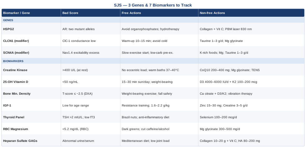
7 Biomarkers Worth Tracking in Schwartz-Jampel Syndrome
Tracking biomarkers in a monogenic condition like SJS is not about questioning the diagnosis — it is about understanding what the mutation is doing downstream in real time, inside a specific body, under specific conditions. The HSPG2 mutation disrupts perlecan, a structural proteoglycan present across basement membranes, cartilage, muscle, and the neuromuscular junction. That disruption sets off consequences in multiple biological systems simultaneously. These seven markers are worth monitoring because each one maps to a system directly compromised by SJS, and because a suboptimal result in any of them comes with a defined intervention pathway — not just a number on a page.
1. Creatine Kinase — Reading Muscle Stress in Real Time
Why it matters
Creatine kinase (CK) is an enzyme released into circulation when muscle cells are mechanically or metabolically stressed. In myotonic disorders, chronically impaired muscle relaxation creates continuous low-grade contractile strain on fiber membranes. Elevated CK is not diagnostic of SJS — it is a signal that the muscle is being pushed beyond its current recovery threshold, whether from myotonia itself, secondary physical stress, or the early stages of myopathic deterioration. SJS type 1B patients in particular may show persistent CK elevation tied to the dystrophic component of their presentation.
Monitoring trends over months matters more than any single reading. A rising trajectory is more informative than a single elevated value.
How to measure it
Total serum CK is a standard blood test available at any lab, typically costing $20–$60. Optimal is generally below 200 U/L in the absence of recent vigorous exercise. In SJS, persistent elevation above 400–500 U/L without recent exercise or intramuscular injections warrants closer attention. Testing every 3–6 months provides a useful trend line.
If the score is bad, the plan without supplements
The first step is eliminating unnecessary mechanical triggers. Eccentric-heavy exercise — downhill running, slow-lowering strength movements — generates the most membrane disruption per unit of work and should be minimized during periods of elevated CK. Low-resistance aquatic activity, passive stretching, and regulated warm heat (warm baths at 37–40°C for 20 minutes) reduce myotonic stiffness without adding contractile damage. Adequate slow-wave sleep is non-negotiable here, since muscle repair is concentrated in that phase. Removing pro-inflammatory foods — highly processed seed oils, refined carbohydrates — reduces the metabolic burden on already-stressed muscle tissue.
If the score is bad, the plan with supplements or equipment
Coenzyme Q10 at 200–400 mg/day has been studied in mitochondrial myopathies with consistent evidence for reducing CK over 3–6 months; direct SJS data is limited, but the mechanism (supporting mitochondrial electron transport in muscle) is applicable. Magnesium glycinate at 300–400 mg/day in divided doses supports membrane stability and may reduce the frequency and intensity of myotonic discharge. A TENS device applied around major muscle groups for 15–20 minutes daily can reduce post-contracture soreness and support local circulation. Consistency over 3–6 months is needed to assess response — neither CK nor its determinants shift rapidly.
2. 25-Hydroxyvitamin D — The Bone and Muscle Foundation
Why it matters
Vitamin D operates at the intersection of two systems that SJS compromises most directly: skeletal mineralization and neuromuscular function. Low vitamin D amplifies the fracture risk already elevated by skeletal dysplasia, worsens muscle weakness, and increases myotonic sensitivity through effects on calcium channel regulation. Vitamin D deficiency is consistently overrepresented in patients with neuromuscular disorders, and this is not coincidental — the vitamin D receptor is expressed in muscle tissue and its activation directly modulates muscle fiber contractility.
A target of 50–80 ng/mL (125–200 nmol/L) is appropriate for most SJS patients. The conventional "sufficient" threshold of 30 ng/mL, as Peter Attia has emphasized, is calibrated for deficiency prevention rather than for functional optimization in patients with active musculoskeletal disease.
How to measure it
The 25-OH Vitamin D serum test is widely available and costs $30–$80 out of pocket. Test at baseline and recheck every 3 months when correcting a deficiency. Always pair with serum calcium and PTH — hypercalcemia is a real risk with aggressive correction and must be monitored.
If the score is bad, the plan without supplements
Daily direct sun exposure of 15–30 minutes between 10am and 2pm — face, arms, and legs exposed — provides meaningful D3 synthesis in moderate skin types at adequate latitudes. In northern climates, darker skin tones, or during winter months, sunlight alone is typically insufficient. Weight-bearing activity upregulates vitamin D metabolic pathways, though it must be adapted to SJS constraints. Dietary sources are minimal contributors (fatty fish, egg yolks) but worth including.
If the score is bad, the plan with supplements or equipment
Vitamin D3 at 4,000–6,000 IU/day paired with vitamin K2 (100–200 mcg MK-7 form) — K2 directs calcium toward bone rather than arterial tissue, making the combination meaningfully safer than D3 alone. Retest at 3 months and adjust dose to reach target. Avoid high-bolus protocols (such as 50,000 IU weekly) without close monitoring. No cycling necessary — vitamin D functions as a hormone precursor requiring stable, sustained levels rather than pulsed stimulation.
3. Bone Mineral Density via DXA — Tracking the Skeletal Foundation
Why it matters
Skeletal dysplasia is one of the defining features of SJS. Vertebral deformities, hip joint abnormalities, and altered long bone architecture create a baseline fracture risk that demands active quantification, not just clinical awareness. Bone mineral density (BMD) measured by DXA scan provides a longitudinal record of whether skeletal integrity is stable, improving, or eroding — information that directly informs decisions about physical activity, pharmacological intervention, and fall prevention.
How to measure it
A DXA scan is the clinical gold standard for BMD and costs approximately $100–$300 depending on coverage. It measures density at the lumbar spine and femoral neck. For children and adolescents — SJS is frequently diagnosed in the first decade of life — Z-scores adjusted for age and sex are the correct metric. Adults use T-scores, with -2.5 or below indicating osteoporosis. Frequency of rescanning depends on clinical status: annually if density is declining or pharmacological intervention is underway; every 2 years if stable.
If the score is bad, the plan without supplements
Weight-bearing physical activity — standing, walking, adapted low-load resistance exercise — provides mechanical signals to bone that stimulate osteoblast activity. This is the most robustly evidence-supported non-pharmacological strategy for preserving BMD. Aquatic therapy is valuable for muscle and cardiorespiratory health but unloads the skeleton and should not replace land-based movement entirely when BMD is a concern. Reducing falls risk through footwear, home modification, and balance training is critically important given the fracture consequences of SJS-related bone architecture.
If the score is bad, the plan with supplements or equipment
Calcium citrate (600–1,200 mg/day in divided doses — citrate form is better absorbed than carbonate and does not require high stomach acid) combined with the vitamin D3/K2 protocol described above is the nutritional foundation. For patients with significant density decline — particularly T-scores below -2.0 — a referral to an endocrinologist for bisphosphonate or other pharmacological therapy is appropriate; that decision is beyond supplement territory. Whole-body vibration platforms at 25–40 Hz, used for 10–20 minutes daily, have documented modest benefits in maintaining BMD in patients with limited mobility; safety in SJS with spinal involvement should be discussed with the managing physician before use.
4. IGF-1 — The Growth Axis Signal
Why it matters
Insulin-like Growth Factor-1 (IGF-1) mediates the tissue-level effects of growth hormone: bone elongation, muscle protein synthesis, and satellite cell activation — the cells responsible for repairing damaged muscle fiber. SJS patients frequently present with short stature, which itself signals that the growth axis has been under strain from early development. Beyond growth, IGF-1 is the primary anabolic signal sustaining muscle mass in adults, and it declines with age. In a condition where muscle tissue is already under chronic contractile stress, a low IGF-1 axis means the repair signal is also diminished.
Low IGF-1 is associated with reduced lean mass, slower tissue recovery, and elevated fracture risk — three concerns that are acutely relevant in SJS.
How to measure it
IGF-1 is a serum test costing $60–$150 out of pocket. Reference ranges are age-adjusted; the functional goal is generally the upper third of the age-appropriate range. Testing every 6–12 months is adequate for monitoring. Pairing with IGFBP-3 (insulin-like growth factor binding protein 3) gives a more complete picture of the overall growth axis.
If the score is bad, the plan without supplements
Resistance training — even adapted, very low-load versions tolerated in SJS — is the most potent physiological stimulus for IGF-1 production. Adequate dietary protein at 1.6–2.2 g/kg/day of body weight provides the amino acid substrate that IGF-1 requires to exert anabolic effects. Sleep quality is directly coupled to growth hormone pulsatility (which drives IGF-1): consistent sleep timing, a cool bedroom (18–20°C), and darkness during sleep all support growth hormone release in its largest overnight pulses.
If the score is bad, the plan with supplements or equipment
Zinc (15–30 mg/day as zinc bisglycinate — a well-absorbed chelated form) is required for IGF-1 receptor signaling and is commonly suboptimal in patients with restricted dietary variety. Creatine monohydrate at 3–5 g/day (no loading phase needed) has an extensive safety record across decades of research and supports both muscle energy metabolism and, modestly, IGF-1-dependent anabolic signaling. Note that creatine may slightly affect serum CK values — correlate both markers when tracking. No cycling is necessary at maintenance doses. Retest IGF-1 at 6 months to assess response.
5. Thyroid Panel — The Myotonia Amplifier
Why it matters
Hypothyroidism — including subclinical hypothyroidism, where TSH is elevated but within the technical lab reference range — is one of the most documented exacerbators of myotonia. Thyroid hormone governs the expression of sodium and potassium channels in muscle membranes. When thyroid output falls, membrane ion channel activity slows, and the threshold for myotonic discharge drops. In SJS, where the baseline myotonic burden is already high due to the perlecan-related neuromuscular junction defect, even a subclinical thyroid problem can produce disproportionate functional worsening.
Thomas Dayspring, a leading lipidologist who has written extensively on metabolic contributors to neuromuscular and cardiovascular dysfunction, has noted that thyroid status remains one of the most consistently missed modifiable contributors to fatigue and muscle symptoms in clinical practice. A complete panel — TSH, free T3, and free T4 — provides meaningful information that TSH alone does not.
How to measure it
A full thyroid panel costs $60–$150 out of pocket. Functional targets differ from standard lab reference ranges: TSH ideally 0.5–2.0 mIU/L, free T3 in the upper half of the reference range, free T4 in the middle of its range. Retest every 6–12 months when stable; more frequently if myotonic severity changes unexpectedly.
If the score is bad, the plan without supplements
Selenium is required for the enzyme (iodothyronine deiodinase) that converts T4 to the active T3 form. Dietary selenium sources — 1–2 Brazil nuts per day (each contains approximately 60–90 mcg), seafood, and organ meats — can substantially support this conversion. Reducing dietary inflammation (minimizing ultra-processed foods, refined carbohydrates, and vegetable seed oils) reduces the inflammatory burden on thyroid tissue. Adequate iodine from whole food sources — occasional seaweed, iodized salt used in moderation — supports thyroid hormone synthesis.
If the score is bad, the plan with supplements or equipment
Selenium supplementation at 100–200 mcg/day as selenomethionine has been studied in Hashimoto's thyroiditis and supports T4-to-T3 conversion in broader thyroid contexts. Do not exceed 400 mcg/day — selenium toxicity is real and potentially serious. If TSH remains persistently above 3.0 mIU/L despite optimized selenium and iodine intake, and free T3 is in the lower half of the reference range, a discussion with an endocrinologist about low-dose thyroid hormone supplementation is warranted — this is not self-managed territory.
6. RBC Magnesium — The Membrane Stability Marker
Why it matters
Magnesium is essential for the function of voltage-gated ion channels across muscle membranes — including the chloride and sodium channels whose dysfunction underlies myotonic conditions. As a natural calcium antagonist, magnesium helps regulate the calcium influx that triggers and sustains muscle contraction. Adequate intracellular magnesium status is associated with more efficient membrane repolarization — the process by which muscle transitions from contracted back to relaxed — which is precisely the process impaired in myotonia.
Serum magnesium is a deeply unreliable marker because the body tightly regulates serum levels at the expense of intracellular stores. A normal serum magnesium reading can coexist with significant intracellular depletion. Red blood cell (RBC) magnesium measures intracellular concentration and is a substantially more accurate reflection of functional status.
How to measure it
RBC magnesium is available through major reference labs (including LabCorp and Quest) and costs approximately $30–$80. The optimal functional range is generally 5.2–6.5 mg/dL within the RBC, though many labs report lower "normal" cutoffs that do not reflect functional adequacy. Test at baseline; recheck 8–12 weeks after beginning supplementation.
If the score is bad, the plan without supplements
Dietary magnesium from whole food sources should be prioritized: dark leafy greens (spinach, Swiss chard), pumpkin seeds, dark chocolate above 70% cacao, avocado, legumes, and whole grains. Reducing alcohol and excess caffeine intake decreases urinary magnesium excretion — both are significant magnesium wasters. Epsom salt baths (magnesium sulfate dissolved in warm bath water) have modest transdermal absorption and provide meaningful symptomatic relief from muscle stiffness; while not a reliable route to correcting intracellular depletion, they are safe and often worth including regularly.
If the score is bad, the plan with supplements or equipment
Magnesium glycinate or magnesium malate at 300–500 mg/day of elemental magnesium in two divided doses (with meals or one dose before sleep). Avoid magnesium oxide — its absorption rate is poor. Magnesium L-threonate crosses the blood-brain barrier more effectively than other forms and may be particularly relevant if neurological or cognitive symptoms accompany the myotonic picture, though it is more expensive. Allow 6–10 weeks for RBC levels to shift meaningfully. No cycling is needed — magnesium requires sustained intake to maintain intracellular stores.
7. Heparan Sulfate Glycosaminoglycans — The Direct Disease Marker
Why it matters
HSPG2 encodes perlecan, a heparan sulfate proteoglycan. Perlecan is a cornerstone of basement membrane architecture across muscle, cartilage, bone, and the neuromuscular junction. When HSPG2 is mutated, perlecan is absent or severely reduced, and the structural and signaling functions it normally performs — anchoring growth factors, organizing the neuromuscular junction, maintaining cartilage matrix integrity — are compromised. Heparan sulfate glycosaminoglycan (HS-GAG) levels in urine or serum are an emerging biomarker that reflects the downstream state of this proteoglycan biology.
This is not yet a standard clinical test, but it represents the closest available direct window into the biology that SJS disrupts at its root.
How to measure it
Urinary GAG analysis (which includes HS-GAG quantification) is available through academic medical centers and specialized metabolic laboratories. Cost ranges from $100–$500 and typically requires a referral from a metabolic geneticist or rare disease specialist. As a more advanced marker, it is most relevant for patients who are engaged with a tertiary care center that has expertise in connective tissue or neuromuscular rare diseases.
If the score is bad, the plan without supplements
Supporting extracellular matrix integrity begins with reducing pro-inflammatory and pro-degradative signaling. A Mediterranean-style dietary pattern — emphasizing whole vegetables, olive oil, oily fish, and minimizing ultra-processed foods — reduces the cytokine environment that accelerates matrix breakdown. Avoiding prolonged joint compression and high-impact loading protects vulnerable matrix tissues from mechanical degradation that perlecan would normally buffer.
If the score is bad, the plan with supplements or equipment
Hydrolyzed collagen peptides at 10–20 g/day (taken with 500 mg vitamin C, which is required for proline and lysine hydroxylation in collagen synthesis) provide substrate for extracellular matrix repair. Vitamin C facilitates the cross-linking steps that give collagen structural integrity. Hyaluronic acid at 80–200 mg/day supports the broader glycosaminoglycan matrix. Evidence for these interventions is indirect for SJS specifically — they support extracellular matrix biology broadly, not perlecan in particular. Sustained use over 3–6 months is appropriate for assessment; cycling is not required.
The Genetic Layer: HSPG2 and Two Modifier Genes That Shape Disease Expression
Knowing you have an HSPG2 mutation explains the diagnosis. Understanding how that mutation interacts with the rest of your genomic background explains the variation. Two patients with identical HSPG2 variants can present with meaningfully different levels of myotonia, skeletal involvement, and functional limitation. Some of that variation is positional within the gene itself — certain domains of perlecan are more critical than others. But some of it reflects the influence of modifier genes: variants that do not cause disease independently but shift the severity of an already-compromised system.
HSPG2: The Primary Gene
HSPG2 (Heparan Sulfate Proteoglycan 2) encodes perlecan — a massive multidomain glycoprotein that anchors basement membranes and coordinates extracellular matrix organization across virtually every connective tissue. Perlecan binds and presents growth factors including FGF-2 and VEGF, organizing their local signaling. At the neuromuscular junction, it tethers acetylcholinesterase (AChE) — the enzyme that clears acetylcholine after neuromuscular transmission. Without perlecan, AChE is poorly anchored, neuromuscular transmission becomes dysregulated, and the result is the myotonic firing pattern characteristic of SJS.
HSPG2 is located on chromosome 1p36.1. SJS follows autosomal recessive inheritance, requiring two pathogenic variants — one from each parent. Over 100 distinct pathogenic HSPG2 variants have been documented. Variants affecting domain III of perlecan (which contains the AChE-binding sites and growth factor interaction regions) tend to produce more severe neuromuscular presentations.
If the gene is bad, the plan without supplements
Because perlecan cannot currently be replaced pharmacologically, the intervention strategy targets the systems perlecan normally scaffolds. This means protecting the neuromuscular junction from additional stressors: avoiding organophosphate pesticide exposure (organophosphates inhibit AChE, compounding the perlecan-related AChE anchoring deficit), being cautious with medications that alter neuromuscular junction function (certain antibiotics, statins, and anesthetic agents require disclosure of SJS status), and maintaining physical capacity through adapted low-impact movement — hydrotherapy, guided passive stretching, resistance exercise within tolerance.
If the score is bad, the plan with supplements or equipment
Extracellular matrix support through hydrolyzed collagen, vitamin C, and hyaluronic acid (as described in the biomarker section) provides substrate for the matrix systems perlecan normally organizes. While no supplement restores perlecan function, abundant matrix substrate may allow residual structural machinery to function more effectively. Low-level laser therapy (photobiomodulation) stimulates fibroblast activity and extracellular matrix protein synthesis — discussed in depth in the complementary approaches section below. At 830 nm wavelength applied to major joints 3–5 times weekly, it represents a plausible adjunct for maintaining the matrix environment that perlecan normally supports.
Modifier Gene 1: CLCN1 — The Chloride Channel Amplifier
CLCN1 encodes the voltage-gated chloride channel (ClC-1) that is predominantly expressed in skeletal muscle. ClC-1 constitutes approximately 75% of the resting muscle membrane conductance and is responsible for the rapid repolarization that follows an action potential — essentially the mechanism that allows muscle to "turn off" promptly after it fires. Pathogenic mutations in CLCN1 cause myotonia congenita, a distinct condition. However, functional variants that reduce ClC-1 conductance without meeting the threshold for a separate diagnosis can shift the myotonic burden in an already-compromised system like SJS significantly upward.
This is the concept of modifier genetics: a variant that is benign in isolation becomes consequential in the context of a primary disease. A patient with SJS who also carries reduced-function CLCN1 variants may experience substantially more severe myotonia than their HSPG2 genotype alone would predict.
If the gene is bad, the plan without supplements
Cold exposure is a well-established trigger for chloride channel-related myotonia — cold reduces ClC-1 conductance further, lowering the threshold for myotonic discharge. Maintaining warm muscle temperature through layered clothing, a thorough 10–15 minute pre-activity warm-up (gentle whole-body movement before any exertion), and avoiding sudden cold immersion significantly reduces episode frequency. Graduated warm-to-cool transitions rather than abrupt temperature changes apply to showers, swimming environments, and outdoor activity planning.
If the score is bad, the plan with supplements or equipment
Taurine — an amino acid that modulates chloride channel conductance and is naturally abundant in muscle tissue — has theoretical relevance here. At 1–3 g/day, it is well-tolerated with a long safety record, and early research in myotonic conditions (primarily animal models and limited human case series) suggests potential for reducing myotonic discharge amplitude. Direct SJS evidence does not yet exist; this is an early-evidence, low-risk option. Magnesium, again, plays a supporting role through its effects on membrane excitability broadly. In cases where CLCN1 modifier status is confirmed on genetic testing and myotonia is severe, a specialist conversation about mexiletine — a sodium channel blocker used in myotonia congenita — is appropriate, though this requires physician management.
Modifier Gene 2: SCN4A — The Sodium Channel Excitability Variable
SCN4A encodes Nav1.4 — the primary voltage-gated sodium channel in skeletal muscle. Nav1.4 initiates and propagates action potentials in muscle fibers. Gain-of-function pathogenic variants in SCN4A cause sodium channel myotonias and periodic paralyses — conditions characterized by excess membrane excitability. Sub-pathogenic functional variants in SCN4A that increase channel opening probability or slow inactivation can have the same directional effect at lower magnitude: they lower the threshold at which muscle fires, compounding the myotonic propensity already present from HSPG2 dysfunction.
Patients with SJS and SCN4A modifier variants may show cold-aggravated myotonia, post-exertion stiffness that exceeds typical SJS severity, and episodic weakness — features that standard SJS management may not fully address because their additional genetic contributor is not recognized.
If the gene is bad, the plan without supplements
Exercise pacing is the most evidence-supported non-pharmacological tool for sodium channel-influenced myotonia. Beginning any movement sequence slowly — the first 4–8 repetitions at lower intensity — allows the warm-up effect to normalize sodium channel inactivation kinetics before demanding activity begins. This is called the "warm-up phenomenon" and is well-documented in sodium channel myotonias. Avoiding high-carbohydrate meals immediately before exertion also matters: postprandial insulin-driven potassium flux into cells alters extracellular potassium concentration, which modulates sodium channel resting state and can trigger episodes in susceptible individuals.
If the score is bad, the plan with supplements or equipment
Maintaining potassium in the high-normal dietary range — through avocado, banana, potato skin, leafy greens, and legumes — supports extracellular potassium concentration, which influences sodium channel resting potential. Magnesium again contributes here through its effects on ion channel gating broadly. For confirmed pathogenic SCN4A variants (not merely modifier-level variants), mexiletine under specialist supervision is the pharmacological reference standard. For modifier-level SCN4A involvement, the dietary and lifestyle strategies above, combined with the broader magnesium and taurine protocol, represent the current practical ceiling of self-directed intervention.
Neuromuscular Function: 10 Insights That Challenge What You Think Is Fixed
The Huberman Lab podcast series featuring Dr. Andy Galpin — a muscle physiologist at California State University, Fullerton — covers neuromuscular science in a level of mechanistic depth that is rare in public health content. While no episode addresses SJS specifically, the framework Galpin presents dismantles the assumption that muscle dysfunction is a static state. For anyone managing a myotonic condition, ten insights stand out.
1. Muscle Stiffness Has Two Separate Components
Galpin distinguishes structural stiffness from excitability-driven stiffness. In SJS, both are present — the first from connective tissue and skeletal architecture, the second from impaired membrane repolarization. Only the excitability component is modifiable through training, temperature regulation, and nutritional status. This reframe shifts the therapeutic goal from stretching tissue out to lowering the electrical threshold that keeps it contracted.
2. Low-Load Resistance Exercise Preserves the Neural Drive
Even sub-threshold resistance training — loads far below those used in typical strength programs — maintains the neural drive to muscle and slows the atrophy cascade. For SJS patients, adapted resistance work performed within tolerance is categorically better than avoiding all load. The neural signaling benefits occur even when mechanical stimulus is modest.
3. Post-Exercise Protein Timing Matters in Compromised Muscle
Muscle protein synthesis is elevated for 2–3 hours following exercise. Consuming 20–40 g of complete protein (from meat, fish, eggs, or whey) within that window maximizes the repair signal — particularly relevant given that SJS muscle undergoes more contractile stress per movement than typical muscle and generates a greater repair demand per session.
4. Cold Immersion Has a Complicated Relationship with Myotonic Conditions
Huberman has discussed cold exposure extensively as a recovery and alertness tool. Galpin's nuance is essential: cold reduces muscle excitability in some contexts (post-exercise) and can paradoxically increase it in others (at rest or in predisposed individuals). For myotonic conditions, cold water immersion may acutely worsen stiffness and trigger episodes. Contrast therapy — brief cold exposure followed by longer warm application — is a safer framing for SJS patients than cold immersion protocols alone.
5. Slow-Wave Sleep Is Where Muscle Repair Happens
The largest growth hormone pulses occur during slow-wave sleep and drive downstream IGF-1-mediated muscle repair. For someone whose muscle fibers sustain chronic contractile stress, sleep architecture — not just sleep duration — is a biological lever. Consistent sleep timing, a room temperature of 18–20°C, and darkness during sleep all protect slow-wave sleep quality. This is not a wellness cliché in the SJS context; it is mechanistically relevant.
6. Zone 2 Cardio Supports Connective Tissue, Not Just Cardiovascular Fitness
Sustained moderate aerobic activity — walking, swimming, cycling at a conversational intensity — improves mitochondrial density across all tissues, including connective tissue fibroblasts. For SJS, gentle zone 2 work provides a matrix-supportive stimulus beyond cardiovascular benefit: it promotes collagen turnover and supports the vascular supply to joint tissues that perlecan would normally help organize.
7. Neural Adaptations Precede Structural Ones
Early improvements in movement quality following any new physical stimulus are predominantly neural — the motor cortex and spinal motor neurons learn to recruit existing muscle more efficiently before any fiber-level change occurs. For SJS patients beginning adapted exercise programs, perceived improvements in coordination and ease of movement are real physiological adaptations, not subjective placebo effects.
8. Hydration Affects Neuromuscular Performance at 1–2% Deficit
Even mild dehydration — 1–2% of body weight — measurably impairs neuromuscular signal transmission. For myotonic conditions where membrane ion channel function is already compromised, this is not a trivial margin. Consistent fluid intake at approximately 30–35 ml/kg/day, with attention to electrolyte balance (sodium, potassium, magnesium), supports the ionic gradients that muscle membranes depend on.
9. Gut Inflammation Communicates with Muscle Physiology
Galpin references emerging research on the gut-muscle axis: systemic inflammatory signals originating from gut dysbiosis impair muscle satellite cell activation and slow fiber repair. While no SJS-specific data exists, reducing gut-derived inflammatory burden through dietary fiber diversity, fermented foods, and minimizing ultra-processed food has a plausible upstream benefit for anyone whose muscle is already under stress.
10. Consistency Beats Intensity in Limited-Recovery Contexts
The cumulative adaptation signal from consistent, tolerable, moderate-intensity movement over months exceeds the adaptive signal from sporadic high-intensity sessions — particularly when recovery capacity is constrained. For SJS, daily gentle movement executed consistently produces better neuromuscular maintenance outcomes than infrequent demanding sessions that exceed recovery capacity and elevate CK.
Complementary Approaches With Clinical Support
The following modalities have evidence aligned with the key symptom domains of SJS — myotonia, bone fragility, chronic pain, and reduced functional mobility. None substitute for medical management, and none will alter the HSPG2 gene. What they offer is a set of well-defined, applicable protocols that address the experiential reality of living with SJS's consequences.
Progressive Muscle Relaxation
Progressive muscle relaxation (PMR) is a structured practice in which specific muscle groups are sequentially tensed and released, originally developed by Edmund Jacobson. In the context of myotonia, the approach may initially seem counterintuitive — but the controlled tensing and deliberate release trains the neuromuscular system to access relaxation pathways that myotonic physiology chronically bypasses. The practice also activates the parasympathetic nervous system, reducing sympathetic tone, which is a known amplifier of myotonic stiffness through autonomic effects on motor neuron excitability.
Randomized controlled trials in chronic neuromuscular pain populations have demonstrated that 8 weeks of regular PMR practice produces significant reductions in perceived muscle tension, pain intensity, and sleep disruption. The biological mechanism — reducing autonomic arousal and facilitating voluntary access to muscle relaxation pathways — is directly applicable to the myotonic component of SJS, even though SJS-specific trial data does not yet exist.
In practical terms: 15–20 minutes of PMR performed before sleep is the most accessible protocol. Begin with distal muscle groups (hands, feet) and progress proximally. Avoid holding tension in areas of severe myotonic stiffness for more than 3–5 seconds. Use an audio guide for consistency during the first 4–6 weeks. Establishing the practice initially with a physiotherapist familiar with myopathic conditions allows the protocol to be adapted for individual SJS constraints.
Massage Therapy
Sustained myotonia results in accumulated metabolic byproducts — lactate, protons, adenosine — in muscle tissue, impaired local microcirculation, and progressive secondary tension in connective tissue sheaths around affected muscles. Skilled manual therapy addresses each of these through improved lymphatic drainage, increased local circulation, and mechanical separation of fascial adhesions. In myotonic conditions specifically, technique selection is critical: vigorous effleurage can trigger myotonic episodes, while slow, sustained myofascial release and deep longitudinal pressure applied at sub-provocative intensity does not.
Systematic reviews of manual therapy in neuromuscular disorders report consistent modest improvements in range of motion and pain perception when therapy is delivered by practitioners familiar with myopathic presentations. The key finding across studies is that patient-practitioner communication about myotonic triggers, combined with appropriate technique selection, determines outcome more than modality alone.
For SJS: seek a massage therapist with neuromuscular experience or who is willing to adapt standard protocols with physiotherapy guidance. Sessions of 45–60 minutes targeting key regions of secondary stiffness — cervical, thoracic paraspinals, hip flexors, and shoulder girdle, given spinal involvement common in SJS — performed twice monthly at minimum. Warm packs applied to target areas for 10 minutes before hands-on work reduce contractile threshold and allow more effective tissue release without provoking sustained contraction.
Biofeedback
Surface electromyographic (sEMG) biofeedback uses real-time monitoring of muscle electrical activity to teach voluntary reduction of baseline muscle tone. Electrodes placed over target muscles feed signal data to a display, and patients learn — through visual or auditory feedback — to reduce their baseline activation levels. In myotonic conditions, this translates to training the neuromuscular system to reduce the resting electrical activity that precedes myotonic discharge, increasing the margin before the myotonic threshold is crossed.
A study published in Applied Psychophysiology and Biofeedback (2007) demonstrated that sEMG biofeedback training significantly reduced resting muscle activity in patients with chronic muscle tension disorders over 8–12 sessions. Neuromuscular biofeedback has also been used in myotonia congenita — the most closely studied related condition — with reported reductions in episode frequency. The transferability to SJS is reasonable given the shared membrane excitability mechanism.
Practical application: 8–12 sessions with a practitioner certified by the Biofeedback Certification International Alliance (BCIA), each approximately 45 minutes. Between-session practice using a portable sEMG device reinforces the learning. The therapeutic goal is not elimination of muscle activity but reduction of resting tone — a distinction that takes several sessions to internalize. Three to six months of consistent practice is needed to assess durable benefit.
Low-Level Laser Therapy (Photobiomodulation)
Photobiomodulation (PBM) delivers near-infrared light at 630–1000 nm to biological tissue. At the cellular level, photons are absorbed by cytochrome c oxidase in the mitochondrial inner membrane, increasing electron transport efficiency, ATP production, and reducing reactive oxygen species. In fibroblasts, PBM stimulates collagen synthesis and glycosaminoglycan production — processes directly relevant to SJS, where extracellular matrix biology is structurally compromised. In muscle tissue, it has been consistently shown to reduce post-exercise CK elevation and accelerate recovery from mechanical damage.
A systematic review published in Photomedicine and Laser Surgery (2014) found consistent evidence that PBM reduces muscle damage markers including CK and accelerates recovery across multiple myopathic and injury contexts, with effects being clearly dose-dependent within the therapeutic window. Under-dosing is a common reason for absent response in clinical practice.
Protocol for SJS: 830 nm wavelength, 30–50 mW/cm² power density applied to major muscle groups and affected joint areas — hips, knees, and spinal regions — for 60–120 seconds per site, 3–5 times per week. Clinical-grade class IIIb units (such as those used in physiotherapy settings) reach therapeutic dosing reliably; higher-end consumer LED panels can approximate some benefit at lower cost. Always protect eyes during application. Initial guidance from a physiotherapist or PBM-certified practitioner is recommended to establish appropriate sites and duration for SJS-specific anatomy.
Mindfulness-Based Stress Reduction (MBSR)
MBSR is an 8-week structured program combining body scan meditation, seated mindfulness practice, and mindful movement developed by Jon Kabat-Zinn at the University of Massachusetts. Its relevance to SJS operates at two levels: first, chronic pain management through central nervous system modulation — MBSR reduces activity in the anterior insula and default mode network, areas that amplify nociceptive signals — and second, the development of a non-reactive relationship with the sensory experience of myotonia, which reduces the autonomic stress response that worsens muscle excitability.
A landmark randomized controlled trial in patients with chronic musculoskeletal conditions demonstrated clinically meaningful reductions in pain intensity, pain-related disability, and psychological distress following the 8-week MBSR program. Neuroimaging studies have confirmed that these are structural changes in how the brain processes pain signals, not simply subjective tolerance shifts.
The 8-week standardized MBSR curriculum — available in person through hospital-based programs or online through the original UMass Medical School format — is the evidence-based starting point. Body scan practices are particularly relevant to myotonic conditions: they develop the capacity to observe the sensation of muscle tension without reacting to it, which directly reduces the autonomic amplification of myotonic episodes. The practical protocol is 20–30 minutes daily of formal practice during the 8-week program, followed by 10–15 minutes daily of informal practice thereafter. Group instruction during the formal program significantly outperforms self-directed apps for initial learning.
Conclusion
Schwartz-Jampel Syndrome sits at a difficult intersection: the primary cause is genetic and currently non-correctable, yet the biological terrain surrounding that cause is measurable and partially modifiable. The seven biomarkers in this article — from creatine kinase to RBC magnesium to heparan sulfate glycosaminoglycans — provide a structured way to monitor how SJS is affecting your physiology in real time, and what to do when each signal is off. The genetic section adds a layer of precision: understanding whether modifier genes in CLCN1 or SCN4A are amplifying the myotonic picture can meaningfully change the intervention strategy.
None of this replaces the relationship with a physician who understands rare neuromuscular disease. What it does is give you more specific, more actionable input to bring to that relationship. The next useful step is choosing one or two markers to measure, establishing a baseline, and reviewing what you find with a knowledgeable clinician. Small, sustained, well-targeted adjustments consistently outperform dramatic but uninformed overhauls — particularly in conditions where the margin for recovery is limited and the cost of getting it wrong is high.
Musculoskeletal: Bone Conditions Muscle Conditions Spine Conditions
Neurological: Nerve Conditions
Endocrine & Metabolic: Thyroid Conditions
Autoimmune: Connective Tissue Conditions