This article contains AI generated content.
· UpdatedSepticemia Genes And Biomarkers — 5 Genes And 7 Biomarkers To Track
Introduction
Septicemia — blood poisoning triggered when a pathogen enters the bloodstream and ignites a systemic inflammatory response — moves fast. Within hours, what begins as an infection can cascade into organ failure, coagulation collapse, and septic shock. If you have survived it, watched a family member fight through it, or are managing a condition that raises your infection risk, you already know that the standard medical conversation tends to center on antibiotics and vital signs, with little room for the deeper biological variables that actually determine who deteriorates and who recovers.
The frustrating part is that those variables are increasingly measurable. Both the inflammatory signals circulating in your blood and the genetic variants shaping your immune response tendencies are now accessible through clinical and direct-to-consumer testing in ways that were not available a decade ago. Generic advice about immune health — eat vegetables, sleep more, reduce stress — is not wrong, but it is not targeted enough for a condition as high-stakes as septicemia.
What tends to matter more in this context is specificity: knowing which biomarkers are rising before symptoms overtake the clinical picture, understanding which genes make the immune response faster or slower, more explosive or more muted, and translating that information into concrete, practical steps. The evidence for this approach is not theoretical. It is built on clinical trials, human genetic studies, and biomarker research that has quietly reshaped how critical care specialists think about both early sepsis detection and post-sepsis recovery.
This article takes two complementary angles. The primary section covers the seven most informative biomarkers for septicemia monitoring — what they reveal, how to measure them affordably, and what can be done when a result is concerning. The second covers five genes with meaningful human evidence in sepsis susceptibility and severity, including specific supplement and lifestyle plans for each risk variant. Beyond those two core frameworks, the article draws on relevant insights from the Huberman Lab podcast and four evidence-reviewed complementary modalities that matter specifically in post-sepsis recovery.
Summary
This article maps the biology of septicemia risk and recovery across two scientific frameworks. On the biomarker side, you will find 7 key blood markers — procalcitonin, serum lactate, CRP, interleukin-6, ferritin, D-dimer, and presepsin — with cost ranges, normal thresholds, and specific plans for each abnormal result, both with and without supplementation. On the genetic side, 5 genes — TLR4, TNF-α, IL-6, MBL2, and CD14 — are examined for how each variant changes infection risk and what practical steps can compensate. After the two core strategies, the article covers 10 evidence-backed insights from the Huberman Lab on immune resilience and inflammatory control, then reviews four complementary modalities — mindfulness, microbiome restoration, breathing therapy, and music therapy — that have real human trial evidence in ICU recovery and post-sepsis immune reconstitution.
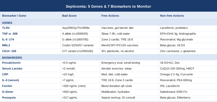
7 Biomarkers to Track for Septicemia Risk and Recovery
Biomarker monitoring in septicemia is not limited to the ICU. For post-sepsis survivors managing prolonged recovery, for high-risk patients with chronic illness or immunosuppression, and for clinicians trying to catch deterioration before it becomes irreversible, a targeted panel of blood markers provides information that temperature, heart rate, and blood pressure simply cannot. The seven biomarkers below represent the most informative, best-supported options for both early detection and longitudinal monitoring.
1. Procalcitonin (PCT)
Why it matters: Procalcitonin is a precursor to calcitonin that healthy tissue produces in negligible quantities. When bacteria trigger a systemic inflammatory response, PCT levels surge — sometimes by a factor of thousands — while remaining low or minimally elevated in viral infections and non-infectious inflammatory states. This selectivity makes PCT one of the most specific early indicators of bacterial sepsis currently available.
Serial PCT measurements over 24 to 48 hours are more informative than a single value. A falling PCT suggests effective antimicrobial treatment; a plateau or rise signals inadequate response or an uncontrolled infectious source. Multiple meta-analyses support PCT-guided antibiotic de-escalation as a strategy that reduces antibiotic exposure without increasing mortality.
How to measure it: Standard blood draw processed at most hospital and reference laboratories. Cost: $30–$80 with insurance in the U.S., $50–$150 out-of-pocket. Point-of-care PCT analyzers are increasingly available in emergency settings for faster turnaround.
Reference ranges: Below 0.1 ng/mL (low bacterial infection risk); 0.1–0.25 ng/mL (borderline); above 0.5 ng/mL (high suspicion); above 2 ng/mL (sepsis likely).
If PCT is elevated — the plan without supplements
A rising PCT outside a hospital setting is a medical emergency signal, not a self-management opportunity. Seek immediate medical evaluation. In post-sepsis outpatient monitoring, an upward PCT trend during follow-up warrants urgent clinical reassessment: review for occult infection sources, evaluate antibiotic adequacy, and reassess fluid status and systemic response. Serial testing every 24–48 hours during active treatment provides the most actionable trajectory data.
If PCT is elevated — the plan with supplements
Vitamin D deficiency has been associated in multiple observational studies with higher PCT levels during infection and worse sepsis outcomes. Optimizing serum 25-OH-D to 50–80 ng/mL through vitamin D3 supplementation (2,000–5,000 IU daily with K2 MK-7 at 100 mcg) may support baseline PCT regulation. Test 25-OH-D quarterly and adjust dose accordingly. Zinc (15–30 mg daily with food, as zinc bisglycinate or zinc picolinate) supports antimicrobial innate immune function, and deficiency has been linked to exaggerated inflammatory signaling. These are supportive baseline measures; they do not replace medical management of active sepsis.
2. Serum Lactate
Why it matters: Lactate accumulates when cells shift from aerobic to anaerobic metabolism — a reliable cellular signal that tissues are not receiving adequate oxygen. In septicemia, elevated lactate indicates tissue hypoperfusion: even when blood pressure appears acceptable, high lactate reveals that perfusion is failing at the cellular level. The Surviving Sepsis Campaign guidelines treat lactate as a core diagnostic and management variable, recommending measurement within one hour of suspected sepsis presentation.
Lactate above 4 mmol/L in the context of infection is now considered a criterion for septic shock regardless of blood pressure status. Lactate-guided resuscitation has been shown in randomized trials to improve outcomes compared to resuscitation guided by oxygen delivery targets alone.
How to measure it: Measured from arterial or venous blood in emergency and hospital settings; handheld point-of-care lactate meters (such as the Lactate Pro or StatStrip) are increasingly available for rapid testing in clinic or pre-hospital settings. Cost: $20–$60 when ordered separately; often included in critical care panels. Home monitoring is emerging but not yet standard practice.
Reference ranges: Below 2 mmol/L (normal); 2–4 mmol/L (elevated, warrants clinical attention); above 4 mmol/L (severe, meets septic shock criteria).
If lactate is elevated — the plan without supplements
In acute sepsis, high lactate requires immediate medical intervention: IV fluids, oxygen supplementation, source control, and antibiotics. In the post-sepsis recovery setting, persistent mild lactate elevation (2–3 mmol/L) may reflect mitochondrial dysfunction — a recognized post-sepsis complication affecting a significant proportion of ICU survivors. Non-supplement interventions include graduated aerobic exercise (starting at very low intensity, 10–15 minutes walking daily), strict pacing strategies to avoid metabolic overload, and sleep optimization, which is essential for mitochondrial biogenesis and recovery.
If lactate is elevated — the plan with supplements or equipment
Coenzyme Q10 (100–300 mg daily in divided doses with fat-containing meals) supports mitochondrial electron transport chain function and has evidence in mitochondrial dysfunction states. D-Ribose (5 g twice daily) provides substrate for ATP regeneration and has been studied in post-viral and post-critical illness fatigue with mitochondrial involvement. For post-sepsis patients with significant mitochondrial impairment, hyperbaric oxygen therapy (HBOT) — typically 20–40 sessions at 1.5–2.0 ATA — has early-stage evidence for improving mitochondrial function and cellular oxygenation. CoQ10: continuous use with 25-OH-D monitoring; HBOT in discrete 30–40 session treatment blocks with clinical reassessment between courses.
3. C-Reactive Protein (CRP)
Why it matters: CRP is synthesized by the liver in response to IL-6 signaling and rises within hours of an inflammatory trigger. While less pathogen-specific than PCT — it rises in both bacterial and viral infection, autoimmune flares, and tissue injury — CRP offers an accessible, affordable, and meaningful measure of systemic inflammation load. Serial CRP is especially valuable during sepsis recovery: a failure to normalize CRP after completing antibiotic therapy is a clinical warning sign for retained infection focus, inadequate source control, or an inflammatory complication such as metastatic abscess or endocarditis.
How to measure it: Standard blood panel, universally available. High-sensitivity CRP (hs-CRP) is preferred for cardiovascular risk and low-grade chronic inflammation monitoring; standard CRP is used in acute infectious settings. Cost: $15–$40 typically; often included in comprehensive metabolic panels. Recommended to track as a series with 2–4 week intervals during post-sepsis recovery.
Reference ranges: Below 1 mg/L (low inflammatory burden); 1–10 mg/L (moderate elevation); above 10 mg/L (active infection or significant inflammation likely); above 100 mg/L (severe bacterial infection, sepsis-consistent).
If CRP is persistently elevated — the plan without supplements
Persistently elevated CRP in a post-sepsis patient warrants imaging — CT abdomen-pelvis, transthoracic echocardiogram — to rule out retained infection focus before attributing elevation to non-infectious inflammation. Once infectious causes are excluded, dietary approaches with meaningful CRP evidence include elimination of ultra-processed foods, refined carbohydrates, and industrial seed oils, all of which drive sustained IL-6 and CRP production. Mediterranean dietary pattern adoption has been associated with measurably lower hs-CRP in large observational studies. Brief cold water exposure (10–15 minutes at 15–17°C, 3–4 times per week) activates anti-inflammatory pathways through norepinephrine-mediated NF-κB suppression.
If CRP is persistently elevated — the plan with supplements or equipment
Omega-3 fatty acids at therapeutic doses (EPA+DHA combined: 2–4 g daily with a fat-containing meal, taken as triglyceride-form fish oil or algal oil) have among the most robust supplement evidence for CRP and broader inflammatory marker reduction. Re-test hs-CRP at 8–12 weeks to assess response. Curcumin with piperine (500–1,000 mg curcuminoid complex twice daily) has multiple RCT-level evidence for CRP reduction; use enhanced bioavailability forms. Side effects: mild GI discomfort at high doses; avoid combining with anticoagulants without medical oversight. Berberine (500 mg twice daily with meals) reduces NF-κB pathway activation — cycle 8 weeks on, 4 weeks off to prevent receptor downregulation.
4. Interleukin-6 (IL-6)
Why it matters: IL-6 is one of the earliest pro-inflammatory cytokines released during bacterial infection, rising within 30–90 minutes of pathogen exposure — well before PCT and CRP. This kinetic advantage makes IL-6 one of the most sensitive early warning markers for impending septic cascade. Mechanistically, IL-6 is also the upstream driver of CRP production, acute-phase reactant synthesis, fever generation, and neutrophil activation, making it a central hub in the inflammatory architecture of septicemia. The success of IL-6 receptor antagonists such as tocilizumab in treating cytokine storm syndromes underscores the clinical weight of this pathway.
How to measure it: Specialized laboratory testing; not yet universally available as a point-of-care assay but increasingly offered by reference laboratories. Processing time from a standard blood draw: 24–48 hours at most academic medical centers. Cost: $60–$150 out-of-pocket. Increasingly included in sepsis biomarker panels at academic and advanced care centers. Consider ordering alongside PCT for high-stakes diagnostic situations.
Reference ranges: Below 7 pg/mL (healthy adults); 7–50 pg/mL (mild elevation); above 100 pg/mL (significant systemic inflammation); above 1,000 pg/mL (cytokine storm territory, requiring immediate specialist involvement).
If IL-6 is elevated — the plan without supplements
In active sepsis with very high IL-6 levels, anti-IL-6 therapy is a physician-managed decision. Outside acute care, persistently elevated IL-6 in post-sepsis patients suggests chronic inflammatory dysregulation requiring structured intervention. Lifestyle measures with the strongest IL-6 evidence include time-restricted eating (16:8 or 18:6 windows), Zone 2 aerobic training (3–5 sessions weekly at moderate intensity, maintaining conversational pace), and sleep quality optimization. Notably, sleep architecture matters as much as duration — fragmented sleep with reduced deep sleep stages directly and measurably elevates IL-6, even when total sleep time appears adequate.
If IL-6 is elevated — the plan with supplements or equipment
Resveratrol (500 mg daily standardized extract with a fat-containing meal) inhibits NF-κB and STAT3 signaling — two of the central transcription factors driving IL-6 production. Human studies show IL-6 reductions with consistent use. Cycle: 8–12 weeks on, 4 weeks off. Tocotrienols (the less common vitamin E isomers, 150–300 mg daily) have shown anti-IL-6 effects in small human trials, particularly in the context of chronic inflammation. Palmitoylethanolamide (PEA, 600–1,200 mg daily) modulates endocannabinoid signaling in ways that reduce mast cell activation and inflammatory cytokine release, including IL-6. Red light photobiomodulation (660–850 nm, 10–20 minutes daily over relevant body areas) has emerging clinical evidence for systemic cytokine burden reduction.
5. Ferritin and Hyperferritinemia
Why it matters: Ferritin is primarily known as an iron storage protein, but it is also an acute-phase reactant that rises sharply during infection and inflammatory activation. In severe sepsis and septic shock, ferritin levels can reach extreme heights — sometimes exceeding 10,000 ng/mL — a pattern associated with macrophage activation syndrome (MAS) and hemophagocytic lymphohistiocytosis (HLH), both potentially life-threatening inflammatory states that can be triggered or worsened by septicemia. Even moderately elevated ferritin (above 500 ng/mL) in a post-sepsis context can signal persistent macrophage hyperactivation and is worth tracking over months of recovery.
How to measure it: Standard blood test, included in most iron status panels. Cost: $20–$50. Critically, ferritin should always be interpreted alongside serum iron, total iron-binding capacity (TIBC), and transferrin saturation to distinguish iron overload from inflammation-driven elevation — both produce high ferritin through different mechanisms.
Reference ranges: 20–200 ng/mL (women); 20–300 ng/mL (men). Functional medicine practitioners often flag values above 100–150 ng/mL in women and 150–200 ng/mL in men as worth investigating in the presence of inflammatory conditions.
If ferritin is critically elevated in acute sepsis
Ferritin above 1,000–10,000 ng/mL during active sepsis should prompt evaluation for HLH and MAS by hematology. This is a medical emergency requiring specialist input, not a self-management scenario.
If ferritin is persistently mildly elevated — the plan without supplements
Regular blood donation (every 8–12 weeks for eligible individuals) is one of the most effective and underutilized strategies for reducing ferritin when true iron overload contributes to the elevation. Research in cardiovascular and metabolic contexts consistently confirms its clinical benefit. Dietary adjustments — reducing red meat and supplemental iron while increasing polyphenol-rich foods consumed with meals (coffee, tea, dark chocolate, legumes) that mildly compete with non-heme iron absorption — gradually reduce ferritin over months.
If ferritin is persistently mildly elevated — the plan with supplements or equipment
IP6 (inositol hexaphosphate, 1–2 g on an empty stomach before meals) acts as an iron chelator and has evidence for reducing ferritin stores over time. Lactoferrin (200–600 mg daily) modulates iron homeostasis and exerts direct anti-inflammatory effects that may reduce the inflammatory component of elevated ferritin. Cycle both 8–12 weeks with repeat ferritin testing. Important caveat: when ferritin elevation is driven purely by inflammation rather than iron overload, iron-chelating strategies may not be appropriate — always verify with a full iron panel and discussion with a physician before initiating.
6. D-Dimer
Why it matters: D-dimer is a fibrin degradation product released when the body breaks down clots. In septicemia, coagulation activation is not a complication — it is a hallmark feature. The pathogen-driven inflammatory cascade triggers widespread systemic clotting, and when this process is not controlled, it progresses to disseminated intravascular coagulation (DIC) — one of the most feared consequences of septic shock. Elevated D-dimer in sepsis signals activation of the entire coagulation-fibrinolysis axis and predicts organ injury severity and mortality risk more accurately than many standard laboratory markers.
How to measure it: Standard coagulation test available at all clinical laboratories. Cost: $30–$80. Most informative when ordered alongside PT/INR, aPTT, and fibrinogen for full coagulation status characterization. Age-adjusted D-dimer cutoffs (age × 10 ng/mL for patients over 50) are increasingly used to improve specificity.
Reference ranges: Below 500 ng/mL or 0.5 µg/mL FEU (normal, lab-specific ranges apply); above 500 ng/mL warrants clinical investigation in the context of infection or post-sepsis monitoring.
If D-dimer is elevated — the plan without supplements
Elevated D-dimer in a confirmed or suspected sepsis context requires immediate evaluation for thrombotic complications including pulmonary embolism and deep vein thrombosis. Non-pharmacological coagulation risk management in the post-sepsis recovery phase includes: aggressive early mobilization as soon as clinically stable (even seated leg exercises during bed rest reduce coagulation activation significantly), robust hydration, and elimination of known pro-coagulant exposures including smoking and prolonged immobility. Physical therapy referral is appropriate for any post-sepsis patient with mobility limitation.
If D-dimer is elevated — the plan with supplements or equipment
Nattokinase (2,000–4,000 FU daily, away from meals) is a fibrinolytic enzyme derived from fermented soybeans with multiple small human trials demonstrating D-dimer reduction. Critical safety note: nattokinase has meaningful anticoagulant activity and must not be combined with pharmaceutical anticoagulants (warfarin, heparin, DOACs) without direct medical supervision. Cycle: 8–12 weeks on with repeat D-dimer testing before re-initiation. Lumbrokinase (30–60 mg twice daily) is an earthworm-derived fibrinolytic enzyme with slightly stronger evidence in post-COVID and cardiovascular thrombotic states, increasingly discussed alongside nattokinase in functional cardiology contexts. Both carry bleeding risk and require medical oversight.
7. Presepsin (sCD14-ST)
Why it matters: Presepsin is a relatively new biomarker — a soluble fragment of the CD14 co-receptor shed from monocytes and macrophages during phagocytosis of bacteria. It rises faster than both PCT and CRP in the early hours of bacterial infection and has shown strong diagnostic accuracy for sepsis in growing clinical evidence. Unlike PCT, presepsin reflects the direct cellular phagocytic response to pathogens rather than a systemic hormonal cascade, making it mechanistically complementary. Studies comparing presepsin to PCT suggest that combining both markers substantially improves early sepsis detection accuracy over either alone.
How to measure it: Requires specialized laboratory analysis; not yet universally available but increasingly offered by academic and advanced reference laboratories. A point-of-care PATHFAST presepsin assay is established in some European and Japanese centers. Cost: $80–$150. Given its comparative novelty, presepsin is most valuable as an addition to standard biomarker panels in high-stakes diagnostic situations or for close post-sepsis monitoring.
Reference ranges: Below 317 pg/mL (generally considered normal in most studies); above 600 pg/mL (associated with sepsis); above 1,000 pg/mL (severe sepsis with poor prognostic implications in multiple cohorts).
If presepsin is elevated — the plan without supplements
A standalone elevated presepsin in the clinical context of infection suspicion should prompt comprehensive sepsis workup including blood cultures, CBC with differential, comprehensive metabolic panel, and PCT. In outpatient post-sepsis monitoring, persistently elevated presepsin may suggest low-grade ongoing bacteremia or persistent macrophage dysregulation — circumstances warranting infectious disease specialist review and possible repeat imaging to rule out occult infection sources.
If presepsin is elevated — the plan with supplements
Given presepsin's direct relationship to macrophage activation and phagocytic activity, supporting healthy macrophage regulation is the relevant biological target. Beta-glucan (500 mg daily from standardized yeast or oat-derived sources) has human evidence for macrophage priming and functional enhancement without excessive inflammatory activation. Elderberry standardized extract (500–1,000 mg daily during active infection risk periods) supports innate immune function at the mucosal level. These are immune support measures; neither has direct presepsin-lowering evidence in controlled trials as of current literature, and both are best understood as complementary rather than primary interventions.
With the seven core biomarkers covered, it is worth stepping back to consider the upstream question: why do some individuals respond to an identical pathogen load with a runaway inflammatory cascade, while others mount a controlled, effective response? Part of the answer lies in the genetics of immune recognition and cytokine regulation.
The Genetic Side: 5 Genes That Shape Sepsis Susceptibility
Genetic variants do not determine outcomes, but they do shape immune response tendencies in ways that are increasingly well characterized for septicemia. The five genes below have meaningful human evidence — not just animal models or in vitro data — for their role in sepsis susceptibility, inflammatory severity, and recovery. The most useful application of this genetic information is as a layer of context on top of biomarker data: a high-risk genetic profile combined with elevated IL-6 and PCT tells a different story than either variable alone.
Gene 1: TLR4 (Toll-Like Receptor 4)
TLR4 is the primary pattern recognition receptor for lipopolysaccharide (LPS) — the endotoxin present on the outer membrane of gram-negative bacteria. When TLR4 detects LPS, it triggers the innate immune cascade that drives both bacterial clearance and, if unchecked, the cytokine storm of septic shock. Loss-of-function variants in TLR4 — particularly Asp299Gly (rs4986790) and Thr399Ile (rs4986791) — impair LPS recognition and have been associated in multiple human studies with increased susceptibility to gram-negative bacteremia and septic shock. Carriers of these variants may fail to mount adequate early innate immune responses to gram-negative bacteria, allowing infection to establish and escalate more readily before the adaptive immune system can contribute.
If TLR4 variant is present — the plan without supplements
Prevention is the core strategy. Prioritize aggressive infection management at every stage: prompt wound care, timely antibiotic treatment for confirmed bacterial infections, and up-to-date vaccination including pneumococcal, meningococcal, and annual influenza vaccines. Since gut-derived gram-negative bacteria are a major source of LPS translocation into the bloodstream — especially during illness, surgery, or intestinal injury — maintaining excellent gut barrier integrity is directly relevant. Practical means: a diverse, fiber-rich diet (30+ g daily from varied plant sources), minimal alcohol (a direct gut barrier disruptor), and cautious NSAID use. These are ongoing lifestyle habits, not temporary interventions.
If TLR4 variant is present — the plan with supplements
Lactoferrin (300–600 mg daily) binds to LPS directly, neutralizing its pro-inflammatory potential and reducing bacterial translocation across mucosal barriers — a mechanism directly relevant to TLR4-LPS biology. Phosphatidylcholine (1–3 g daily) supports gut mucosal integrity and the protective mucus layer over intestinal epithelium. Multi-strain probiotics emphasizing Lactobacillus rhamnosus and Bifidobacterium longum have human evidence for reducing gut permeability and systemic LPS burden. Cycle probiotics 8 weeks on, assess, then continue or rotate strains. Side effects: initial bloating common; generally well tolerated after the first 10–14 days.
Gene 2: TNF-α (rs1800629, -308G/A Promoter Variant)
Tumor Necrosis Factor-alpha is a central cytokine in the inflammatory cascade of septicemia. The -308G/A variant (rs1800629) in the TNF-α promoter region is associated with increased TNF-α transcriptional activity: carriers of the A allele tend to produce higher TNF-α in response to infection. While elevated TNF-α improves bacterial killing in the early infection phase, it also dramatically raises the risk of excessive inflammatory escalation, cytokine storm, and organ-level damage during septicemia. Multiple meta-analyses have associated this variant with increased sepsis severity and mortality, making it one of the better-replicated genetic risk signals in sepsis biology.
If TNF-α variant is present — the plan without supplements
The core strategy is reducing baseline inflammatory tone so that when an infection triggers TNF-α production, the body is starting from a lower inflammatory setpoint. TNF-α levels follow circadian biology closely — they are significantly elevated by sleep deprivation, circadian rhythm disruption, and chronic psychological stress. Structural interventions: consistent 7–9 hours of sleep at regular times; cold water immersion (3–4 sessions per week at 10–15°C for 10–15 minutes) which activates norepinephrine-mediated NF-κB suppression; and time-restricted eating within an 8–10 hour window, which supports circadian-aligned inflammatory gene expression patterns.
If TNF-α variant is present — the plan with supplements
EPA and DHA from fish or algal oil (3–4 g combined daily, triglyceride form) directly suppress TNF-α transcription and release through competitive eicosanoid pathway modulation — this is one of the most robustly documented supplement-to-cytokine relationships in the human literature. Andrographis paniculata standardized extract (300–400 mg twice daily) has human evidence for TNF-α modulation during infectious and inflammatory states. Cycle: use during active infection risk periods or ongoing for high-risk genetic profiles; 8-week cycles with 4-week breaks. Palmitoylethanolamide (PEA, 600–1,200 mg daily) reduces mast cell and microglial TNF-α release through PPAR-α activation. Side effects: generally minimal across all three; fish oil may cause mild GI discomfort at high doses and has additive anticoagulant effects with blood thinners.
Gene 3: IL-6 (rs1800795, -174G/C Promoter Variant)
The IL-6 promoter variant rs1800795 influences how much IL-6 is produced under inflammatory stimulation. The G allele is consistently associated with higher IL-6 output in response to infection and inflammatory triggers — a pattern that parallels the TNF-α -308 story. Higher IL-6 production can accelerate early immune response (potentially beneficial) while simultaneously raising risk of inflammatory overshoot and organ-level damage during severe infection (potentially harmful). The clinical net effect depends heavily on the type and burden of infecting organism. Cohort studies examining this variant in sepsis have produced mixed direction of effect, but consistently show that it is biologically active in the context of bacterial infection.
If IL-6 rs1800795 G allele is present — the plan without supplements
Lifestyle strategies overlap substantially with the TNF-α section: circadian-aligned eating, sleep architecture optimization, and structured aerobic exercise. One nuance worth knowing: strength training acutely raises IL-6 during and immediately after sessions, but produces long-term reductions in baseline IL-6 over weeks of consistent training — a counterintuitive but well-documented adaptation. Anti-inflammatory dietary patterns — low refined carbohydrate, high polyphenol, omega-3 enriched, and diverse in plant fiber — reduce chronic IL-6 stimulation at the gene expression level over months of consistent adherence.
If IL-6 rs1800795 G allele is present — the plan with supplements
Resveratrol (500 mg daily with a fat-containing meal, standardized trans-resveratrol) inhibits both NF-κB and STAT3 — two of the three major transcription factor pathways driving IL-6 gene expression. Cycle 8–12 weeks on, 4 weeks off. Magnesium glycinate (300–400 mg nightly) has measurable IL-6-reducing effects in human studies, likely through influence on inflammatory gene expression and mitochondrial function. Tart cherry extract (480 mg standardized anthocyanin extract daily) has IL-6-reducing evidence in athletic recovery and osteoarthritis contexts. Side effects for all three: minimal; resveratrol has mild potential for interaction with CYP3A4-metabolized medications at higher doses.
Gene 4: MBL2 (Mannose-Binding Lectin 2)
Mannose-binding lectin activates the lectin pathway of complement — one of the three routes through which complement can be initiated on the surface of pathogens. MBL recognizes specific sugar patterns on bacterial, fungal, and viral surfaces, binding and triggering complement activation that promotes opsonization and phagocytosis. Loss-of-function variants in MBL2 — codon 52 (rs5030737), codon 54 (rs1800450), and codon 57 (rs1800451) — lead to low or absent circulating MBL, significantly impairing this arm of first-line complement defense. Human studies have consistently associated MBL deficiency with increased risk of septicemia, meningococcal disease, and invasive bacterial infection, particularly in immunocompromised adults and during early childhood.
If MBL2 loss-of-function variant is present — the plan without supplements
Vaccination is disproportionately important for MBL-deficient individuals, because the pathogens most covered by MBL-mediated complement activity — meningococcus, pneumococcus, Haemophilus influenzae type b — are precisely those targeted by available vaccines. Ensure coverage includes MenACWY, MenB, PCV20 or PPSV23, and Hib as appropriate for age and risk. Avoid immunosuppressant medications unless medically necessary, and disclose MBL deficiency status to any treating physician before initiating immunosuppressive therapy. Early and aggressive antibiotic treatment at the first sign of confirmed bacterial infection — rather than watchful waiting — is warranted for MBL-deficient individuals.
If MBL2 loss-of-function variant is present — the plan with supplements
Beta-glucan (500–1,000 mg daily, yeast-derived standardized extract) activates macrophages and natural killer cells through Dectin-1 receptors — a complement-independent innate immune activation pathway that can partially compensate for reduced lectin-pathway complement activity. Vitamin D3 (2,000–4,000 IU daily) supports cathelicidin and defensin antimicrobial peptide production, providing a layer of bacterial defense that operates entirely outside complement pathways. Colostrum (2–4 g daily) delivers immunoglobulins including IgG and secretory IgA that support mucosal defense when complement-mediated opsonization is impaired. Monitor serum 25-OH-D quarterly; target 50–70 ng/mL.
Gene 5: CD14 (rs2569190, -159C/T Promoter Variant)
CD14 functions as a co-receptor alongside TLR4 in the recognition of LPS from gram-negative bacteria. Beyond its membrane-bound form, CD14 is also shed into circulation as soluble CD14 — and presepsin (sCD14-ST), covered in the biomarker section, is a direct fragment of sCD14. This creates a direct genetic-to-biomarker connection: individuals with CD14 variants that alter expression levels will have different baseline presepsin biology and potentially different early sepsis biomarker trajectories. The -159C/T promoter variant (rs2569190) influences CD14 expression and soluble CD14 concentration. Evidence on direction of effect is more nuanced than for TLR4 or TNF-α — some studies show increased, others show altered susceptibility depending on infecting organism type — making this variant most informative as part of a broader genetic immune profile rather than in isolation.
If CD14 variant is present — the plan without supplements
Given the mechanism, the most rational focus for CD14 variant carriers is reducing chronic LPS translocation from the gut — the most significant modifiable driver of chronic CD14 and TLR4 pathway activation outside of acute infection. Gut microbiome diversity is the primary lever: dietary fiber from at least 30 different plant sources per week provides the substrate for LPS-sequestering microbiome species and short-chain fatty acid producers that strengthen gut barrier function. Alcohol reduction is high priority — even moderate alcohol consumption measurably increases gut permeability and LPS translocation within hours. Judicious antibiotic use (necessary courses used fully, unnecessary courses declined) preserves the microbiome diversity that is central to gut barrier integrity.
If CD14 variant is present — the plan with supplements
Zinc carnosine (75 mg twice daily) has human RCT evidence for reducing intestinal permeability and LPS translocation — mechanistically directly relevant to the CD14-LPS recognition axis. L-glutamine (5 g twice daily in divided doses, mixed in water away from meals) is the primary fuel for enterocytes and supports tight junction protein expression. Sodium butyrate (1–2 g daily) is absorbed by colonocytes and upregulates tight junction proteins including claudin and occludin, reducing paracellular LPS leakage into systemic circulation. Cycle zinc carnosine in 8–12 week courses; L-glutamine and butyrate can be maintained continuously with periodic gut health reassessment.
Immune Resilience and Infection Biology: 10 Key Insights from the Huberman Lab
Andrew Huberman has covered immune function, inflammatory biology, and the nervous system's role in infection resistance across multiple podcast episodes. While no single episode is dedicated exclusively to septicemia, the underlying science is directly applicable to both sepsis susceptibility and post-sepsis recovery. The following ten insights represent the most impactful takeaways for individuals tracking the biomarkers and genetics covered in this article.
1. The Vagus Nerve Is a Direct Inflammatory Suppressor
The cholinergic anti-inflammatory reflex — the pathway through which vagal nerve activation suppresses macrophage TNF-α and IL-6 production — is one of the most significant connections between the nervous system and sepsis biology. Practices that increase vagal tone (slow diaphragmatic breathing, cold exposure, and meditation) act directly on the same cytokine pathways that drive septic cascade. This is not metaphorical — it operates through documented acetylcholine-mediated suppression of NF-κB transcription in macrophages.
2. Sleep Is the Most Powerful Immune Modulator Available Without a Prescription
Even a single night of sleep restriction below 6 hours measurably reduces NK cell activity and raises baseline IL-6 and TNF-α. Huberman emphasizes that for post-sepsis patients — or anyone seeking to reduce sepsis vulnerability — consistent 7–9 hour sleep is the foundation from which every other intervention operates. Sleep also drives growth hormone release overnight, which promotes immune cell reconstitution and tissue repair following severe infection.
3. Cold Exposure Activates Anti-Inflammatory Gene Programs
Brief cold water immersion activates norepinephrine release, which suppresses NF-κB — the master transcription regulator of TNF-α, IL-6, and IL-1β production. Huberman references research suggesting that approximately 11 minutes of cold exposure per week, distributed across 3–4 sessions, is sufficient to produce measurable anti-inflammatory and autonomic nervous system effects without excessive physiological stress load.
4. Zone 2 Cardio Reduces Baseline Systemic Inflammation Over Weeks
Chronic low-grade inflammation is a risk amplifier for sepsis severity at the individual level. Huberman summarizes research showing that 150–200 minutes weekly of Zone 2 aerobic exercise — conversation-pace cardio that maintains aerobic metabolism without crossing into anaerobic zones — measurably lowers hs-CRP, IL-6, and TNF-α over 8–12 weeks of consistent practice. This reduction in inflammatory baseline directly reduces the setpoint from which a septic cascade would escalate.
5. Morning Sunlight Anchors the Immune-Circadian Interface
Circadian rhythm drives expression of nearly every immune gene, including those encoding TNF-α, IL-6, and toll-like receptors. Huberman emphasizes morning sunlight exposure (10–30 minutes within two hours of waking) as the primary circadian anchor that keeps immune gene expression patterns aligned with appropriate inflammatory rhythms — high cortisol and anti-inflammatory tone in the morning, immune activation and recovery processes overnight.
6. Gut Microbiome Diversity Is Upstream of Immune Regulation
Multiple Huberman episodes cover the gut-immune axis, highlighting that microbiome diversity directly supports regulatory T cell populations that prevent immune overactivation during infection. The practical takeaway: 30+ different plant species weekly, inclusion of fermented foods (kefir, kimchi, tempeh), and avoidance of unnecessary antibiotics are not generic wellness advice — they directly support the regulatory immune balance that determines whether an infection triggers a controlled or runaway inflammatory response.
7. Chronic Stress Cortisol Dysregulation Impairs Both Immune Activation and Resolution
Huberman covers the cortisol-immune tradeoff: cortisol that is appropriately high in the morning and low by evening supports healthy immune function, while chronic daytime cortisol elevation (from sustained psychological stress or circadian disruption) suppresses both NK cell activity and antimicrobial peptide production. For individuals with TNF-α or IL-6 high-producer genotypes, addressing chronic stress is not optional — it directly influences the inflammatory gene expression that makes their immune response more likely to overshoot.
8. Nasal Breathing Provides Direct Antibacterial Defense
Huberman discusses the production of nitric oxide in the nasal passages during nasal breathing — a molecule with documented direct antibacterial and antiviral properties that protects against respiratory pathogen entry. Since respiratory tract infections are a common precursor to bacteremia in at-risk individuals, optimizing nasal breathing through addressing chronic nasal obstruction and avoiding habitual mouth breathing is a practical upstream bacteremia risk reduction strategy.
9. Social Connection Has Measurable Immunological Consequences
Loneliness and social isolation reliably increase inflammatory markers including IL-6 and CRP, and measurably impair NK cell function in human studies. For post-sepsis survivors managing prolonged recovery — a period often associated with significant social withdrawal — social reintegration is not purely a mental health consideration. It is an immunological one, with direct downstream effects on the inflammatory biomarkers covered in this article.
10. Deliberate Breathing Protocols Can Acutely Modulate Cytokine Response
Huberman covers the striking human trial in which participants trained in Wim Hof breathing techniques showed dramatically blunted cytokine responses — including TNF-α and IL-6 — when administered standardized bacterial endotoxin compared to untrained controls. This protocol involves cycles of voluntary hyperventilation followed by breath retention, producing alkalotic shifts and sympathetic activation that acutely suppress inflammatory cytokine release. For individuals with TNF-α or IL-6 high-producer genotypes, this represents a potentially important tool for managing acute inflammatory overshoots — though it should be practiced under guidance and avoided in cardiac, pulmonary, or seizure-risk conditions.
Complementary Approaches for Post-Sepsis Recovery and Immune Resilience
The following four evidence-reviewed modalities address different aspects of post-sepsis biology: psychological trauma processing, gut immune reconstitution, respiratory recovery, and nervous system regulation. Each has human clinical evidence — not just theoretical plausibility — for its relevant mechanisms in ICU recovery or sepsis-related immune reconstitution.
Mindfulness Meditation and MBSR
Post-sepsis syndrome — characterized by cognitive impairment, PTSD symptoms, immune dysregulation, and persistent fatigue — affects a substantial proportion of ICU survivors. Mindfulness-based stress reduction (MBSR) has a well-developed evidence base for reducing inflammatory biomarkers including CRP and IL-6, and for addressing the psychological trauma component of critical illness recovery. Meta-analyses of MBSR trials in inflammatory conditions have found meaningful reductions in CRP and IL-6 with consistent practice — directly applicable to post-sepsis inflammatory dysregulation.
The standard MBSR protocol is an 8-week structured program involving 2.5 hours of group practice per week plus daily home practice. It combines body scan meditation, seated attention practice, and gentle mindful movement. Post-sepsis adaptations for cognitive fatigue — shorter sessions of 10–15 minutes initially, progressing gradually — are common in rehabilitation settings and equally evidence-supported.
In practice, MBSR is most accessibly delivered through hospital wellness programs, academic medical centers, or validated online programs. Daily consistency matters more than session duration in the evidence base — even 10-minute daily body scan practice produces measurable HRV improvement and cortisol normalization within 4 weeks of consistent practice.
Microbiome-Directed Therapies
The gut microbiome is now recognized as a central regulator of systemic immune function, and its disruption — near-universal after the broad-spectrum antibiotics and physiological stress of septicemia — is a key driver of post-sepsis immune dysregulation and delayed recovery. Critical care research has shown that sepsis causes rapid and profound microbiome disruption, with loss of diversity and pathobiont overgrowth that can persist for months after hospital discharge.
Targeted microbiome restoration in post-sepsis patients includes: high-fiber dietary rebuilding (30+ plant species weekly, diverse prebiotic fiber types including inulin, pectin, and resistant starch), multi-strain probiotic supplementation emphasizing Lactobacillus rhamnosus GG and Bifidobacterium longum at minimum 50 billion CFU daily, and in more challenging cases, spore-forming probiotic species (Bacillus subtilis, Bacillus coagulans) that survive the disrupted post-antibiotic gut environment more reliably than fragile Lactobacillus strains.
Realistically, microbiome restoration following septicemia is a months-long process requiring patience and sequential layering. A practical protocol: begin with multi-strain probiotics and dietary fiber expansion in weeks 1–4 post-discharge; add prebiotic supplementation (inulin 10–15 g daily or partially hydrolyzed guar gum 5–10 g daily) as tolerated from weeks 4–8; introduce fermented foods from week 6 onward at gradual quantities. Microbiome testing at 3 months post-sepsis (Genova GI-MAP or equivalent) can objectively assess recovery trajectory and guide further targeted intervention.
Breathing-Based Therapies
Respiratory dysregulation is common after septicemia, arising from both direct lung involvement and from autonomic nervous system disruption that persists well beyond the acute phase. Breathing-based interventions address both dimensions: improving respiratory muscle function and lung capacity while simultaneously activating the vagal anti-inflammatory pathways that suppress cytokine burden. Randomized trials of inspiratory muscle training in critically ill patients have shown significant improvements in respiratory muscle strength and reduced time to liberation from mechanical ventilation.
The most evidence-supported protocol for post-sepsis respiratory and autonomic recovery combines diaphragmatic breathing training (10–15 minutes daily at 5–6 breaths per minute — the physiological resonance frequency that maximally activates vagal tone) with inspiratory muscle training using a threshold resistance device starting at 30% of maximal inspiratory pressure, progressing weekly. The extended exhale technique — inhale 4 counts, exhale 6–8 counts — can be practiced anywhere without equipment and has the most immediate parasympathetic activating effect.
Practically, breathing practice can begin as early as 2 weeks post-hospital discharge for most patients. Start with 5-minute sessions twice daily and progress over 4 weeks to 15-minute sessions. Threshold inspiratory training should initially be supervised by a physiotherapist when significant respiratory muscle weakness is present. Daily diaphragmatic breathing has no meaningful side effects and is appropriate even for severely deconditioned post-sepsis patients who cannot yet engage in aerobic exercise.
Music Therapy
ICU survivors of septicemia frequently experience significant psychological trauma including PTSD, depression, and delirium aftereffects that complicate both psychological recovery and immune reconstitution. Music therapy in ICU and post-ICU settings has been examined in multiple randomized trials for effects on anxiety, pain perception, cortisol levels, and sedation requirements. Meta-analyses of music interventions in ICU settings consistently find significant reductions in patient anxiety, sedative medication needs, and cortisol levels — all of which have downstream consequences for immune function and recovery trajectory.
The protocol with the strongest ICU evidence involves patient-preferred music delivered via headphones for 30–60 minutes daily, most commonly during rest periods and before procedures. Slow, personally meaningful music (generally 60–70 BPM or below) produces the most consistent autonomic calming and cortisol-reducing effects. Post-ICU, structured music therapy sessions with a trained music therapist can extend these benefits into emotional processing and nervous system regulation work.
For post-sepsis patients at home, using dedicated playlists of personally meaningful, slow-tempo music for 20–30 minutes before sleep supports both cortisol normalization and sleep quality improvement — with direct benefits to immune reconstitution. Music therapy practitioners are increasingly available in rehabilitation and hospital outpatient settings; referral is particularly appropriate for post-sepsis patients with documented psychological sequelae or prolonged delirium recovery.
Progressive Muscle Relaxation
Progressive muscle relaxation (PMR) involves the systematic deliberate tensing and releasing of major muscle groups throughout the body, producing deep physiological relaxation that measurably reduces cortisol, blood pressure, and inflammatory cytokine burden. For post-sepsis patients managing chronic fatigue, anxiety, and inflammatory dysregulation — particularly those who cannot yet engage in aerobic exercise — PMR offers a structured, teachable relaxation technique that can be practiced independently within days of learning. Human trials have shown meaningful reductions in IL-6 and CRP with regular PMR practice in inflammatory conditions, and it is specifically recommended in post-ICU rehabilitation guidelines for managing the psychological and physiological sequelae of critical illness.
A standard PMR session involves 15–20 minutes of systematically tensing each major muscle group for 5–7 seconds, followed by full release and 20–30 seconds of focused attention on the relaxation sensation. Muscle groups are worked sequentially from feet to face. The tension-release contrast deepens the relaxation response and builds body awareness — particularly relevant after ICU stays involving prolonged immobility where proprioceptive disconnection is common.
In practice, initiate PMR at home or in rehabilitation within the first 2–4 weeks after hospital discharge. Daily practice — ideally at the same time each day, with before-sleep practice most effective for sleep quality improvement — produces cumulative benefits over 4–8 weeks. Audio-guided sessions have been validated against in-person delivery and are widely available free of charge. Post-sepsis patients with significant muscle deconditioning should begin with lower body only before progressing to full-body sessions.
Conclusion
Septicemia demands more precision than generic immune health advice can offer. The biology of who becomes critically ill after an infection, why the inflammatory cascade in some individuals spirals while others contain it, and what accelerates recovery after the acute phase — all of these questions have increasingly specific, measurable, and actionable answers. The seven biomarkers in this article give you a concrete monitoring framework for both early detection and post-sepsis recovery tracking. The five genes provide a layer of upstream context about your individual immune response tendencies. Together, they represent a more complete picture of septicemia risk than vital signs and standard bloodwork panels offer.
The smartest next step depends on where you are. If you have survived sepsis, bring a targeted biomarker panel to your next physician appointment and ask specifically about post-sepsis syndrome screening, including cognitive assessment and inflammatory markers at 3 months post-discharge. If you are managing a chronic condition that elevates infection risk, discuss genetic immune profiling with a clinician who can contextualize results. If you are supporting someone in post-sepsis recovery, the microbiome restoration, breathing, and sleep strategies in this article are low-risk, evidence-supported starting points that can be implemented now, alongside ongoing medical care. Better information, applied carefully and consistently, leads to better decisions — and that is where meaningful outcomes are made.
Infectious Cardiovascular Digestive Mental Health
Autoimmune: Inflammatory Conditions
Infectious: Bacterial Infections