This article contains AI generated content.
· UpdatedSimple Bone Cyst — 4 Genes And 6 Biomarkers To Track
Introduction
A simple bone cyst — also called a unicameral bone cyst — is a fluid-filled cavity that forms inside bone, most often in children and adolescents between the ages of five and fifteen. It typically shows up as an incidental finding on an X-ray, or after a fracture that happened with surprisingly little force. Either way, the moment it appears, a cascade of questions follows: Why did this form? Will it grow? Will it heal on its own, and if not, what actually makes a lasting difference? The answers provided in standard orthopedic consultations tend to be reasonable but incomplete.
The default approach — injections, possibly surgical grafting, follow-up imaging, and guidance to protect the bone from impact — is clinically sound. But it says very little about why the cyst formed in that particular bone in that particular person, or why some cysts resolve after a single treatment while others persist for years despite repeated intervention. The biology underneath is more specific than management protocols typically acknowledge. There is a distinct molecular environment inside and around simple bone cysts: elevated inflammatory mediators, an imbalance between bone-resorbing and bone-forming signals, abnormal matrix enzyme activity, and disrupted fluid dynamics driven partly by identifiable proteins. Some of these signals can be measured. Some of the genetic variants that predispose a person to this environment can also be identified.
This article takes a more precise look at that biology. Rather than offering general bone health advice, it examines the specific pathways that researchers have mapped in simple bone cysts — the biomarkers in blood and cyst fluid, the genetic variants that shape bone metabolism, and the complementary evidence-based interventions that may meaningfully support healing. The goal is not to replace orthopedic care. Simple bone cysts require orthopedic supervision, and intervention is often necessary. But understanding the molecular context of a specific cyst — or a child's — makes it possible to ask better questions and support healing more deliberately.
Two major lenses run through what follows. The first focuses on six trackable biomarkers that illuminate cyst activity: from the inflammatory cytokine IL-6 and the RANKL/OPG balance driving bone resorption, to vitamin D status and matrix-degrading enzymes. The second covers four genetic variants that explain individual susceptibility and point toward personalized compensating strategies. Beyond those, a section on emerging bone biology research challenges some assumptions in conventional bone management, and three evidence-based complementary modalities round out the picture. Better information about a complex condition rarely produces miracles — but it consistently produces better decisions.
Summary
This article examines the molecular biology of simple bone cysts through two practical frameworks. The biomarker section identifies six measurable signals — including PTHrP, the RANKL/OPG ratio, bone-specific alkaline phosphatase, IL-6, 25-OH vitamin D, and MMP-2 — that map cyst activity, reveal where healing is stalling, and point to targeted interventions both with and without supplements. The genetics section then covers four key variants — in VDR, COL1A1, MMP2, and TNFRSF11B — that explain why some people's biology is more cyst-permissive than others, with concrete plans to compensate. Further sections cover 10 research insights that challenge conventional bone management thinking, and three complementary modalities with genuine clinical evidence for bone healing and procedure-related stress reduction. Each section is built around practical plans, not general advice.
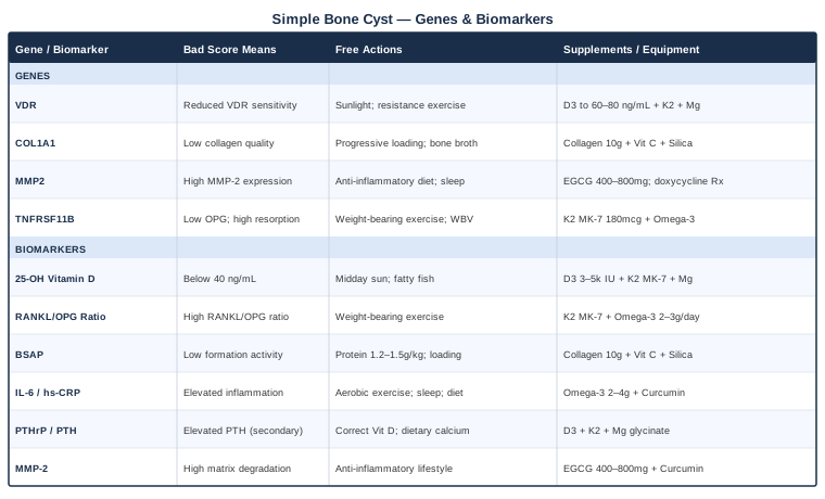
6 Biomarkers to Track for Simple Bone Cysts
Biomarker tracking for simple bone cysts is not yet standard orthopedic practice. Most management decisions rely almost entirely on imaging — X-ray and occasionally MRI — to assess cyst size, cortical thinning, and fracture risk. Imaging tells the structural story. It says very little about the biological forces driving that structure. The six biomarkers below address the functional level: what is actually happening in the cellular environment inside and around the cyst, and whether the conditions for healing are present.
Some of these biomarkers require nothing more than a routine blood draw. Others need more specialized laboratory requests. One or two are primarily relevant in research contexts or during orthopedic procedures. Together, they provide a picture that imaging alone cannot.
1. 25-OH Vitamin D
Vitamin D is the most accessible biomarker on this list, and arguably the most impactful lever in simple bone cyst management. The active form — 1,25-dihydroxyvitamin D — is a master regulator of calcium and phosphate metabolism, osteoblast differentiation, immune modulation in bone tissue, and critically, the RANKL/OPG balance that determines the ratio of bone destruction to bone formation. The vitamin D receptor (VDR) is expressed not only in osteoblasts but in macrophages and T-lymphocytes — the immune cells that populate the inflammatory cyst lining. Vitamin D deficiency does not simply slow bone mineralization; it impairs the immune resolution of the cyst membrane itself.
In pediatric orthopedics, vitamin D insufficiency correlates with slower healing after steroid injections and higher recurrence rates in cyst management. This makes biological sense: simple bone cysts form during periods of rapid growth — ages five through fifteen — when vitamin D demand is high and dietary and sunlight sources are often inadequate. Restoring adequate vitamin D doesn't cure a cyst, but it removes one of the most modifiable barriers to healing.
How to measure it
The test is serum 25-hydroxyvitamin D (25-OH D3), available through routine blood panels worldwide. Cost ranges from $30 to $60 depending on the country and ordering pathway. Most labs flag deficiency at below 20 ng/mL and sufficiency above 30 ng/mL. However, bone health specialists — including those in Peter Attia's clinical framework — generally target 40–60 ng/mL (100–150 nmol/L) as a functional optimum for tissue repair and immune function, meaningfully above the statistical "sufficiency" threshold. Retest every 3–6 months during active correction, then annually once stable.
If the score is bad, the plan without supplements
Sunlight exposure is the most effective and natural vitamin D source. Ten to thirty minutes of midday sun on the arms and legs — without sunscreen — on most days significantly raises levels in fair-skinned individuals. For darker skin tones or higher latitudes (above 40° north or south), the required exposure time may be two to three times longer. Dietary sources — fatty fish (salmon, mackerel), cod liver oil, egg yolks — provide meaningful but smaller contributions. Building consistent sun habits over 8–12 weeks and retesting is a reasonable first approach before adding supplements.
If the score is bad, the plan with supplements or equipment
Vitamin D3 (cholecalciferol) is the supplement form. For children, corrective doses of 1,000–2,000 IU/day are standard under medical guidance; for adults, 3,000–5,000 IU/day typically brings levels into the functional target range within 8–12 weeks. Vitamin D3 must be combined with vitamin K2 (MK-7 form): D3 increases calcium absorption, and without K2 to direct that calcium into bone matrix, it can accumulate in soft tissues. A dose of 90–180 mcg MK-7 daily alongside D3 completes the pairing. Magnesium (200–400 mg/day as glycinate or malate) is equally essential — it is required for the hepatic and renal hydroxylation steps that activate D3, and deficiency silently blocks supplementation from working.
Frequency and cycling: Daily, year-round. No cycling required at standard doses. Adjust seasonally based on retesting. Side effects: Well tolerated at these doses. Long-term supplementation above 10,000 IU/day without monitoring carries hypercalcemia risk — the reason regular testing matters.
2. RANKL/OPG Ratio
The RANKL/OPG axis is the central molecular switch governing the balance between bone resorption and bone formation. RANKL (Receptor Activator of Nuclear Factor Kappa-B Ligand) activates osteoclasts — the specialized cells that dissolve bone matrix. OPG (osteoprotegerin) acts as a decoy receptor that intercepts RANKL before it reaches its receptor, effectively suppressing osteoclast activity. When RANKL dominates, bone is progressively destroyed. When OPG dominates, bone formation can proceed unimpeded.
Multiple studies on simple bone cyst tissue have found significant RANKL elevation in the stromal cells lining the cyst wall, with relative OPG deficiency — a local molecular environment that continuously activates osteoclasts, perpetuating the cavity. Research published in pediatric orthopedic journals has confirmed elevated RANKL in cyst membranes compared to adjacent normal bone, suggesting the RANKL/OPG imbalance is not incidental to cyst biology but central to it. While the most pronounced imbalance is local (within the cyst microenvironment), serum measurements provide a useful systemic window and baseline for tracking the effects of interventions.
How to measure it
Both serum RANKL and serum OPG can be measured through ELISA-based assays available at specialty labs (LabCorp, Quest, or equivalent). They are not part of standard bone panels but can be requested specifically. Combined cost ranges from $100 to $250. The ratio matters more than absolute values: a high RANKL/OPG ratio indicates a pro-resorptive systemic state. Pairing these markers with a standard bone resorption marker (CTX, C-terminal telopeptide) provides additional confirmation of osteoclast activity.
If the score is bad, the plan without supplements
Weight-bearing and resistance exercise is the most powerful non-supplement intervention for improving the RANKL/OPG balance. Mechanical loading stimulates osteoblasts and osteocytes to upregulate OPG secretion through the Wnt/β-catenin signaling pathway — directly improving the ratio. For patients with simple bone cysts who must avoid high-impact activities due to fracture risk, low-impact alternatives are appropriate: stationary cycling, walking on flat surfaces, swimming with resistance components, and gentle progressive resistance exercises prescribed by a physical therapist. Even partial mechanical stimulation produces measurable OPG responses. Whole-body vibration platforms — used in bone research for disuse osteoporosis — offer an alternative mechanical stimulus where direct loading is contraindicated.
If the score is bad, the plan with supplements or equipment
Vitamin K2 (MK-7 form, 90–180 mcg/day) has the clearest human evidence for OPG upregulation. Multiple randomized trials have shown MK-7 supplementation increasing circulating OPG and reducing RANKL-driven bone resorption markers. A 2013 study by Knapen et al. in Osteoporosis International demonstrated that MK-7 (180 mcg/day over three years) improved bone strength indices in postmenopausal women, with measurable effects on the OPG/RANKL balance. Omega-3 fatty acids (EPA/DHA, 2–3g/day from high-quality fish oil) also reduce RANKL expression through NF-κB pathway suppression, supported by clinical trial data in inflammatory bone and joint conditions.
Frequency and cycling: Both K2 and omega-3s can be taken daily without established cycling requirements. K2 is fat-soluble — always take with a meal containing fat for absorption. Side effects: K2 MK-7 is well tolerated at these doses. Individuals on warfarin must consult their physician before use, as K2 affects vitamin K-dependent clotting factors. High-dose fish oil may modestly reduce platelet aggregation — relevant if surgery is planned in the near term.
3. Bone-Specific Alkaline Phosphatase (BSAP)
Bone-specific alkaline phosphatase is an enzyme produced exclusively by osteoblasts during active bone matrix mineralization. It is the most direct available marker of bone formation activity — a signal that osteoblasts are actively working to build new bone matrix. In the context of a simple bone cyst, BSAP is essentially measuring whether the healing response is being mounted. The cyst continuously drives osteoclast-mediated bone resorption; BSAP tells you whether the counter-response — osteoblast-driven matrix formation — is keeping pace.
Children physiologically have higher BSAP than adults because growing bone requires continuous formation. Within age-appropriate reference ranges, however, a child with an active cyst showing low-normal BSAP may be indicating an insufficient bone-forming response. This is particularly useful after treatment: a rising BSAP in the 6–12 weeks following steroid injection or bone grafting is one of the earliest laboratory signals of successful healing — potentially visible before structural changes appear on imaging.
How to measure it
BSAP is measured from a blood draw and is available at most clinical labs, sometimes as part of a bone turnover marker panel. Cost ranges from $50 to $100. It must be interpreted against age- and sex-specific reference ranges, since normal BSAP in a growing child is significantly higher than in an adult and does not in itself indicate pathology. Pairing BSAP with the bone resorption marker CTX (C-terminal telopeptide) provides a coupled picture: high CTX plus low-normal BSAP indicates a destructive imbalance; rising BSAP alongside declining CTX indicates healing.
If the score is bad, the plan without supplements
Osteoblast activity is fueled by adequate protein intake — the amino acids glycine and proline form the repeating units of type I collagen, the organic scaffold that BSAP helps mineralize. Ensuring adequate dietary protein (1.2–1.5g/kg body weight per day) from whole food sources, particularly those rich in collagen amino acids (eggs, poultry, slow-cooked meats, bone broth, gelatin-based foods), directly supports the biosynthetic demands of active BSAP-producing osteoblasts. Simultaneously, consistent weight-bearing and resistance exercise — even gentle, physical-therapist-guided protocols — mechanically activates osteoblast signaling and reliably increases BSAP over 6–12 weeks.
If the score is bad, the plan with supplements or equipment
Hydrolyzed collagen peptides (10g/day) provide the specific amino acid substrate — high in glycine, proline, and hydroxyproline — that osteoblasts require for type I collagen synthesis. A 2019 randomized controlled trial in Nutrients showed that collagen peptide supplementation improved bone formation markers including BSAP in subjects at risk for low bone mineral density. Vitamin C (250–500 mg/day) is an essential cofactor for the prolyl and lysyl hydroxylase enzymes that stabilize the collagen triple helix — without it, collagen cannot properly form or mineralize regardless of substrate availability. Silica (as orthosilicic acid, 5–10 mg/day) has emerging human evidence for collagen stimulation in bone tissue.
Frequency and cycling: All three are safe for daily continuous use. No cycling required. Side effects: Collagen peptides and silica are well tolerated. High-dose vitamin C above 2g/day increases kidney stone risk in susceptible individuals — the doses suggested here are well below that threshold.
4. Interleukin-6 (IL-6)
IL-6 is one of the most important inflammatory cytokines identified in simple bone cyst biology. Studies examining aspirated cyst fluid — including work from Japanese orthopedic research groups — have consistently found significantly elevated IL-6 inside cysts compared to surrounding normal bone tissue. IL-6 has a dual role in the cyst environment: it promotes osteoclast differentiation and activation (amplifying bone resorption), and it suppresses osteoblast function (reducing bone formation). This double action makes it a particularly potent driver of the destructive imbalance that maintains the cyst.
At the systemic level, chronic low-grade IL-6 elevation — driven by poor sleep, a diet high in refined seed oils and ultra-processed foods, physical inactivity, or gut dysbiosis — amplifies the local inflammatory milieu inside the cyst through circulating cytokine spillover. Serum IL-6, or the more accessible surrogate marker hs-CRP (high-sensitivity C-reactive protein), provides a window into this systemic background. Reducing systemic IL-6 may not resolve the cyst directly, but it removes one of the forces that actively hinders resolution.
How to measure it
High-sensitivity CRP (hs-CRP) is the most accessible marker: widely available, inexpensive ($20–$50), and consistently correlated with IL-6 activity. Serum IL-6 itself can be ordered directly for $50–$150 but is less standardized across labs and more vulnerable to preanalytical variability. Both should be measured during a period of clinical health, away from recent infection or injury, which cause transient elevations unrelated to the chronic inflammatory state of interest. For tracking purposes, hs-CRP at baseline and every 3–6 months during intervention is a practical and cost-effective approach.
If the score is bad, the plan without supplements
Regular moderate aerobic exercise is one of the most reliably effective lifestyle interventions for chronic IL-6 and CRP reduction. Twenty to thirty minutes of moderate-intensity aerobic activity (sufficient to elevate heart rate to 60–70% of maximum) four to five days per week reduces chronic IL-6 through improved insulin sensitivity, reduced visceral adipose signaling, and direct anti-inflammatory effects of myokines released during exercise. Sleep is equally essential: clinical studies consistently show that shortened or fragmented sleep (below seven hours per night) raises IL-6 and CRP. For pediatric patients, ensuring adequate, high-quality sleep is a non-negotiable foundation.
Dietary pattern also produces measurable effects. A dietary shift toward more vegetables, fatty fish, olive oil, legumes, and fiber — and away from ultra-processed foods, refined sugars, and high-linoleic seed oils (sunflower, soybean, corn oil) — produces significant hs-CRP and IL-6 reductions within 4–12 weeks in controlled trials.
If the score is bad, the plan with supplements or equipment
Omega-3 fatty acids (EPA+DHA combined, 2–4g/day from high-quality fish oil) are the most evidence-backed supplement for IL-6 reduction. A meta-analysis in Clinical Nutrition confirmed significant reductions in serum IL-6 and CRP with omega-3 supplementation across multiple inflammatory conditions. Liposomal or phospholipid-bound curcumin (500–1,000 mg/day) — the active component of turmeric — has also shown significant IL-6 reductions in randomized controlled trials. Standard curcumin has poor bioavailability; formulations with piperine, in phospholipid complexes (Meriva), or liposomal delivery dramatically improve absorption and clinical effect.
Frequency and cycling: Both omega-3s and curcumin can be used daily without cycling requirements. Fat-soluble — take with meals. Side effects: High-dose fish oil may modestly reduce platelet aggregation. Curcumin at very high doses may cause mild GI discomfort. Both are generally well tolerated at these therapeutic doses. If surgery is scheduled in the near term, discuss fish oil timing with the orthopedic team.
5. Parathyroid Hormone-Related Protein (PTHrP)
PTHrP binds the PTH receptor and triggers the same downstream signaling as parathyroid hormone — with significant effects on bone remodeling. In the normal skeleton, PTHrP plays essential roles in growth plate regulation and bone development. In the pathological context of simple bone cysts, however, PTHrP expression has been identified in the stromal cells lining the cyst wall, where it appears to amplify RANKL signaling and sustain the osteoclast activation that maintains the cyst cavity. Research from Japanese orthopedic groups has described PTHrP immunoreactivity in the cyst lining tissue, positioning it as a local amplifier of the destructive environment rather than merely a bystander.
Clinically, PTHrP is most commonly measured to evaluate hypercalcemia of malignancy — but its relevance to simple bone cyst biology is genuine. In the broader assessment of parathyroid axis function in patients with recurrent or poorly resolving cysts, measuring intact PTH (the standard test), calcium, phosphate, and vitamin D together provides a practical surrogate picture without requiring PTHrP specifically in most cases.
How to measure it
Serum PTHrP is available through major labs (LabCorp, Quest, or equivalent) at $100–$200. Intact PTH is a more standard and less expensive test ($30–$80), clinically accessible in any blood panel. For most practical purposes, an initial panel of intact PTH, serum calcium, serum phosphate, magnesium, and 25-OH vitamin D covers the parathyroid axis comprehensively. PTHrP is most meaningful in cases of hypercalcemia, unusual cyst behavior, or when the treating team is specifically investigating PTHrP-driven cyst dynamics.
If the score is bad, the plan without supplements
Elevated intact PTH in the absence of primary hyperparathyroidism most commonly reflects secondary hyperparathyroidism driven by vitamin D deficiency and/or inadequate calcium intake. The parathyroid glands respond to falling serum calcium by secreting PTH — which then activates osteoclasts to pull calcium from bone, worsening the cyst environment. Correcting vitamin D (as described in biomarker #1) typically normalizes PTH within 8–16 weeks. Ensuring adequate dietary calcium through whole food sources — dairy, leafy green vegetables, fortified plant milks, sardines with bones — ensures the parathyroid system is not chronically stimulated to draw calcium from the skeleton.
If the score is bad, the plan with supplements or equipment
When vitamin D deficiency is the driver, the D3 + K2 + magnesium protocol from biomarker #1 applies directly and typically normalizes secondary hyperparathyroidism as vitamin D levels correct. Magnesium deserves specific emphasis in this context: magnesium is required not only for vitamin D activation but also for PTH secretion regulation at the parathyroid cell level. Magnesium glycinate or malate at 200–400 mg/day, taken in the evening, is the most bioavailable and GI-tolerated form.
Frequency and cycling: Magnesium can be used continuously. Some practitioners alternate between 400 mg/day during active correction and 200 mg/day for maintenance. Side effects: Magnesium oxide forms cause loose stools in many individuals — glycinate and malate forms are significantly better tolerated. At standard doses, magnesium is safe for children and adults alike.
6. Matrix Metalloproteinase-2 (MMP-2)
Matrix metalloproteinases are enzymes that degrade components of the extracellular matrix — the structural scaffolding of bone and soft tissue. MMP-2 and MMP-9 have been detected at elevated levels in simple bone cyst fluid compared to surrounding healthy tissue, where they contribute to ongoing degradation of the bone matrix immediately adjacent to the cyst wall. This enzymatic activity is part of what allows the cyst to maintain its cavity against the body's natural healing responses and, in some cases, to expand. MMP-2 in particular targets type IV collagen and fibronectin — components of the basement membrane and periosteal matrix that are essential for structural containment of the cyst.
In most clinical settings, MMP-2 is not measured as part of routine bone cyst management. It surfaces primarily in research aspirates obtained during therapeutic procedures. However, understanding MMP-2's role matters practically, because several accessible interventions have well-characterized MMP-inhibitory activity supported by human clinical data — making this a biomarker that informs actionable decisions even when it cannot be directly measured in most settings.
How to measure it
Serum MMP-2 is available through specialized labs at approximately $150–$400. It is not a standard clinical test for simple bone cyst monitoring and reflects systemic, not local, enzyme activity. The most meaningful measurement context is analysis of cyst fluid aspirated during therapeutic procedures — this requires a treating team willing to send the aspirate for cytokine and enzyme profiling, which is primarily a research-context request. For most patients, hs-CRP and IL-6 serve as accessible proxies for the broader pro-inflammatory, matrix-degrading environment that co-elevates MMPs: addressing inflammation systemically suppresses MMP expression through shared NF-κB and AP-1 transcription factor pathways.
If the score is bad, the plan without supplements
The NF-κB inflammatory pathway drives transcription of multiple MMP genes, including MMP-2. Anything that durably suppresses NF-κB — and there is robust evidence here — reduces MMP expression over time. The same anti-inflammatory lifestyle package described under IL-6 (moderate exercise, sleep optimization, reduction of refined seed oils and ultra-processed foods, Mediterranean-style dietary pattern) measurably reduces MMP gene expression. This is not hypothetical: clinical trials in inflammatory joint disease and cardiovascular contexts have demonstrated reductions in circulating MMP-2 and MMP-9 following sustained lifestyle changes, with effects visible within 8–16 weeks.
If the score is bad, the plan with supplements or equipment
EGCG (epigallocatechin-3-gallate), the primary catechin in green tea, has well-documented MMP-2 and MMP-9 inhibitory activity through AP-1 transcription factor suppression. Standardized green tea extract at 400–800 mg/day (standardized to 45–50% EGCG) is supported by human tissue and clinical studies across cancer biology, arthritis, and connective tissue conditions. Curcumin — already discussed under IL-6 — also demonstrates MMP-inhibitory properties in multiple human cell-line and animal studies, providing meaningful overlap between the two supplements and making the combination a practical double-purpose intervention for both inflammatory cytokines and matrix-degrading enzyme activity.
Frequency and cycling: EGCG is commonly used in 8–12 week cycles with 4-week breaks, as very high doses have been linked to rare liver enzyme elevations in case reports. At 400 mg/day, most practitioners use it continuously with periodic liver function monitoring every 3–6 months. Do not take on an empty stomach. Side effects: High-dose EGCG (above 800 mg/day) may cause nausea or, rarely, mild transaminase elevation. Standard doses in healthy individuals are well tolerated. Curcumin is safe at 500–1,000 mg/day; higher doses may cause mild GI discomfort.
These six biomarkers together build a functional map of simple bone cyst activity that imaging cannot provide. Some are as easy to measure as a standard blood draw. Others require specific requests or are most relevant during orthopedic procedures. But together, they tell a biological story — and that story points directly toward interventions rather than watchful waiting alone.
The Genetic Landscape Behind Simple Bone Cysts
Genetics does not cause simple bone cysts through a single deterministic mechanism. There is no "simple bone cyst gene" in the way there is a gene for sickle cell anemia. What genetic variants do in this context is shift the biological environment: reducing vitamin D receptor sensitivity, lowering collagen synthesis capacity, increasing matrix metalloproteinase expression, or blunting the natural OPG response to RANKL. Any one of these shifts, alone, is not enough to cause a cyst. But the combination — particularly against a background of vitamin D insufficiency, chronic low-grade inflammation, or inadequate mechanical bone loading — creates conditions in which cyst formation and persistence become more likely.
The four variants below represent the most relevant and well-studied genes in this molecular context. If genetic testing is accessible through platforms like 23andMe, AncestryDNA, or clinical panels, these variants provide context that meaningfully refines the intervention strategy. If testing is not available, the interventions listed here are broadly beneficial for bone health regardless of genotype — knowing the variant simply helps calibrate dose and priority.
1. VDR — Vitamin D Receptor Gene
The VDR gene encodes the intracellular receptor through which vitamin D exerts its biological effects in every tissue that expresses it — including osteoblasts, osteoclasts, macrophages, and T-lymphocytes. Three single nucleotide polymorphisms are most studied: BsmI (rs1544410), FokI (rs2228570), and TaqI (rs731236). Specific combinations of these variants reduce the sensitivity or transcriptional activity of the VDR, meaning that a given circulating vitamin D level produces less biological effect at the cellular level. An individual with unfavorable VDR combinations may need significantly higher serum 25-OH D3 levels to achieve the same downstream signaling that someone with optimal VDR would achieve at a lower level.
This is directly relevant to simple bone cysts because both the bone-forming and immune-resolution functions of vitamin D — the functions most needed for cyst healing — depend on functional VDR signaling. Studies linking VDR polymorphisms to lower bone mineral density, delayed fracture healing, and greater inflammatory bone disease risk are consistent across multiple ethnic populations.
If the gene is bad, the plan without supplements
Maximizing vitamin D from sunlight and diet becomes disproportionately important for individuals with impaired VDR function. Sunlight exposure produces photoproducts beyond 25-OH D3 — including nitric oxide and other signaling molecules — that may have additional bone-supportive effects independent of the VDR pathway. Consistent midday sun exposure on maximal skin surface area, without sunscreen, is the priority.
Resistance exercise directly upregulates VDR expression in bone cells through mechanical loading and growth factor signaling, partially compensating for genetically reduced receptor sensitivity. Even moderate resistance training has been shown in human studies to increase VDR protein levels in muscle and bone tissue — a meaningful non-pharmacological offset to unfavorable genotypes.
If the score is bad, the plan with supplements or equipment
Individuals with unfavorable VDR variants typically require higher vitamin D3 supplementation to achieve the same functional effect. Targeting serum 25-OH D3 in the upper portion of the optimal range — 60–80 ng/mL rather than the standard 40–60 ng/mL target — is a common clinical approach in these cases, always with physician oversight and regular monitoring. Magnesium (200–400 mg/day as glycinate or malate) remains essential as a cofactor for D3 activation that is commonly deficient in the population.
Frequency and cycling: D3 and magnesium are taken daily, year-round. Retest serum 25-OH D3 every 3 months during dose adjustment, then every 6 months once stable. Side effects: D3 is safe at these doses with monitoring. K2 co-supplementation and periodic calcium checks mitigate hypercalcemia risk.
2. COL1A1 — Collagen Type I Alpha 1
COL1A1 encodes the alpha-1 chain of type I collagen — the most abundant structural protein in bone and the organic scaffold upon which bone mineral is deposited. The most studied COL1A1 variant is the Sp1 polymorphism (rs1800012) in the gene's promoter region. The s allele reduces COL1A1 transcription, resulting in lower-quality type I collagen. This variant is robustly associated with reduced bone mineral density and elevated fracture risk in large population studies, including the Rotterdam Study and multiple European cohorts.
In simple bone cyst biology, impaired collagen production matters across three interconnected levels. First, the bone matrix surrounding the cyst depends on high-quality collagen for mechanical strength — reduced collagen quality increases cortical vulnerability and pathologic fracture risk. Second, healing after aspiration or grafting depends on osteoblast-driven collagen synthesis to fill the cavity with new matrix. Third, early-stage cyst formation may itself reflect areas of weaker matrix architecture — the kind that COL1A1 variants produce — as a predisposing factor. Research in orthopedic genetics has linked COL1A1 Sp1 variants to a wider range of bone fragility conditions, suggesting a shared vulnerability mechanism.
If the gene is bad, the plan without supplements
Mechanical loading is the most powerful stimulus for COL1A1 gene expression in osteoblasts. Studies in exercise physiology consistently show that progressive resistance training increases collagen synthesis in bone, tendon, and ligament regardless of underlying genetic background — the mechanical signal overrides a portion of the transcriptional disadvantage. Physical-therapist-guided exercise protocols that incorporate gentle progressive loading of the affected limb (within fracture risk parameters) are both safe and directly beneficial at the gene expression level.
Diet contributes meaningfully: glycine and proline — the most abundant amino acids in type I collagen and the building blocks encoded by COL1A1 — must come from diet or supplementation. Whole food sources include slow-cooked meats, bone broth, gelatin, poultry skin, and eggs. Building dietary collagen amino acid intake provides the raw material regardless of transcription rate.
If the score is bad, the plan with supplements or equipment
Hydrolyzed collagen peptides (10–15g/day) provide the specific amino acid profile — particularly the glycine-proline-hydroxyproline tripeptide sequences — that most effectively stimulates osteoblast collagen production. Research by Dr. Keith Baar (UC Davis) has further shown that taking 15g of vitamin C-enriched gelatin or collagen 45–60 minutes before mechanical loading exercise synergistically increases collagen synthesis beyond either intervention alone — a practical combination protocol with direct application to bone healing contexts.
Vitamin C (500 mg/day) is required as a cofactor for prolyl hydroxylase and lysyl hydroxylase — enzymes that stabilize the collagen triple helix during synthesis. Without adequate vitamin C, collagen formed even from adequate substrate cannot properly cross-link or mineralize. Silica as orthosilicic acid (5–10 mg/day) has emerging human data for collagen stimulation and is a low-cost addition.
Frequency and cycling: Daily, continuous use. No cycling requirement. Side effects: Collagen peptides and vitamin C are safe at these doses for children and adults.
3. MMP2 — Matrix Metalloproteinase 2 Gene
MMP2 codes for the enzyme matrix metalloproteinase-2, one of the primary enzymes responsible for degrading type IV collagen, gelatin, and basement membrane components of the extracellular matrix. Two promoter polymorphisms are most studied: -1306C>T (rs243865) and -735C>T (rs2285053). The C allele at position -1306 is associated with higher MMP-2 promoter transcriptional activity and therefore greater enzyme production. Since elevated MMP-2 is a documented feature of active simple bone cyst fluid — where it contributes to ongoing matrix degradation in the cyst wall — individuals who genetically produce more MMP-2 enter the cyst microenvironment at a disadvantage from the start.
The clinical implications are specific: individuals with the -1306CC genotype may produce a more destructive cyst environment, may show faster cyst expansion, and may have a slower post-treatment resolution due to the sustained matrix-degrading activity that higher MMP-2 expression produces. When MMP genetics are combined with the TNFRSF11B OPG variants (below), the compounding effect on cyst biology is significant.
If the gene is bad, the plan without supplements
MMP-2 transcription is driven by the AP-1 and NF-κB transcription factor systems — the same inflammatory pathway drivers that respond most predictably to lifestyle interventions. The entire anti-inflammatory lifestyle package (moderate exercise, sleep optimization, reduction of refined seed oils, Mediterranean-style diet) durably suppresses both NF-κB and AP-1 activity and produces measurable reductions in MMP gene expression. This has been demonstrated in human tissue contexts in arthritis, cardiovascular, and inflammatory joint research, with effects on MMP-2 specifically documented at the mRNA level.
If the score is bad, the plan with supplements or equipment
EGCG at 400–800 mg/day (green tea catechin extract, standardized) directly inhibits AP-1 transcription factor activity, suppressing MMP-2 and MMP-9 expression. This mechanism has been validated in multiple human cell culture and translational studies across oncology, rheumatology, and connective tissue contexts. Sub-antimicrobial doxycycline (20 mg twice daily, available by prescription) is the most mechanistically specific MMP inhibitor available clinically — it chelates the zinc ion in MMP catalytic domains, inhibiting all MMPs at this dose without antibiotic effect. It is FDA-approved for this use in periodontal disease and has theoretical direct relevance to MMP-driven cyst biology, though no clinical trial in simple bone cysts has tested it directly.
Frequency and cycling: EGCG: 8–12 weeks on, 4 weeks off. Monitor liver enzymes periodically at extended use. Doxycycline: as prescribed, typically 3–6 months for established indications. Not appropriate for young children. Side effects: EGCG — nausea if taken on empty stomach. Doxycycline — photosensitivity, possible GI effects, requires physician prescription and monitoring.
4. TNFRSF11B — The OPG Gene
TNFRSF11B is the gene encoding osteoprotegerin — the natural RANKL decoy receptor that prevents osteoclast over-activation. The rs2073618 polymorphism (C allele) in TNFRSF11B has been associated with lower OPG levels and greater bone resorption in population studies of osteoporosis and inflammatory bone disease. In the context of simple bone cysts, this variant matters precisely because the cyst's pro-resorptive environment depends on inadequate OPG to contain local RANKL activity. A person who genetically produces less OPG is less well equipped to neutralize the RANKL excess found in the cyst lining — resulting in more sustained osteoclast activity, less natural containment of the cyst, and potentially a less favorable response to treatments that do not specifically address the OPG deficiency.
Large population studies have linked this TNFRSF11B variant to faster bone mineral density loss and increased fracture risk, supporting its mechanistic relevance in bone resorption contexts broadly. For simple bone cyst management, identifying this variant provides one of the strongest rationales for specific OPG-supportive interventions.
If the gene is bad, the plan without supplements
Mechanical loading is the most reliable stimulus for OPG upregulation. Osteoblasts and osteocytes under physical stress increase OPG secretion via Wnt/β-catenin signaling — directly compensating for the genetically reduced OPG baseline. For patients who cannot safely load the affected bone, whole-body vibration (WBV) platforms provide an alternative mechanical stimulus: research in disuse osteoporosis and pediatric bone conditions has shown measurable bone density improvements with 5–10 minutes per day of low-magnitude WBV, without the joint impact of traditional exercise.
If the score is bad, the plan with supplements or equipment
Vitamin K2 (MK-7 form, 180 mcg/day) is the most clinically supported supplement for OPG upregulation. The Knapen et al. trial in Osteoporosis International (2013) demonstrated that MK-7 at this dose over three years increased OPG and measurably improved bone strength indices in postmenopausal women. The mechanism — K2's activation of osteocalcin and stimulation of OPG gene expression — is well-characterized in bone cell biology. Adding omega-3s (2–3g/day EPA/DHA) compounds the effect through independent NF-κB-mediated OPG support.
Frequency and cycling: MK-7 is taken daily, with fat-containing meals, continuously. No cycling requirement. Side effects: Excellent tolerability at standard doses. Critical note for individuals on warfarin or other vitamin K-antagonist anticoagulants: K2 supplementation requires coordination with prescribing physicians due to drug-nutrient interaction effects on INR.
What Bone Biology Research Gets Right That Most Doctors Don't Discuss
A body of research increasingly points to systemic biological optimization as a meaningful parallel to local orthopedic intervention in bone healing conditions. The summary below draws on insights from researchers including Dr. Rhonda Patrick (FoundMyFitness), Dr. Andrew Huberman (Huberman Lab), and translational bone biologists like Dr. Keith Baar (UC Davis) — whose combined work on vitamin D metabolism, collagen synthesis timing, mechanical signaling, and gut-bone crosstalk challenges the standard "wait and see" framing for bone healing conditions. These ten findings are among the most impactful and least commonly communicated to patients.
1. Vitamin D is an immune regulator inside the cyst — not just a "bone vitamin"
The VDR is expressed on macrophages and T-lymphocytes within the cyst lining membrane. This means vitamin D directly modulates the immune cells responsible for maintaining (or resolving) the inflammatory cyst tissue. Deficiency doesn't just slow mineralization — it prevents immune resolution of the cyst membrane itself. Most patients are told vitamin D matters for calcium absorption. That's accurate but incomplete.
2. Magnesium deficiency silently blocks vitamin D from working
Approximately 45–50% of the US population has suboptimal magnesium intake. Without adequate magnesium, the hepatic 25-hydroxylase and renal 1α-hydroxylase enzymes cannot properly convert ingested D3 into its active form. People who supplement vitamin D without addressing magnesium may see deceptively low 25-OH D3 levels or inadequate clinical response despite adequate dosing. Magnesium is the missing piece in most vitamin D protocols.
3. Collagen synthesis timing changes everything
Dr. Keith Baar's research has demonstrated that consuming 15g of vitamin C-enriched gelatin 45–60 minutes before a loading exercise session produces a substantially greater increase in circulating hydroxyproline (collagen marker) than gelatin or exercise alone. The anabolic window around mechanical loading is when the bone-forming machinery is most receptive to collagen precursors. Pre-exercise collagen + vitamin C, combined with physical therapy loading sessions, represents a precision protocol for bone matrix formation that standard rehabilitation does not leverage.
4. The gut microbiome directly regulates RANKL and OPG
Short-chain fatty acids (SCFAs) — particularly butyrate, produced by gut bacteria fermenting dietary fiber — have been shown in both animal models and human microbiome studies to increase OPG secretion and reduce RANKL expression in bone tissue. This gut-bone axis is a newer but increasingly robust field. Adequate dietary fiber (25–35g/day) and prebiotic foods (oats, garlic, leeks, legumes, resistant starch) support the bacterial communities that produce these bone-protective SCFAs. The implication: gut health is bone health.
5. The growth hormone/IGF-1 axis explains spontaneous cyst resolution at puberty
Simple bone cysts often resolve spontaneously as children approach and pass through puberty — a phenomenon widely observed but rarely explained in plain language to families. The likely driver is the adolescent surge in growth hormone and IGF-1, which dramatically increases osteoblast activity and bone formation capacity, overwhelming the cyst's resorptive environment. Adequate sleep (during which 70% of growth hormone is secreted in pulsatile surges) and adequate dietary protein are the primary lifestyle levers for maximizing this growth hormone axis during the critical healing window.
6. Omega-6 to omega-3 ratio matters more than omega-3 dose alone
The inflammatory signaling that underlies cyst biology is shaped more by the ratio of omega-6 to omega-3 in the diet than by omega-3 intake in isolation. A diet dominated by refined linoleic acid (from corn, sunflower, soybean, and canola oils) creates a pro-inflammatory lipid signaling environment that fish oil cannot fully offset. Reducing omega-6 dietary sources — primarily by replacing refined seed oil cooking with olive oil, butter, or coconut oil — while adding omega-3s, produces larger reductions in IL-6 and CRP than adding omega-3s to a high seed-oil diet alone.
7. Mechanical signaling cannot be replaced by any supplement
No supplement, protein, or drug replicates the osteogenic signal of mechanical bone loading. Piezoelectric currents generated in bone matrix under physical stress activate osteocyte and osteoblast signaling through pathways (mechanosensing via primary cilia, YAP/TAZ signaling, Wnt activation) that are not accessed biochemically. For patients with simple bone cysts where loading is restricted due to fracture risk, whole-body vibration, aquatic resistance exercise, and carefully designed physical therapy protocols provide partial mechanical input with greatly reduced impact. These are not optional — they are the primary healing stimulus.
8. Periosteal bone formation is the primary healing mechanism
Healing in simple bone cysts occurs primarily through periosteal new bone formation at the cyst margins — cortical thickening and trabecular infilling from the edges inward — not through direct transformation of the cyst lining. This has been confirmed on sequential imaging studies. The practical implication: interventions that support periosteal osteoblast activity (vitamin D, K2, protein, mechanical loading) are more directly aligned with the actual healing mechanism than those targeting systemic bone turnover markers alone.
9. Stress hormones actively worsen the RANKL/OPG balance
Cortisol — the primary stress hormone — directly increases RANKL expression and reduces OPG at the cellular level. Chronic psychological stress in parents or patients facing ongoing orthopedic monitoring, repeated procedures, or fracture anxiety translates into a biochemical environment that actively worsens the molecular driver of cyst biology. This is not a minor effect: studies in stress physiology have shown significant, dose-dependent cortisol effects on the RANKL/OPG ratio. Stress management is not "soft" medicine in this context — it has a specific molecular consequence.
10. Sleep quantity and quality are underrated bone healing inputs
Growth hormone — essential for osteoblast activity in growing children and for bone regeneration in adults — is predominantly secreted during slow-wave (deep) sleep, in pulsatile surges that account for the majority of daily GH output. Sleep deprivation or fragmented sleep significantly reduces anabolic bone metabolism and elevates cortisol, compounding the RANKL/OPG effect described above. For pediatric patients recovering from cyst-related fractures or procedures, optimizing sleep duration (8–10 hours for school-age children) and sleep quality (consistent bedtime, dark environment, no screens 60 minutes before sleep) is a zero-cost, high-return intervention.
Complementary Approaches with Clinical Evidence for Bone Healing
The three approaches below are drawn from the evidence-based complementary medicine landscape and selected for their specific relevance to simple bone cyst management — either for stimulating bone tissue healing, reducing the systemic inflammatory environment, or managing the psychological burden that comes with ongoing orthopedic monitoring, procedures, and fracture risk. None of them replaces orthopedic care. Each can meaningfully complement it.
Low-Level Laser Therapy (Photobiomodulation)
Low-level laser therapy (LLLT), also known as photobiomodulation, delivers specific wavelengths of red and near-infrared light (typically 630–850 nm) to biological tissue at power densities low enough to avoid thermal damage. The primary mechanism of action is absorption by cytochrome c oxidase in the mitochondrial respiratory chain, which accelerates ATP synthesis and activates intracellular signaling cascades involved in cell proliferation, differentiation, and tissue repair. In osteoblasts specifically, LLLT has been shown in multiple human and animal studies to increase alkaline phosphatase activity, stimulate collagen synthesis, and accelerate bone matrix mineralization. These are precisely the osteoblast functions needed to fill and structurally resolve a bone cyst cavity after treatment.
Human clinical evidence for LLLT in bone healing is most developed in the dental and maxillofacial surgery context — particularly for alveolar bone defects and post-extraction socket healing — but the cellular mechanisms are tissue-generic. A randomized controlled trial published in Photomedicine and Laser Surgery found significantly accelerated mandibular bone healing in patients receiving LLLT compared to sham treatment after surgical bone procedures. For long bone cysts in the humerus or femur — the most common sites — LLLT applied over the affected segment after orthopedic procedures provides a scientifically grounded rationale for supporting the osteoblast-driven repair phase.
In practice, LLLT for bone healing uses devices in the 630–850 nm range at power densities of 50–200 mW/cm². Treatment sessions typically last 10–20 minutes and are applied two to three times weekly. Clinical devices are available in physical therapy offices; home-use near-infrared panels (devices from companies such as Joovv or PlatinumLED) provide broader daily coverage. There are no known serious adverse effects at appropriate power levels — eye protection during direct LED exposure is the main precaution. For pediatric patients with active cysts, it is prudent to discuss LLLT application with the treating orthopedic team before beginning, to confirm it does not interfere with active treatment protocols or post-procedural healing parameters.
Mindfulness-Based Stress Reduction (MBSR)
MBSR is a structured 8-week program developed at the University of Massachusetts Medical School by Dr. Jon Kabat-Zinn. It integrates body scan meditation, breath awareness practice, and mindful movement in sessions of 20–45 minutes daily, supplemented by a weekly group class. Its relevance to simple bone cysts operates through two distinct pathways. First, systemic inflammation reduction: consistent mindfulness practice has been shown in multiple randomized trials and meta-analyses to reduce serum IL-6 and cortisol — the same molecular markers that directly worsen the RANKL/OPG imbalance at the center of cyst biology. This is not an indirect or speculative connection; it is a specific biochemical effect with measured magnitude. Second, psychological burden management: living with an active bone cyst — particularly as a parent of a child with one — involves sustained uncertainty, procedural anxiety, and the stress of repeated imaging and follow-up. That chronic stress has real biological consequences.
The landmark meta-analysis by Goyal et al. published in JAMA Internal Medicine (2014) confirmed moderate-sized reductions in anxiety, depression, and pain with mindfulness meditation programs, effects durable at follow-up assessments. For simple bone cyst patients undergoing procedures, the cortisol-lowering and anxiety-reduction effects are particularly relevant: high procedural cortisol acutely blunts immune function and healing for hours after an injection or aspiration. Pre-procedural MBSR practice attenuates this cortisol response, potentially improving the post-procedure biological environment.
Practically, MBSR is accessible through in-person programs at hospitals and community centers, online-based versions (MBSR Online through the Brown University Mindfulness Center), and app-based formats (Insight Timer, Calm, Ten Percent Happier) for self-guided practice. For pediatric patients, child-adapted mindfulness programs — the MindUP curriculum, designed for school-age children — use age-appropriate language and shorter session formats (5–15 minutes). The primary caution is for individuals with significant trauma histories, where body-scan practices may be counterproductive; in those cases, guided imagery (below) is a better-matched alternative.
Guided Imagery
Guided imagery is a structured mind-body technique in which a facilitator, therapist, or recorded audio program leads the listener through detailed, positive sensory and narrative visualization — often including direct mental images of healing processes within the body. It is particularly well-matched to pediatric contexts, where imaginative engagement is natural and where the active participation in one's own recovery has documented psychological benefits. For simple bone cyst patients — who frequently face multiple procedures, prolonged watchful waiting, and the stress of fracture vigilance — guided imagery offers support at both the procedural and recovery levels.
Clinical evidence for guided imagery in pediatric orthopedic and surgical contexts is meaningful. Research by Dr. Belleruth Naparstek and colleagues, along with subsequent pediatric surgery studies, has demonstrated significant reductions in procedure-related anxiety, pain medication requirements, and post-procedure nausea with pre-procedure guided imagery. A 1997 controlled trial in Alternative Therapies in Health and Medicine found measurable reductions in cortisol and anxiety with guided imagery in pre-surgical patients. These effects directly address the cortisol-RANKL link described in the research insights section: blunting the procedural cortisol spike protects the post-procedure molecular environment. There is also a growing body of work in sports medicine rehabilitation suggesting that active mental imagery of movement and tissue repair accelerates actual tissue recovery — an effect thought to occur through motor cortex and autonomic nervous system mechanisms.
In practice, guided imagery for simple bone cyst patients is most useful in two contexts: the immediate pre- and post-procedure period (injections, aspirations, anesthesia), and as a regular daily practice of imaging bone healing during the recovery phase. Audio programs developed by Belleruth Naparstek (Health Journeys series) include tracks specifically designed for surgical healing and bone recovery. Sessions typically run 20–25 minutes and can be done lying down in a quiet space. There are no meaningful contraindications at this level of use. For children, a parent participating alongside can significantly improve adherence and provide a shared calming ritual around otherwise anxiety-provoking medical events.
Conclusion
Simple bone cysts occupy an uncomfortable middle space: common enough to feel routine from the medical side, yet biologically specific enough that the generic management framework often leaves patients and families without a clear picture of what is actually driving the cyst or what they can do to support healing beyond waiting. The molecular environment of a simple bone cyst is not random. It has identifiable features — RANKL/OPG imbalance, elevated inflammatory cytokines, matrix metalloproteinase activity, impaired vitamin D signaling — that are trackable and, in several meaningful ways, modifiable.
The six biomarkers in this article build a functional map that imaging alone cannot provide. The four genes explain individual vulnerability and point toward targeted compensating strategies. The research insights offer a framework for supporting healing that most orthopedic consultations do not have time to cover. The complementary approaches add evidence-based options for stimulating tissue repair and managing the psychological environment around ongoing medical care.
The next step does not require overhauling anything. Start with what is most accessible: a blood panel that includes 25-OH vitamin D, hs-CRP, and BSAP. Consider genetic testing if it is available and affordable. Discuss the biomarker picture with the treating orthopedic team as a supplement to imaging — not a replacement for it. Optimize the modifiable variables: sleep quality, dietary protein, anti-inflammatory eating, and physical activity within the constraints the cyst currently allows. Cysts do heal. The biology that makes that more or less likely is increasingly well understood — and increasingly something that individuals and families can take an informed role in shaping.
Musculoskeletal Endocrine & Metabolic
Musculoskeletal: Bone Conditions
Autoimmune: Inflammatory Conditions