This article contains AI generated content.
· UpdatedSinding-Larsen-Johansson Syndrome: 4 Genes and 6 Biomarkers to Track
Introduction
If you or your child has been diagnosed with Sinding-Larsen-Johansson syndrome, you have probably already heard the standard advice: rest, ice, stretch, and wait for the growth spurt to pass. That guidance is not wrong, but it is incomplete. It treats the condition as a uniform experience when in reality some adolescents recover in weeks while others struggle for months, and a few go on to develop persistent patellar tendon problems well into adulthood.
Sinding-Larsen-Johansson (SLJ) syndrome is an osteochondrosis affecting the inferior pole of the patella, typically appearing in athletically active young people between the ages of 10 and 14. During rapid growth, the patellar tendon pulls repeatedly on an immature bone, creating micro-damage faster than the tissue can repair itself. What is less commonly discussed is that the speed and quality of that repair process are not the same in every child. Genetics, nutrition, inflammation status, and hormonal environment all shape how resilient that tissue is and how quickly it bounces back.
Generic protocols ignore those individual variables. A child with low vitamin D, elevated baseline inflammation, and a collagen gene variant that produces slightly stiffer tendons faces a meaningfully different recovery challenge than a child without those factors, even if the imaging looks identical.
This article approaches SLJ syndrome from two complementary angles. The first looks at six trackable biomarkers — measurable signals in blood and tissue that reflect how well the body is managing inflammation, growth, collagen synthesis, and bone remodeling. The second examines four genes whose variants consistently appear in research on tendon and bone health, with practical notes on how to work with each one. Neither replaces clinical care, but both give you sharper questions to bring to your doctor and a clearer picture of where the body may need extra support.
6 Biomarkers That Matter in Sinding-Larsen-Johansson Syndrome
Biomarkers are not a cure and they are not diagnostic tools for SLJ specifically. What they offer is a window into the biological processes that drive recovery — or slow it down. The six below were selected because they are either directly involved in the physiology of tendon-to-bone attachment and growth plate stress, or because they reflect systemic conditions that worsen outcomes in musculoskeletal injuries. Most can be ordered through a standard blood panel.
1. 25-Hydroxyvitamin D (25-OH Vitamin D)
Why it matters
Vitamin D is not simply a bone mineral. Its receptor is expressed in chondrocytes, osteoblasts, and tendon fibroblasts, meaning it actively regulates the cells most involved in growth plate function and patellar tendon remodeling. Deficiency is strongly associated with impaired periosteal bone apposition — exactly the tissue under mechanical stress in SLJ syndrome. Studies in adolescent athletes have found that vitamin D insufficiency is significantly more prevalent in those with apophyseal injuries than in injury-free peers, though research specific to SLJ as a distinct entity is limited.
How to measure it
A serum 25-OH vitamin D test is widely available and inexpensive ($25–$60 depending on lab). Optimal range for musculoskeletal health is generally considered 40–60 ng/mL, though many labs flag anything above 20 ng/mL as "normal." For an active growing adolescent with a patellar apophysitis, aiming toward the higher end of the functional range is more defensible.
If the score is low — the plan without supplements
Daily midday sun exposure of 10–20 minutes on arms and legs (without sunscreen for that period) produces meaningful vitamin D synthesis in most latitudes from spring through autumn. Dietary sources including fatty fish, egg yolks, and fortified dairy can help but are rarely sufficient alone to correct a deficiency.
If the score is low — the plan with supplements or equipment
Vitamin D3 (cholecalciferol) at 2,000–4,000 IU daily is a well-tolerated starting point for adolescents with confirmed insufficiency, always taken with a meal containing fat. Co-supplementing with vitamin K2 (MK-7 form, 100–200 mcg/day) helps direct calcium toward bone rather than soft tissue. Retest at 8–12 weeks. Avoid megadosing without professional supervision; toxicity is rare but real above 10,000 IU/day sustained. Cycling is not needed at maintenance doses, but a higher loading protocol should be supervised and time-limited.
2. High-Sensitivity C-Reactive Protein (hs-CRP)
Why it matters
Inflammation is the mechanism through which micro-damage at the inferior patellar pole either resolves cleanly or becomes chronic and destructive. CRP is the most widely used systemic marker of low-grade inflammation. Elevated hs-CRP does not cause SLJ syndrome, but a chronically inflamed metabolic environment significantly slows collagen synthesis, impairs chondrocyte differentiation, and reduces the pain threshold in already-sensitized tissue. In adolescents, dietary patterns, sleep quality, and body composition are the primary drivers of baseline hs-CRP.
How to measure it
hs-CRP is a standard blood test, typically $20–$50. Target: below 1.0 mg/L for low cardiovascular and metabolic risk; below 0.5 mg/L is achievable in lean, well-rested adolescents. Values above 3.0 mg/L indicate elevated systemic inflammation worth investigating.
If the score is elevated — the plan without supplements
The most impactful free interventions are sleep (8–10 hours for adolescents), removing ultra-processed food and excess sugar from the diet, increasing vegetables and whole food carbohydrates, and reducing sedentary screen time. These alone can reduce hs-CRP by 30–50% within 8 weeks in motivated teenagers.
If the score is elevated — the plan with supplements or equipment
Omega-3 fatty acids (EPA+DHA combined 2–3 g/day with meals) have solid evidence for reducing hs-CRP in adolescents. Curcumin with piperine (500 mg curcumin, 5 mg piperine twice daily with meals) has shown anti-inflammatory effects in musculoskeletal contexts, though evidence in adolescents is thinner. Cycle curcumin 8 weeks on, 4 weeks off. Side effects at these doses are mild (occasional GI upset). A red-light therapy device (660–850 nm, 10 minutes over the knee, 4–5 times per week) is a promising adjunct with emerging evidence for reducing local tendon inflammation; cost for a basic panel runs $80–$200.
3. Insulin-Like Growth Factor 1 (IGF-1)
Why it matters
IGF-1 is the primary growth factor responsible for longitudinal bone growth and periosteal expansion during puberty — the exact mechanism that makes growth plates vulnerable to apophyseal injuries in the first place. Beyond explaining susceptibility, IGF-1 levels also correlate with tendon collagen synthesis capacity. Children with low IGF-1 relative to their growth velocity may have growth plates that are outpacing their tendon's ability to remodel. Conversely, very high IGF-1 in a child doing heavy loading may actually accelerate the traction forces on the patellar enthesis.
How to measure it
Serum IGF-1 with an age- and sex-adjusted reference range, $60–$120. Results must be interpreted in the context of Tanner stage and chronological age. The test is not routinely ordered for SLJ but is worth requesting if recovery is prolonged or if there are other signs of growth dysregulation.
If the score is suboptimal — the plan without supplements
Sleep is the most powerful natural driver of IGF-1 in adolescents, as growth hormone peaks in the first hours of deep sleep. Prioritize consistent sleep timing and minimize evening light exposure. Protein adequacy (at least 1.2–1.5 g/kg body weight per day from whole food sources) is essential; IGF-1 drops significantly in protein-restricted adolescents.
If the score is suboptimal — the plan with supplements or equipment
Zinc (10–15 mg/day elemental zinc with food, not on an empty stomach) supports IGF-1 signaling and is frequently suboptimal in athletic adolescents eating processed diets. Cycle 8 weeks on, 2 weeks off. Avoid exceeding 25 mg/day without monitoring copper levels, as zinc competitively inhibits copper absorption. Collagen peptides (10–15 g/day, ideally taken 30–60 minutes before activity with vitamin C) support tendon matrix synthesis but do not directly raise IGF-1; they are a complement rather than a driver.
4. CTX-1 (C-Terminal Telopeptide of Type I Collagen)
Why it matters
CTX-1 is a breakdown product of type I collagen, the primary structural protein in bone and tendon. Elevated CTX-1 means collagen is being degraded faster than it is being replaced — a scenario directly relevant to the enthesopathy in SLJ syndrome. In actively growing adolescents, CTX-1 is naturally higher than in adults, so pediatric reference ranges must be used. The marker becomes informative when it is disproportionately elevated relative to age-matched norms or when combined with low bone formation markers.
How to measure it
Serum or urine CTX-1, $60–$100. Best collected in a fasting morning state, as CTX-1 varies significantly with food intake and time of day. Ideally paired with a bone formation marker such as P1NP (procollagen type 1 N-terminal propeptide) to assess the balance between breakdown and synthesis.
If the score is elevated — the plan without supplements
Load management is the primary intervention: reducing high-impact jumping and sprinting temporarily lowers the mechanical stimulus driving collagen breakdown at the patellar enthesis. This is not the same as full rest; controlled low-load tendon exercise (isometric quad holds, slow eccentric movements) has been shown to promote collagen synthesis rather than breakdown, making it a more useful protocol than complete immobilization.
If the score is elevated — the plan with supplements or equipment
Collagen peptides (10–15 g/day, with 50 mg vitamin C to support hydroxylation) directly supply the substrate for type I collagen synthesis. Silicon (as orthosilicic acid, 10 mg/day) has early evidence supporting its role in collagen cross-linking. Gelatin-based protocols before tendon loading sessions (developed by Keith Baar at UC Davis) are gaining traction in sports medicine. Cycle collagen supplementation continuously during the recovery period, then reassess. Side effects are minimal; some children find large collagen doses mildly bloating when taken in protein shakes.
5. Serum Magnesium (or Red Blood Cell Magnesium)
Why it matters
Magnesium is a cofactor in over 300 enzymatic reactions, including those governing bone mineralization, muscle relaxation, and the inflammatory response. Low magnesium correlates with higher baseline pain sensitivity and impaired muscle recovery — both relevant to the clinical picture of SLJ syndrome, where persistent quadriceps tightness amplifies patellar tendon tension. Standard serum magnesium tests often miss deficiency because magnesium is tightly regulated in blood; red blood cell (RBC) magnesium is a more accurate reflection of tissue stores.
How to measure it
Serum magnesium is widely available and free in many panels; RBC magnesium is more informative and runs $40–$80. Optimal serum range: 0.85–1.05 mmol/L. RBC magnesium optimal: 5.0–6.5 mg/dL.
If the score is low — the plan without supplements
Dietary improvement is the baseline: dark leafy greens, nuts, seeds, legumes, and whole grains are the top food sources. Reducing carbonated soft drinks is important because phosphoric acid competes with magnesium absorption.
If the score is low — the plan with supplements or equipment
Magnesium glycinate or magnesium malate (200–300 mg elemental magnesium per day with dinner or before bed) are better tolerated than magnesium oxide, which has poor absorption and causes diarrhea. Avoid magnesium citrate at higher doses for the same reason. Topical magnesium oil applied to the quadriceps and knee area (5–10 sprays, 5 nights per week) is a low-evidence but low-risk adjunct that some athletes report reduces local muscle stiffness. No cycling needed at standard doses; reduce if loose stools develop.
6. Omega-3 Index (EPA + DHA as % of Red Blood Cell Fatty Acids)
Why it matters
The omega-3 index, popularized by researchers William Harris and Douglas Von Schacky, measures the percentage of EPA and DHA in red blood cell membranes. It reflects the anti-inflammatory status of the body's cell membranes over the prior 3–4 months. A low omega-3 index (below 4%) is associated with slower resolution of inflammatory processes, reduced tendon healing efficiency, and higher pain sensitivity. Most adolescents eating Western diets have an omega-3 index of 3–5%, well below the optimal 8–12% associated with anti-inflammatory benefits.
How to measure it
Omega-3 index testing is available from specialized labs ($65–$120) and is not yet standard in most routine panels. Home finger-prick tests exist (OmegaQuant is a validated option). This is one of the more useful advanced tests for any adolescent athlete with slow musculoskeletal recovery.
If the score is low — the plan without supplements
Two to three servings of fatty fish per week (salmon, sardines, mackerel, herring) consistently raises the omega-3 index over 12–16 weeks. Reducing vegetable seed oil consumption (sunflower, soybean, corn oil) lowers competing omega-6 load and allows omega-3 to be more effective.
If the score is low — the plan with supplements or equipment
Fish oil at 2–4 g combined EPA+DHA per day (not total fish oil — check the label for actual EPA+DHA content) is well supported and safe for adolescents. Retest omega-3 index at 16 weeks to confirm response. Algae-based omega-3 is a valid alternative for vegetarians. Side effects at standard doses are mild: fishy aftertaste (mitigated by freezing capsules), occasional loose stools at higher doses. No cycling needed during the recovery period; maintain consistently.
With a clearer picture of what the blood can reveal, it is worth zooming in on the genetic layer, which explains why some children are more vulnerable in the first place and why their tissues may need more targeted support.
Genes That Shape Tendon and Bone Resilience in Growing Athletes
Genetic testing for musculoskeletal conditions is still an emerging field, but several gene variants have robust associations with tendon injury susceptibility, collagen quality, and bone metabolism. None of these variants are destiny — most have modest effect sizes — but knowing your profile helps you understand where the body's weak links are and what compensations are most worthwhile.
COL1A1 (Collagen Type 1 Alpha 1) — rs1107946 and rs2070739
COL1A1 encodes the primary structural component of type I collagen, which forms the backbone of both bone and tendon. The rs1107946 variant (G allele) has been associated with reduced tendon stiffness and slightly greater injury risk in several studies of tendinopathy. Its counterpart variant is linked to stiffer tendons that tolerate high loads better but may be less elastic during growth spurts. At the patellar enthesis in a rapidly growing adolescent, this distinction matters — a stiffer tendon transmitting more force into an immature apophysis increases the mechanical stress driving SLJ pathology.
If the gene variant is unfavorable — plan without supplements
Slow eccentric loading programs (e.g., decline squat protocols, 3 sets of 15 repetitions at low speed, 3 days per week) are the most evidence-supported approach to stimulating collagen synthesis and improving tendon architecture independent of supplementation. Load management — avoiding ballistic jump training until symptoms resolve — is non-negotiable. Sleep prioritization drives nighttime growth hormone release, which directly stimulates collagen gene expression.
If the gene variant is unfavorable — plan with supplements
Collagen peptides (10–15 g with 50 mg vitamin C, 30–60 minutes before tendon loading sessions) provide the precursor amino acids specifically needed for type I collagen synthesis. This timing was studied by Shaw et al. and is now a standard recommendation in sports medicine tendon protocols. Cycle continuously during the active recovery phase (12–16 weeks minimum). Vitamin C at adequate levels (100–200 mg/day from food or supplements) is essential for collagen hydroxylation; deficiency severely impairs collagen cross-linking even when precursors are available.
COL5A1 (Collagen Type 5 Alpha 1) — rs12722
COL5A1 regulates the diameter of collagen fibrils and is a key determinant of tendon mechanical properties — specifically its stiffness-to-elasticity ratio. The TT genotype of rs12722 has been associated in multiple studies on tendinopathy susceptibility with altered tendon compliance. Children with this variant who are in a rapid growth phase may have tendons that are mechanically less well-matched to the growth demands being placed on the patellar enthesis.
If the gene variant is unfavorable — plan without supplements
Proprioceptive training (balance board work, single-leg stance exercises) reduces the risk of repeated micro-trauma by improving neuromuscular control around the knee. Hip and ankle mobility work addresses the kinetic chain factors that amplify patellar tendon stress during running and jumping. These are permanent lifestyle additions for high-risk individuals, not just rehabilitation tools.
If the gene variant is unfavorable — plan with supplements
Silicon as orthosilicic acid (ch-OSA, 10 mg/day) has emerging evidence for supporting collagen fibril cross-linking in connective tissue. It is available in several commercial forms and is generally well tolerated. Cycle 12 weeks on, 4 weeks off. Glycine supplementation (3–5 g/day, commonly added to water or smoothies) supports collagen synthesis as glycine is the most abundant amino acid in collagen and is conditionally limiting during high growth velocity.
VDR (Vitamin D Receptor) — BsmI (rs1544410) and FokI (rs2228570)
The VDR gene encodes the receptor through which vitamin D exerts its effects at the cellular level. Even with adequate serum vitamin D, certain VDR polymorphisms — particularly the BB genotype of BsmI — reduce receptor sensitivity, effectively lowering the biological response per unit of vitamin D. This is clinically significant because it means a child can test "normal" on vitamin D blood work and still have suboptimal signaling in their chondrocytes and osteoblasts.
If the gene variant is unfavorable — plan without supplements
Maximizing dietary calcium from whole food sources (dairy if tolerated, fortified plant milks, sardines with bones, leafy greens) reduces the dependence on vitamin D for calcium absorption and supports bone matrix mineralization independently. Regular weight-bearing activity is one of the most potent stimuli for bone mineralization regardless of vitamin D receptor sensitivity.
If the gene variant is unfavorable — plan with supplements
Higher vitamin D3 targets (aiming for serum 25-OH vitamin D in the 50–70 ng/mL range rather than the standard 30+ threshold) may be warranted to overcome reduced receptor sensitivity, under physician supervision with periodic testing. Magnesium is required for vitamin D activation in the liver and kidneys; ensuring magnesium sufficiency is particularly important for individuals with VDR variants. Vitamin K2 (MK-7, 100–200 mcg/day) ensures that elevated vitamin D-driven calcium absorption is directed into bone, not vascular tissue.
MMP3 (Matrix Metalloproteinase 3) — rs679620
MMP3 is an enzyme responsible for breaking down extracellular matrix components in tendon and cartilage. The rs679620 AA genotype produces higher MMP3 activity, meaning more collagen is degraded during normal tissue turnover. In a healthy adult tendon under normal load, this is kept in balance. In a growing adolescent with a stressed patellar enthesis, elevated MMP3 activity tips the balance toward breakdown over synthesis, potentially prolonging the inflammatory-degenerative cycle of SLJ.
If the gene variant is unfavorable — plan without supplements
Load management is again the primary lever: reducing MMP3 activation by controlling the inflammatory burden at the tissue level is partly mechanical. Reducing systemic inflammatory triggers (diet, sleep, stress) lowers the cytokine signals (notably IL-1β and TNF-α) that upregulate MMP3 expression. Cold water immersion of the knee (10–15°C for 10–12 minutes after activity, 3–4 times per week) can reduce local inflammatory signaling without the systemic effects of NSAIDs.
If the gene variant is unfavorable — plan with supplements
Curcumin with piperine (500 mg twice daily with meals) downregulates NF-κB signaling, one of the primary pathways through which MMP3 is induced. Cycle 8 weeks on, 3 weeks off. Boswellia serrata extract (300–400 mg of standardized extract twice daily, 65%+ boswellic acids) inhibits the 5-LOX pathway and has shown musculoskeletal anti-inflammatory effects in adult studies. Use with caution and medical guidance in adolescents; cycle with physician oversight.
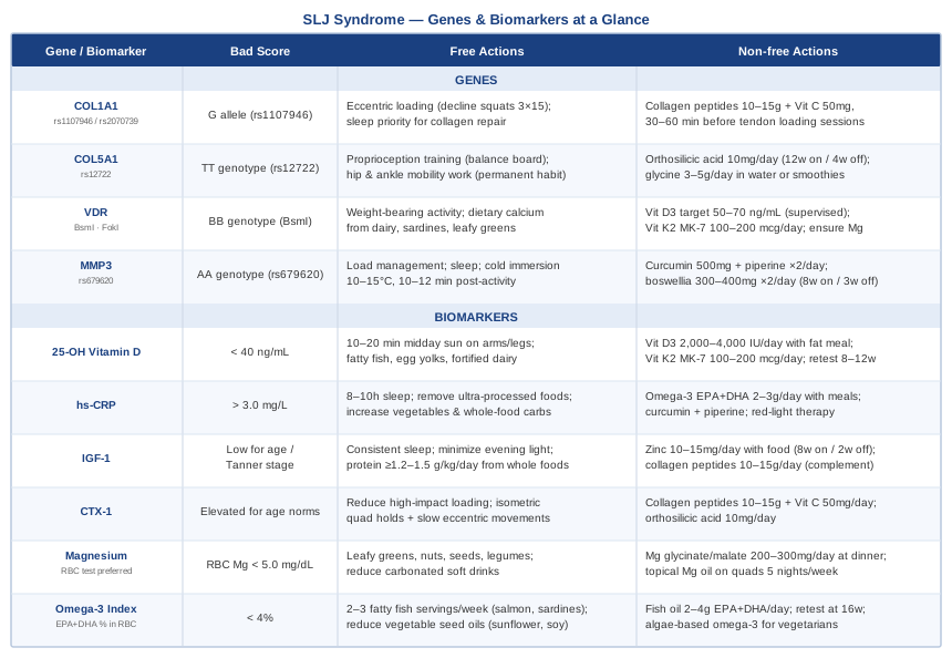
Summary Table: Genes and Biomarkers at a Glance
What Andrew Huberman's Podcast Reveals About Tendon and Connective Tissue Recovery
Andrew Huberman's Huberman Lab podcast has addressed connective tissue biology, injury recovery, and growth factor signaling across multiple episodes — particularly in his discussions with orthopedic scientists and in his deep dives on tissue repair. The perspective offered is grounded in peer-reviewed science and frequently challenges the passive "rest and wait" orthodoxy.
1. Tendons adapt much more slowly than muscles — and most people ignore this
Muscles respond to training within days; tendons take months because their blood supply is poor and their cell turnover is slow. In SLJ syndrome, this mismatch is exactly the problem: the muscle gets stronger and generates more force faster than the tendon-to-bone junction can adapt. Training must be paced to the tendon, not the muscle.
2. Isometric contractions reduce tendon pain better than rest
Huberman has highlighted work showing that sustained isometric contractions (e.g., wall sits, Spanish squat holds at 45–60% of maximum voluntary contraction, held for 30–45 seconds) acutely reduce tendon pain and stimulate collagen synthesis. This is more useful than complete immobilization during recovery.
3. Collagen synthesis has a precise timing window around loading
Based on Keith Baar's research discussed in Huberman's episodes, collagen peptide consumption combined with vitamin C, taken 30–60 minutes before tendon loading exercise, produces significantly higher tendinous collagen synthesis than supplementing at other times. This is not a vague general recommendation — it requires specific timing.
4. Sleep is when most tendon repair happens
Growth hormone, which peaks during slow-wave sleep, is the primary driver of IGF-1 secretion and downstream collagen gene expression. An adolescent averaging 6–7 hours of sleep instead of 8–10 is significantly compromising their tissue repair capacity regardless of what else they do.
5. Inflammation is not always the enemy — but chronic low-grade inflammation is
Huberman distinguishes between acute inflammation (necessary for repair signaling) and chronic low-grade systemic inflammation (which impairs healing and sensitizes pain pathways). NSAIDs taken repeatedly during SLJ recovery may blunt the acute repair signal. This is an area of genuine clinical debate and warrants discussion with the treating physician.
6. Cold and heat have opposite effects on connective tissue healing
Cold water immersion (10–15°C) acutely reduces inflammation and pain but may blunt the inflammatory repair signal if applied immediately after loading. Heat applied to tendons increases collagen extensibility and local blood flow. Huberman suggests sequencing these deliberately: avoid icing immediately after rehabilitation exercises; save cold for more than 4–6 hours post-exercise or pre-exercise for pain management.
7. Vitamin D's role goes well beyond bone density
Huberman has emphasized that vitamin D receptor expression in musculoskeletal tissues means that deficiency impairs not just bone density but tendon and muscle fiber type composition, inflammatory resolution, and even pain modulation. Testing and correcting vitamin D status is not optional for athletes with tendon problems.
8. The quadriceps-patellar chain must be trained, not just stretched
Passive stretching of a tight quadriceps temporarily reduces the tensile load on the patellar tendon but does not address the underlying force production during running and jumping. Huberman and his guests consistently emphasize that strength work through the full kinetic chain — including hip extensors and ankle dorsiflexors — redistributes load away from the patellar enthesis more sustainably.
9. Stress and cortisol directly impair connective tissue repair
Chronically elevated cortisol inhibits collagen synthesis and slows tendon healing. In competitive adolescents under academic and athletic pressure, managing psychological stress is a genuine recovery variable, not a soft add-on.
10. The return-to-sport decision should be guided by tissue capacity, not pain alone
Pain resolution precedes tissue healing by weeks to months in tendon apophysitis. Returning to full activity when pain subsides, without confirming that tendon loading capacity has been restored, is the primary reason for re-injury. Loading programs should be progressive and monitored, not self-directed based on symptom absence.
Complementary Approaches With Clinical Support
Low-Level Laser Therapy (Photobiomodulation)
Low-level laser therapy (LLLT), also called photobiomodulation, uses red and near-infrared light (630–1000 nm) to stimulate mitochondrial activity in cells, reduce local inflammation, and accelerate tissue repair. For patellar tendon pathology, its relevance lies in its ability to reach deep tissue at the enthesis and modulate the inflammatory signaling driving apophysitis without systemic side effects.
A systematic review published in the Lancet (2009) found that LLLT significantly reduced pain and improved function in tendinopathy compared to placebo. Evidence specific to pediatric apophysitis is limited, but the mechanism is directly relevant. Doses of 4–8 J/cm² at 810–904 nm over the patellar enthesis appear to be both safe and effective in comparable conditions.
For practical application in SLJ, a consumer-grade photobiomodulation device (660 nm and 850 nm combined, $100–$250) applied to the inferior patella for 8–10 minutes, 4–5 times per week for 6–8 weeks, is a realistic protocol. Keep device moving or use a panel at a fixed distance of 5–10 cm. Do not apply over active growth plates in young children without guidance; for adolescents with SLJ, the target is the tendon-to-bone junction, not the growth plate itself.
Massage Therapy
Massage therapy targeting the quadriceps and iliotibial band indirectly reduces patellar tendon tension by decreasing the resting tone of muscles that transmit force through it. Tight rectus femoris and vastus lateralis are almost universally present in adolescents with SLJ and amplify the tractional forces at the inferior patella.
A randomized controlled trial on adolescents with patellofemoral pain (a closely related condition) found that manual therapy combined with exercise produced greater pain reduction and functional improvement than exercise alone. While not SLJ-specific, the anatomical overlap makes manual therapy a well-justified adjunct. Transverse friction massage directly at the proximal patellar tendon, applied by a trained physiotherapist, addresses local scar tissue and promotes structured collagen remodeling.
Practically, two sessions per week with a sports physiotherapist during the active pain phase, combined with a daily foam rolling protocol for the quadriceps (2–3 minutes per side, moderate pressure), represents an accessible approach. Parents can be taught gentle effleurage and passive quadriceps stretching for home application between sessions. Massage should feel like meaningful pressure, not acute pain.
Biofeedback
Biofeedback teaches individuals to consciously modify physiological processes — in this context, muscle activation patterns around the knee. Many adolescents with SLJ develop protective movement strategies (altered gait, reduced knee flexion during landing) that redistribute load poorly and increase long-term risk. Biofeedback can retrain landing mechanics and quadriceps activation quality with real-time feedback.
Surface electromyography (sEMG) biofeedback has been studied in patellofemoral rehabilitation with positive outcomes for pain and function. By providing real-time visual or auditory feedback on VMO (vastus medialis oblique) activation during rehabilitation exercises, it improves the quality and coordination of the quadriceps force output, reducing the abnormal loading patterns at the patellar enthesis.
In practical terms, a physiotherapist-led biofeedback program of 6–10 sessions (45–60 minutes each), with home practice using a consumer sEMG device, is feasible for adolescents. The skill transfers: once better neuromuscular patterns are learned, they tend to persist through normal training. Cost is approximately $100–$200 for a supervised program; consumer biofeedback devices run $150–$400.
Conclusion
Sinding-Larsen-Johansson syndrome is almost always self-limiting, but the timeline and quality of recovery are not fixed — they are shaped by measurable biological variables that can be influenced. Tracking six biomarkers (vitamin D, hs-CRP, IGF-1, CTX-1, magnesium, and the omega-3 index) gives you a functional map of where the body's repair machinery is working well and where it needs support. Understanding four relevant gene variants (COL1A1, COL5A1, VDR, MMP3) adds a layer of personalization that explains individual differences in susceptibility and recovery pace.
None of this is a replacement for working with a pediatric sports medicine physician, orthopedic specialist, or physiotherapist. But it is the kind of information that turns a passive waiting game into an active, evidence-informed recovery. The next practical step is straightforward: ask your doctor for a panel that includes 25-OH vitamin D, hs-CRP, and magnesium — that alone costs under $100 in most settings and often reveals something actionable within the first appointment.
Musculoskeletal: Bone Conditions Tendon & Ligament Conditions Sports Injuries
Autoimmune: Inflammatory Conditions