This article contains AI generated content.
· UpdatedSkeletal Fluorosis: 5 Genes And 6 Biomarkers To Track
Introduction
If you or someone close to you has been diagnosed with skeletal fluorosis—or if long-term high fluoride exposure has been part of your history—you've probably already noticed how quickly medical guidance runs out of detail. Reduce exposure. Take pain relief. See a specialist. What those conversations rarely address is why some people develop significant bone damage while others living in the same fluoride-heavy environments remain largely unaffected for decades. That gap isn't a mystery without an answer. It reflects the role individual biology plays in disease risk.
Skeletal fluorosis develops when fluoride accumulates in bone over years, gradually altering the mineral matrix of hydroxyapatite, disrupting the balance between bone-building and bone-breakdown, and eventually changing bone architecture in ways that cause pain, stiffness, and in advanced cases, severe deformity. But the speed and severity of that trajectory varies based on factors most clinical assessments don't look at: how efficiently the kidneys excrete fluoride, whether the vitamin D receptor gene is functioning properly, whether bone remodeling pathways can resist disruption, and what the nutritional baseline looks like across years of exposure.
Generic health advice misses that layer entirely. Tracking the right biomarkers gives you a real window into it. Specific blood and urine markers can show whether bones are under active stress right now, whether the body is handling fluoride load well or accumulating it, and whether the key nutritional and hormonal pathways supporting bone health are working. Alongside that, understanding the genetic variants that shape individual vulnerability can explain patterns in your history and point toward more targeted interventions.
This article covers both dimensions with enough depth to be useful. Six specific biomarkers are worth tracking in anyone with meaningful fluoride exposure—each one is explained in terms of what it reveals, how to measure it affordably, and what to do when the number is off. Five key genetic variants follow, each one linked to individual differences in fluoride sensitivity and bone response. Alongside those two frameworks, a section draws on one of the most research-dense independent reviews of fluoride toxicity ever published, and a final section looks at complementary modalities with meaningful clinical evidence for bone health. The goal throughout is not to offer a shortcut or a cure—fluorosis is a slow-moving, exposure-driven condition—but to replace vague guidance with something genuinely actionable.
Summary
Here is what this article covers and why it matters if you are tracking or managing skeletal fluorosis:
- Six biomarkers reveal active bone stress, fluoride accumulation, and nutritional gaps: urinary fluoride, bone-specific alkaline phosphatase (BALP), CTX (bone resorption marker), PTH, 25-OH vitamin D, and eGFR. Each section includes how to measure it, cost ranges, and what to do if results are abnormal—with and without supplements. - Five genetic variants (VDR, COL1A1, RANKL/OPG, SLC26A1, CYP27B1) explain why some people are far more vulnerable to skeletal fluorosis than others—and how to partially compensate for each with targeted lifestyle and supplementation strategies. - A deep dive into the landmark book The Case Against Fluoride by Paul Connett, James Beck, and Spedding Micklem covers 10 insights that challenge mainstream fluoride safety thinking—including why individual nutrition changes disease risk dramatically and why the official margin of safety is narrower than most practitioners believe. - Complementary approaches with clinical evidence for bone health in fluorosis: yoga, Ayurvedic herbal therapy (including tamarind, with its studied effect on urinary fluoride excretion), photobiomodulation, tai chi, and microbiome-directed strategies.
If you've been told your fluoride exposure was high, or your bones are showing unusual changes, these are the numbers and genes worth understanding first.
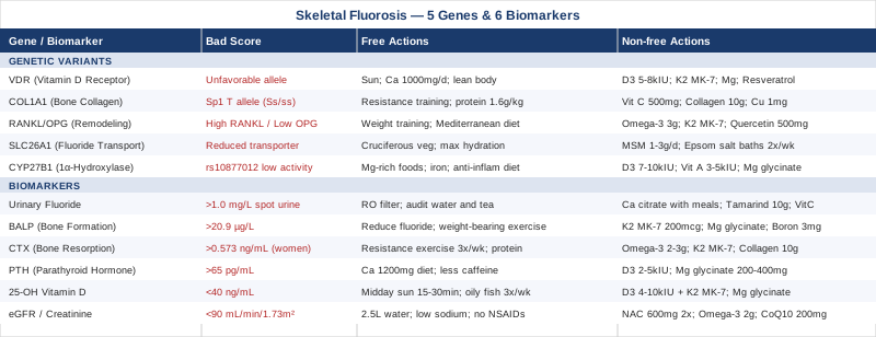
6 Biomarkers Worth Tracking in Skeletal Fluorosis
Biomarkers don't diagnose skeletal fluorosis on their own, but they do something just as valuable: they show you the biological terrain the condition is operating in. Bone turnover, fluoride clearance, hormonal regulation, vitamin D metabolism—each of these can be measured, trended over time, and responded to. In a condition driven by slow, cumulative exposure, that kind of longitudinal tracking is often the difference between catching early changes and dealing with advanced disease.
The six markers below are selected for clinical relevance, measurability, and actionability. Most are accessible through standard labs; a few require specialty ordering. Costs listed are approximate USD ranges for self-pay in the United States.
Biomarker 1: Urinary Fluoride
Why it matters: Urinary fluoride is the most direct available measure of both ongoing fluoride exposure and the body's clearance of it. The kidneys are the primary route for fluoride excretion in humans—roughly 50 percent of ingested fluoride is excreted through urine in healthy adults. Because fluoride doesn't have a useful blood steady-state marker (it distributes quickly into bone), urine is the next best proxy for body burden monitoring. In populations with endemic skeletal fluorosis, elevated urinary fluoride consistently correlates with disease severity (Krishnamachari, Bulletin of the World Health Organization, 1986).
How to Measure It
A 24-hour urine collection is the gold standard; a spot urine corrected for creatinine is a practical alternative and adequate for monitoring trends. Samples are sent to an environmental or occupational medicine lab—Quest Diagnostics and ARUP Laboratories both offer urinary fluoride panels. Cost: approximately $50–150 USD depending on collection method and lab.
Normal reference values for non-occupationally exposed adults: less than 1.0 mg/L spot urine (creatinine-corrected), or under 1.5 mg/day on 24-hour collection. Values above 3 mg/L in spot urine suggest significant ongoing exposure.
If the Score Is High: The Plan Without Supplements
The first priority is source identification. In many cases this requires systematic auditing: municipal water fluoride level, cooking water, bottled teas (brick tea in particular can contain 2–9 mg/L fluoride), non-stick cookware that degrades at high heat, and in some regions, fluoride-rich well water. High-fluoride toothpaste swallowed even in small amounts contributes to load. Installing a reverse osmosis filter at the point of consumption is one of the few household interventions proven to reduce urinary fluoride; activated carbon alone is insufficient. Industrially exposed workers should review occupational hygiene controls and use PPE consistently.
If the Score Is High: The Plan With Supplements
Calcium binds fluoride in the gastrointestinal tract and reduces absorption when taken with meals (calcium citrate or calcium carbonate, 500 mg with each main meal). This is especially relevant if dietary calcium is low. Vitamin C (500–1000 mg/day) supports renal tubular function and has shown modest benefit in some fluoride-exposure animal models, though human data is limited. Tamarind (Tamarindus indica) extract or whole fruit has been studied in Indian endemic fluorosis populations: a clinical trial by Khandare et al. published in European Journal of Clinical Nutrition (2004) found that schoolchildren given tamarind showed significantly increased urinary fluoride excretion compared to controls, suggesting it may help mobilize fluoride from bone. Dose used: 10g tamarind fruit pulp daily. No significant side effects reported at this dose. This can be taken continuously; it is food-based, not a pharmaceutical. Frequency: daily with meals.
Biomarker 2: Bone-Specific Alkaline Phosphatase (BALP)
Why it matters: Bone-specific alkaline phosphatase is released by osteoblasts—the cells responsible for bone formation. Elevated BALP signals accelerated bone-building activity, which in fluorosis does not mean healthier bone. Fluoride stimulates osteoblasts, but the resulting bone has a disorganized, sclerotic structure—denser in appearance but actually weaker and more brittle. BALP is a more reliable marker of osteoblastic activity than total alkaline phosphatase, which also reflects liver activity and can mislead. Studies in patients with endemic skeletal fluorosis consistently show elevated BALP, and it tends to normalize partially as fluoride exposure is reduced (according to reviews in the Journal of Dental Research and fluoride toxicology literature).
How to Measure It
BALP requires a specific immunoassay—not the standard total ALP included in a comprehensive metabolic panel. Order the bone-specific alkaline phosphatase test explicitly. Available through Quest, LabCorp, and specialty endocrinology labs. Cost: approximately $30–80 USD. Draw is a standard venipuncture; no fasting required. Reference range: roughly 3.7–20.9 µg/L in adult women, somewhat higher in men and adolescents (lab-specific; always compare to the reference interval provided).
If the Score Is High: The Plan Without Supplements
The primary lever for reducing BALP over time is reducing fluoride exposure—addressing the driver directly. Structured weight-bearing exercise (walking, resistance training) within pain tolerance helps regularize bone turnover rather than suppress it. Adequate dietary protein (1.4–1.6 g/kg/day) supports healthy osteoblast function and reduces the disorganized bone formation response. If dairy is not well-tolerated, calcium from whole food sources (sardines, broccoli, sesame seeds) should be consistent and daily.
If the Score Is High: The Plan With Supplements
Vitamin K2 (MK-7 form, 100–200 mcg/day) activates osteocalcin, a protein that directs calcium into bone properly and reduces ectopic mineralization—directly relevant in fluorosis where abnormal mineralization is a hallmark. Take daily without cycling; it's fat-soluble and needs a meal with some fat. Magnesium glycinate (200–400 mg/day) supports normal bone matrix formation and is frequently deficient in people with elevated bone turnover. Vitamin D3 should be optimized before or alongside K2 (see Biomarker 5 below). Boron (3–6 mg/day as boron glycinate) has shown in small trials to support bone mineral density and reduce inflammatory bone loss markers; can be taken daily with no cycle required. Side effects of K2 and magnesium are minimal at these doses; boron at doses above 20 mg/day may cause gastrointestinal upset.
Biomarker 3: Serum CTX (C-Terminal Telopeptide of Type I Collagen)
Why it matters: Where BALP reflects bone formation, CTX reflects bone resorption—the activity of osteoclasts breaking down old bone tissue. In healthy bone remodeling, formation and resorption are coupled. In skeletal fluorosis, this coupling is disrupted: fluoride-stimulated formation produces disorganized bone, while resorption markers can fluctuate depending on disease stage. Elevated CTX in a fluorosis patient signals accelerated degradation of bone collagen, which can indicate the remodeling process is running too fast or in the wrong direction. CTX is also sensitive to fasting status, which makes collection standardization important.
How to Measure It
CTX must be drawn fasting, in the morning (ideally before 10 AM), because it follows a diurnal rhythm and varies significantly with food intake. Available through Quest, LabCorp, and most major labs as serum CTX or beta-CTX. Cost: approximately $50–100 USD. Reference ranges vary by sex and menopausal status—premenopausal women typically below 0.573 ng/mL; men varies by age. Ask your lab for the age-sex-specific reference interval.
If the Score Is High: The Plan Without Supplements
Impact and resistance exercise is the most evidence-backed non-pharmacological intervention for reducing bone resorption markers. Even moderate-intensity resistance training three times per week has been shown to decrease CTX in postmenopausal women in multiple randomized trials. Within fluorosis-related pain constraints, aquatic resistance training or chair-based resistance exercises maintain osteogenic stimulus while reducing joint load. Dietary protein consistently at 1.4–1.6 g/kg/day reduces catabolic signaling at the bone level. Address any vitamin D deficiency first (see Biomarker 5)—its absence accelerates bone resorption.
If the Score Is High: The Plan With Supplements
Omega-3 fatty acids (EPA + DHA combined, 2–3 g/day) have been shown in meta-analyses to reduce bone resorption markers including NTX (closely related to CTX); effect size is moderate but consistent. Take with food; fatty fish at least 3x per week is an effective food-based approach. Vitamin K2 MK-7 (100–200 mcg/day) works through the same osteocalcin mechanism relevant for BALP. Collagen peptides (10 g/day) provide glycine and proline, the structural amino acids for type I collagen; a randomized trial in Nutrients (König et al., 2018) found collagen supplementation plus resistance training produced greater improvements in bone markers than exercise alone. Take daily; no cycling needed. Side effects are minimal.
Biomarker 4: Parathyroid Hormone (PTH)
Why it matters: PTH is secreted by the parathyroid glands in response to low serum calcium. It acts on bone (releasing calcium), the kidneys (retaining calcium and excreting phosphate), and the gut (indirectly, via vitamin D activation). In skeletal fluorosis, PTH dysregulation is common: fluoride can directly disrupt the calcium-phosphate axis, and if vitamin D is also deficient, secondary hyperparathyroidism develops—chronically elevated PTH that drives accelerated bone resorption and further destabilizes already-compromised bone. PTH is a critical bridge biomarker: it integrates kidney function, vitamin D status, and calcium balance into a single number.
How to Measure It
Intact PTH (iPTH) is the standard clinical assay. Available through any major lab, usually as part of a bone health or kidney panel. Fasting not strictly required, but morning draws are preferred. Cost: approximately $40–80 USD. Normal range: 15–65 pg/mL (intact PTH). Values above 65 pg/mL persistently suggest secondary hyperparathyroidism; values below 15 may indicate suppressed parathyroid function, sometimes associated with very high calcium or abnormal PTH-related protein.
If the Score Is Elevated: The Plan Without Supplements
First rule out primary hyperparathyroidism with your physician (a serum calcium drawn at the same time will clarify). For secondary hyperparathyroidism driven by vitamin D deficiency or fluoride-related calcium dysregulation: optimize dietary calcium consistently (at least 1000–1200 mg/day from food). Dairy, canned fish with bones, tahini, and dark leafy greens are the most reliable sources. Reduce phosphate from processed foods, which compete with calcium and worsen PTH dysregulation. Limit caffeine excess, which impairs calcium absorption.
If the Score Is Elevated: The Plan With Supplements
Vitamin D3 (2000–5000 IU/day initially) is the most important correction—secondary hyperparathyroidism is very often a vitamin D-driven phenomenon. Retest in 8–12 weeks and adjust. Magnesium (200–400 mg glycinate form daily) is a required cofactor for PTH secretion and vitamin D activation; magnesium deficiency prevents vitamin D from normalizing PTH even when supplemented. Take both daily; they are synergistic. If PTH remains elevated despite optimized vitamin D (above 50 ng/mL) and calcium, consult an endocrinologist—prescription calcitriol may be considered. No cycling needed for D3 or magnesium at standard doses; retest every 3 months while adjusting.
Biomarker 5: 25-Hydroxyvitamin D (25-OH D)
Why it matters: Vitamin D is not optional in skeletal fluorosis—it is central to the disease's trajectory. Active vitamin D (calcitriol) regulates calcium absorption in the gut, bone mineralization, PTH secretion, and immune modulation. Deficiency in fluorosis-exposed populations dramatically worsens outcomes: it impairs calcium absorption precisely when calcium is needed most, it drives secondary hyperparathyroidism, and it leaves bone matrix poorly mineralized even as fluoride drives excessive formation. Researchers studying endemic fluorosis in India and Africa have consistently found vitamin D deficiency as a compounding variable in the most severe cases. Peter Attia, among others, recommends an optimal range of 40–60 ng/mL—higher than the conventional "sufficient" threshold of 20 ng/mL.
How to Measure It
25-OH vitamin D (total) is the standard serum test. Available universally; cost: $30–60 USD, sometimes covered by insurance with appropriate diagnosis codes. Test in late winter for a baseline (levels are lowest in winter months). Results are in ng/mL (US) or nmol/L (Europe—divide by 2.5 to convert to ng/mL).
If the Score Is Low: The Plan Without Supplements
Daily midday sun exposure (15–30 minutes of skin exposure to arms and legs, without sunscreen, when UV index is above 3) is the most effective non-supplemental source. Oily fish (salmon, sardines, mackerel) three or more times per week provides meaningful dietary vitamin D. Egg yolks, beef liver, and fortified foods contribute smaller amounts. Spending more time outdoors in general, even in low-vitamin D seasons, makes a meaningful difference. Do not rely on food alone to correct significant deficiency.
If the Score Is Low: The Plan With Supplements
Vitamin D3 (cholecalciferol, 4000–10,000 IU/day) depending on baseline level; 5000 IU/day is a reasonable starting point for levels below 30 ng/mL, with retesting at 8 weeks. Always pair D3 with vitamin K2 (MK-7, 100–200 mcg)—vitamin D increases calcium absorption, and K2 ensures that calcium is directed to bone rather than accumulating in soft tissue. Magnesium glycinate (300–400 mg/day) is required for vitamin D conversion in the liver and kidney. Without magnesium, supplemental D3 may not raise levels effectively. Take D3 and K2 with a fat-containing meal for absorption. No cycling required; monitor 25-OH D every 3–6 months to maintain in optimal range. Toxicity (above 150 ng/mL) is rare at these doses but is a reason to test rather than supplement indefinitely without monitoring.
Biomarker 6: eGFR and Serum Creatinine
Why it matters: The kidneys are the primary route of fluoride clearance from the body. In adults with healthy kidney function, roughly 50 percent of absorbed fluoride is excreted in urine; the rest is deposited in bone. When kidney function declines—whether from chronic kidney disease, aging, diabetes, or fluoride-related nephrotoxicity itself—fluoride clearance drops and skeletal accumulation accelerates in a dangerous feedback loop. Patients with chronic kidney disease living in fluoride-exposed areas have significantly higher rates of skeletal fluorosis than those with normal kidney function. Monitoring eGFR is not optional in this context; it defines the body's capacity to manage fluoride exposure at the most fundamental level. According to StatPearls (Fluorosis overview), impaired renal clearance is one of the most consistent risk amplifiers in fluorosis progression.
How to Measure It
eGFR is calculated from serum creatinine and is included in any comprehensive metabolic panel (CMP)—the most widely ordered routine blood test in clinical medicine. Cost as part of a CMP: $15–40 USD. No special preparation required. Normal eGFR: above 90 mL/min/1.73m². Values of 60–89 represent mildly reduced function; below 60 is clinically significant chronic kidney disease requiring attention. Creatinine normal: 0.6–1.2 mg/dL (men), 0.5–1.1 mg/dL (women).
If the Score Is Low: The Plan Without Supplements
Optimize hydration consistently—aim for 2.5–3 liters of fluid daily (preferably filtered water low in fluoride), which supports glomerular filtration and urine fluoride excretion. A low-sodium diet reduces kidney workload directly; processed foods and restaurant meals are the primary sodium sources. Avoid or minimize NSAIDs (ibuprofen, naproxen), which reduce renal blood flow and accelerate GFR decline—this is particularly important in fluorosis patients who may be using them for bone pain. Alcohol moderation, blood pressure management, and avoiding dehydration are foundational.
If the Score Is Low: The Plan With Supplements
N-acetylcysteine (NAC, 600 mg twice daily) has been studied as a nephroprotective agent; it supports glutathione synthesis and has shown benefit in contrast nephropathy and early CKD. Take with food; cycle if using long-term (5 days on, 2 days off is a common approach). Omega-3 fatty acids (EPA + DHA, 2–4 g/day) have a consistent evidence base for reducing kidney inflammation markers in early CKD; a meta-analysis published in Clinical Journal of the American Society of Nephrology (Stulnig et al. and others) supports this use. Coenzyme Q10 (200 mg/day) supports mitochondrial function in renal tubular cells and is reasonable at early GFR decline. None of these replace a nephrology consultation when eGFR falls below 60; the point is to support function in the early-compromise range where intervention has the most leverage.
With these six markers tracked consistently over time, you have a real-time picture of how fluoride is being handled, what it is doing to bone, and what nutritional gaps need closing. The next layer—and it complements biomarker tracking directly—is genetic. Genetic variants don't change, but they do explain why standard interventions work differently for different people.
The Genetic Side: 5 Variants That May Explain Your Individual Risk
Not everyone exposed to elevated fluoride over their lifetime develops skeletal fluorosis at the same rate or severity. Environmental exposure is necessary, but it is not sufficient to explain the pattern of who progresses and who doesn't. Genetic variation across several well-characterized pathways—vitamin D metabolism, bone collagen synthesis, osteoclast signaling, fluoride transport—contributes meaningfully to that difference. The following five variants are the most relevant for someone trying to understand personal fluorosis risk or the response to treatment. Testing for most of these is available through consumer genetic services (23andMe, AncestryDNA) at approximately $100–200 USD, or through clinical nutrigenomics panels at higher cost.
Gene 1: VDR (Vitamin D Receptor)
What it affects: The VDR gene encodes the receptor that vitamin D binds to inside cells—the mechanism by which vitamin D signals bone mineralization, calcium regulation, and immune response. Several common polymorphisms (FokI/rs2228570, BsmI/rs1544410, TaqI/rs731236, ApaI/rs7975232) affect how efficiently the receptor functions. In the context of skeletal fluorosis, research conducted in Indian endemic fluorosis populations has found associations between specific VDR genotypes and disease severity—suggesting that impaired vitamin D signaling amplifies fluoride-induced bone damage. The FokI TT genotype, in particular, is associated with lower receptor transcriptional efficiency.
If the Gene Is Unfavorable: The Plan Without Supplements
Maximize the amount of substrate available for the receptor: daily sun exposure during appropriate UV hours (15–30 minutes midday, arms and legs exposed), dietary vitamin D from oily fish at least three times per week, and consistent calcium intake from whole foods (1000–1200 mg/day). In northern latitudes or low-sun seasons, address dietary calcium compensation. Maintain a healthy body composition—adipose tissue sequesters vitamin D and reduces bioavailability regardless of intake.
If the Gene Is Unfavorable: The Plan With Supplements
VDR polymorphisms reduce receptor efficiency, so higher levels of circulating 25-OH D may be needed to drive the same biological effect. Target 50–70 ng/mL rather than 40–50, and adjust D3 dose (typically 5000–8000 IU/day) accordingly, testing every 8 weeks during optimization. Magnesium glycinate (300–400 mg/day) is essential as a cofactor; without it, VDR expression itself is impaired. Vitamin K2 MK-7 (200 mcg/day) pairs consistently. Some practitioners also suggest resveratrol (250–500 mg/day) as a VDR expression modulator based on in-vitro evidence; human data is limited but side effect risk is low. Frequency: all daily, no cycling needed. Monitor for D3 toxicity (25-OH D above 100 ng/mL) by testing every 3 months.
Gene 2: COL1A1 (Collagen Type I Alpha 1)
What it affects: COL1A1 encodes the primary structural protein of bone matrix. A polymorphism in the Sp1 binding site (rs1800012) reduces transcription of the COL1A1 gene, leading to lower quality bone collagen. This directly affects bone mechanical strength and mineral density. In the setting of skeletal fluorosis—where fluoride disrupts the normal relationship between collagen fibers and mineral crystals—a genetically compromised collagen scaffold creates compounding vulnerability. The Sp1 T allele (ss or Ss genotype) is associated with lower bone mineral density and higher fracture risk across multiple European and Asian populations.
If the Gene Is Unfavorable: The Plan Without Supplements
Resistance training consistently three to five times per week is the most evidence-based intervention for stimulating collagen synthesis in bone. Mechanical loading increases COL1A1 transcription through mechanosensory pathways that partially bypass the Sp1 limitation. Dietary protein at 1.6 g/kg/day provides the raw amino acids (glycine, proline, hydroxyproline) for collagen assembly. Avoid tobacco and excessive alcohol, both of which directly suppress collagen synthesis at the biochemical level.
If the Gene Is Unfavorable: The Plan With Supplements
Vitamin C (500–1000 mg/day) is an essential cofactor for prolyl and lysyl hydroxylases—the enzymes that complete collagen crosslinking. Without adequate vitamin C, synthesized collagen is structurally weak regardless of how much raw material is available. Take with meals; no cycling needed. Collagen peptides (10–15 g/day) provide high concentrations of glycine and proline and have been shown in randomized trials to increase bone mineral density in postmenopausal women when combined with resistance training (König et al., Nutrients, 2018). Copper (1–2 mg/day) is required for lysyl oxidase, the enzyme that crosslinks collagen fibers; deficiency is rare but disrupts the same pathway. Daily supplementation; no cycling required. Copper at doses above 10 mg/day long-term may cause toxicity—stay at 1–2 mg.
Gene 3: RANKL / OPG Pathway (TNFSF11 / TNFRSF11B)
What it affects: RANKL (encoded by TNFSF11) is the signaling molecule that activates osteoclasts—the cells responsible for bone resorption. OPG (osteoprotegerin, encoded by TNFRSF11B) acts as a decoy receptor, blocking RANKL and inhibiting osteoclast activation. The RANKL/OPG ratio therefore controls the rate of bone breakdown. Fluoride disrupts this balance, tending to upregulate RANKL activity and drive excessive bone resorption alongside abnormal formation. Several polymorphisms in both genes (rs9533156 in RANKL, rs2073618 in OPG) affect expression levels. Variants associated with high RANKL or low OPG expression compound fluoride-induced bone remodeling disruption significantly.
If the Gene Is Unfavorable: The Plan Without Supplements
Weight-bearing and resistance exercise is the most consistent non-pharmacological upregulator of OPG expression; multiple intervention studies show that mechanical loading increases OPG in osteoblasts and thereby reduces net bone resorption. Three to four sessions per week of impact loading (within tolerance) or resistance training is the target. Reduce systemic inflammation through dietary patterns—the Mediterranean-style diet consistently lowers inflammatory cytokines that upregulate RANKL signaling. Minimize processed vegetable oils (pro-inflammatory omega-6 load).
If the Gene Is Unfavorable: The Plan With Supplements
Omega-3 fatty acids (EPA + DHA, 3–4 g/day) directly reduce RANKL expression and increase OPG in cell culture and some clinical studies; take with a fat-containing meal for absorption. Vitamin K2 MK-7 (200 mcg/day) has been shown to increase OPG production in osteoblasts—a consistent finding across several human trials. Quercetin (500 mg twice daily) inhibits RANKL-induced osteoclastogenesis in multiple in-vitro studies; human clinical evidence is limited but emerging. Cycle quercetin: 5 days on, 2 days off, or one week on/one week off to avoid tolerance adaptation. Side effects at this dose are minimal—mild digestive discomfort in some individuals. Berberine (500 mg twice daily) has shown RANKL-inhibitory effects in bone cell research; take with meals, cycle 8 weeks on/2 weeks off. Monitor blood glucose if diabetic, as berberine has significant glucose-lowering effects.
Gene 4: SLC26A1 (Sulfate-Fluoride Transporter)
What it affects: SLC26A1 is a member of the solute carrier family of anion transporters, primarily involved in sulfate reabsorption in the kidney and intestine. Because fluoride and sulfate are both monovalent anions of similar size and charge, this transporter may handle fluoride transport as well. Variants that reduce SLC26A1 activity could theoretically impair renal fluoride excretion, increasing the proportion of absorbed fluoride that is retained in bone. Important caveat: this area is largely based on animal models and mechanistic studies; direct human evidence linking SLC26A1 variants to skeletal fluorosis risk specifically is limited and preliminary. It is worth knowing about, but interpret with appropriate caution.
If the Gene Is Unfavorable: The Plan Without Supplements
The primary compensation is maximizing renal health generally (see eGFR discussion in biomarkers) to support whatever transport capacity is present. Increase sulfate dietary sources—cruciferous vegetables (broccoli, cauliflower, Brussels sprouts), garlic, and onion are rich sources—as adequate sulfate competes with other anions for transporter sites. Maximize hydration and urine volume to support fluoride flushing by dilution.
If the Gene Is Unfavorable: The Plan With Supplements
MSM (methylsulfonylmethane, 1–3 g/day) is a bioavailable sulfur/sulfate source used in joint health research; it may support sulfate transporter substrate availability. Take with food; no strong cycling evidence—daily is reasonable. Epsom salt baths (magnesium sulfate transdermal, 2 cups dissolved in warm bath, 20 minutes, 2–3 times per week) provide both magnesium and sulfate through skin absorption; the transdermal route bypasses GI processing. No significant side effects at these concentrations. The evidence base here is indirect—this is reasonable supportive practice while stronger human evidence develops for SLC26A1-specific interventions.
Gene 5: CYP27B1 (1-Alpha Hydroxylase)
What it affects: CYP27B1 encodes the enzyme 1-alpha hydroxylase, located primarily in the kidney, which converts 25-hydroxyvitamin D (the storage form) into calcitriol—the biologically active form. Without this conversion, circulating 25-OH D sits unused regardless of how much is produced or supplemented. The polymorphism rs10877012 is associated with reduced enzyme activity, which means individuals with this variant may require higher circulating 25-OH D to achieve the same downstream biological effect—particularly relevant in fluorosis where vitamin D-dependent calcium regulation is central to bone protection.
If the Gene Is Unfavorable: The Plan Without Supplements
Magnesium sufficiency is the first priority—magnesium is an essential cofactor for CYP27B1 function, and deficiency blunts the enzyme regardless of genetics. Food sources: pumpkin seeds, dark chocolate, almonds, spinach, black beans. Adequate iron status matters as well—iron is involved in cytochrome P450 enzymes including CYP27B1. Reduce chronic inflammation (which suppresses renal CYP27B1 activity through cytokine signaling); anti-inflammatory diet patterns are foundational.
If the Gene Is Unfavorable: The Plan With Supplements
Higher D3 dosing (7000–10,000 IU/day) may be needed to drive sufficient calcitriol production when conversion efficiency is genetically reduced—but this requires close monitoring (test 25-OH D every 8 weeks). If 25-OH D reaches optimal range (50–70 ng/mL) without the expected downstream improvements in PTH and calcium, the physician may consider small-dose prescription calcitriol (active vitamin D directly), bypassing the conversion step entirely—this is a clinical decision, not a self-managed one. Vitamin A (retinol, 3000–5000 IU/day) synergizes with vitamin D at the receptor level and supports CYP27B1 expression; avoid high-dose preformed vitamin A (above 10,000 IU long-term), as it competes with vitamin D at excessive levels. Magnesium glycinate (300–400 mg/day) remains essential here; frequency: daily. Retest PTH and 25-OH D every 3 months during optimization.
The genetic picture and the biomarker picture are most powerful together: knowing your VDR genotype helps you understand why your 25-OH D tracking matters more than the average person, just as knowing your COL1A1 status helps contextualize why your CTX trends the way they do. With both layers understood, the next step is looking at what the most rigorous independent analysis of fluoride toxicity actually concludes—and what it means for how you prioritize.
What the Research Really Says: Key Insights From a Landmark Analysis
The Case Against Fluoride (Chelsea Green Publishing, 2010), by Paul Connett, James Beck, and H. Spedding Micklem, is one of the most thoroughly cited independent scientific analyses of fluoride toxicity ever compiled outside of government or industry-funded institutions. Connett is a retired professor of environmental chemistry; Beck a former professor of radiation biophysics; Micklem a retired professor of biology. Together they reviewed hundreds of peer-reviewed studies to assess whether the margin of safety for fluoride—particularly in drinking water fluoridation programs—holds up under scientific scrutiny. Their findings on skeletal fluorosis specifically are among the most actionable sections of the book.
1. Three Distinct Stages of Skeletal Fluorosis—and a Hidden First Stage
The clinical classification of skeletal fluorosis includes preclinical, clinical (stage I–II), and severe/crippling (stage III) forms. Connett et al. emphasize that preclinical skeletal fluorosis—elevated BALP and altered bone density without obvious symptoms—can be present at fluoride exposures lower than commonly acknowledged. Many people dismissing mild symptoms as "aging" or "arthritis" may be in this preclinical stage without knowing it.
2. The Skeleton Is the Primary Fluoride Storage Depot
Ninety-nine percent of the body's total fluoride burden is stored in calcified tissues—bones and teeth. Blood fluoride levels are therefore a poor measure of body burden; skeletal accumulation can be substantial even with unremarkable blood levels. This is precisely why urinary fluoride and bone marker tracking carry more diagnostic weight than fluoride blood tests in most clinical situations.
3. Nutritional Status Dramatically Modifies Disease Risk
This is one of the book's most important findings: populations exposed to identical fluoride levels show radically different rates of skeletal fluorosis depending on nutritional status—particularly calcium, vitamin D, and protein intake. The authors cite multiple studies from African and Indian endemic fluorosis regions showing that malnourished individuals develop crippling fluorosis at fluoride concentrations that produce only mild changes in well-nourished cohorts. Nutrition is not a secondary consideration; it is a primary disease modifier.
4. Kidney Disease Is a Multiplier, Not a Side Variable
Impaired kidney function reduces fluoride excretion and accelerates skeletal accumulation in a way that official risk thresholds do not adequately account for. Connett et al. argue that fluoride safety guidelines developed for the general population are inadequate for individuals with kidney disease—a point with direct relevance to anyone whose eGFR is declining.
5. The Margin of Safety Is Narrower Than Officially Stated
Official guidance typically positions a large gap between the fluoride level causing dental fluorosis and the level causing skeletal damage. The book argues this gap is far narrower when examining sensitive subpopulations, preclinical endpoints, and cumulative lifetime exposure rather than single-exposure models. This does not mean fluoride is acutely dangerous in all contexts, but it does mean individual monitoring—rather than population-level reassurance—is the appropriate framework for high-exposure individuals.
6. Thyroid Interference Is a Documented Compounding Factor
Fluoride is a known enzyme inhibitor and competes with iodine in thyroid biochemistry. Connett et al. cite a body of evidence showing fluoride can disrupt thyroid hormone synthesis and secretion. Since thyroid hormones directly regulate bone metabolism—hypothyroidism is associated with slowed bone turnover and fracture risk—thyroid status is an underappreciated variable in fluorosis progression. Testing TSH, free T3, and free T4 alongside the six primary biomarkers is worth considering, especially in people with fatigue, cold intolerance, or sluggish metabolism.
7. Fluoride Inhibits Key Enzymes in Bone Matrix Synthesis
Beyond its direct effect on hydroxyapatite crystallization, fluoride inhibits multiple enzymes involved in collagen synthesis and processing. This is particularly relevant for individuals with COL1A1 variants (see genetics section)—the genetic disadvantage and the biochemical inhibition compound in the same pathway.
8. Phosphate Similarity Explains Mineral Disruption
Fluoride is incorporated into bone hydroxyapatite because it displaces hydroxyl ions in the crystal lattice, producing fluorapatite—a structure that is harder but more brittle than normal hydroxyapatite, and less responsive to osteoclastic remodeling. The book explains this chemistry clearly: it's not that fluoride weakens bone in simple terms—it makes it abnormally rigid, which actually increases fracture risk differently than osteoporosis. This distinction matters for how bone density scanning results are interpreted in fluorosis patients.
9. Lifetime Accumulation, Not Current Exposure, Drives Risk
Skeletal fluorosis is not triggered by a single exposure event; it is the product of decades of daily fluoride intake integrated into bone. This means that even if exposure is reduced substantially now, the fluoride already in bone does not leave quickly—the half-life of fluoride in bone is measured in years to decades. Managing the condition means simultaneously reducing ongoing exposure AND supporting the body's slow remobilization and renal clearance of stored fluoride.
10. Individual Variation in Susceptibility Is the Rule, Not the Exception
Perhaps the book's most valuable framing: there is no single fluoride threshold below which everyone is safe and above which everyone is at risk. The interaction of genetics, nutrition, kidney function, age, and cumulative exposure creates enormous individual variability. The implication is direct: population-level public health thresholds are not a substitute for individual assessment in high-exposure individuals. Testing your own biomarkers and understanding your own genetic variants is the appropriate response to that variability.
These insights from Connett et al. are most useful not as an alarm but as a framework for understanding why individual-level monitoring matters—and why optimizing nutrition, kidney function, and vitamin D status are not peripheral interventions but central ones.
Complementary Approaches With Clinical Evidence
The following modalities are selected for meaningful human clinical evidence in the context of bone health, pain management, or fluoride metabolism. They are intended as adjuncts to the biomarker and genetic optimization strategies above—not replacements for them.
Yoga
Skeletal fluorosis causes progressive stiffness of the spine and large joints, along with chronic deep bone pain. This constellation maps closely onto the musculoskeletal conditions where yoga has the most robust evidence: ankylosing spondylitis, low back pain, and osteoporotic bone loss. Yoga does not remove fluoride from bone, but it addresses two of the most functionally limiting aspects of the disease: joint mobility and pain-driven deconditioning. Maintaining range of motion in fluorosis-affected joints is a legitimate therapeutic target that slows the functional decline associated with disease progression.
A randomized controlled trial published in Annals of Internal Medicine (Saper et al., 2017) found yoga to be non-inferior to physical therapy for chronic low back pain over one year, with benefits sustained at follow-up. For spinal skeletal fluorosis specifically, a gentled hatha or Iyengar approach—emphasizing supported postures, active decompression, and gentle traction—is appropriate. Avoid high-force twisting, extreme backbends, or unsupported forward folds until range of motion is assessed by a therapist familiar with the condition.
Practically: a guided program three to four times per week, 30–45 minutes per session, works best when started with an instructor experienced in musculoskeletal conditions. Chair yoga or wall-supported sequences are valuable entry points when mobility is significantly restricted. Progress is measured in weeks to months, not days—consistency over time is the therapeutic mechanism.
Ayurvedic Herbal Therapy
Traditional Ayurvedic medicine has been studied specifically in the context of endemic fluorosis—an area of unusually direct relevance. The tamarind fruit (Tamarindus indica) has been the most investigated Ayurvedic intervention for fluoride excretion. In a clinical study by Khandare et al. (published in European Journal of Clinical Nutrition, 2004), children in an endemic fluorosis area in India who were given tamarind as part of their daily diet showed significantly higher urinary fluoride excretion than controls, and some improvements in fluorosis clinical scores. The proposed mechanism is that tamarind's organic acids form soluble complexes with bone fluoride, mobilizing it for renal excretion. Other Ayurvedic herbs studied in fluorosis models include turmeric (curcumin) for anti-inflammatory and antioxidant protection of fluoride-exposed cells, and neem leaf extract for antioxidant effects in bone.
Curcumin specifically has a meaningful anti-inflammatory evidence base in human clinical trials for musculoskeletal conditions—a 2016 meta-analysis in Journal of Medicinal Food found curcumin supplementation significantly reduced inflammatory biomarkers (CRP, IL-6) in multiple conditions. Dose used in research: curcumin 500 mg twice daily with piperine (black pepper extract) for bioavailability. Take with food.
Practical application: tamarind fruit pulp (10 g/day) incorporated into food or drinks is the studied form; bioavailability may vary across commercial extracts. Curcumin 500 mg twice daily with piperine is a reasonable addition for the anti-inflammatory component. Cycle curcumin: 8 weeks on, 2 weeks off. Caution with curcumin at high doses if on anticoagulants (mild blood-thinning effect). Both are food-origin interventions with a strong safety profile at these doses.
Low-Level Laser Therapy / Photobiomodulation
Photobiomodulation (PBM) uses specific wavelengths of red and near-infrared light (typically 630–850 nm) to stimulate mitochondrial cytochrome c oxidase, reducing oxidative stress, increasing ATP production, and modulating inflammatory signaling in tissue. In bone specifically, PBM has been shown to stimulate osteoblast proliferation, increase bone mineral density, and reduce bone pain in multiple clinical contexts. While no trial has targeted skeletal fluorosis directly, the mechanism of action is directly relevant: fluoride-induced oxidative stress and disrupted bone remodeling are both targets of PBM's established effects.
A systematic review published in Journal of Photochemistry and Photobiology B: Biology (Fukuda et al., 2021) covering multiple randomized trials found that PBM significantly improved bone healing and reduced inflammatory bone loss markers across dental, orthopedic, and osteoporosis contexts. Parameters used in the most effective protocols: 830 nm wavelength, 40–100 mW/cm², 4–6 J/cm² per session, applied 3 times per week.
Practical application: commercial near-infrared panels (630–850 nm, full-body or targeted devices) can deliver protocols similar to those studied. Apply to affected joints and the spine for 10–20 minutes per session at manufacturer-recommended distances. Three sessions per week is a reasonable starting frequency. PBM is generally considered very safe; avoid direct eye exposure at high-powered devices. Evidence is strongest for supplementary pain reduction and bone metabolism support—not a standalone treatment.
Tai Chi
Tai chi's relevance in skeletal fluorosis is dual: it has a meaningful evidence base for preserving bone mineral density in older adults, and it addresses the fall risk and balance impairment that complicates advanced skeletal fluorosis—where abnormal spinal rigidity and pain-related deconditioning increase fracture risk significantly. A meta-analysis in Osteoporosis International (Zou et al., 2019) covering 18 randomized controlled trials found that regular tai chi practice significantly reduced the rate of bone mineral density loss at the hip and spine in older adults, and substantially reduced fall frequency. The slow, deliberate weight-shifting movements provide continuous osteogenic loading at low joint stress—a valuable combination when high-impact activity is not well-tolerated.
A standard Yang-style tai chi protocol (Yang 24 form) practiced 3–5 times per week at 30–45 minutes per session represents the most-studied approach. Group classes are preferable to video-only learning for alignment feedback; online classes are a reasonable second option. Supervised programs of at least 12 weeks are needed before bone-level outcomes can be expected; balance improvements appear sooner, often within 6–8 weeks.
Practical application for skeletal fluorosis: start with chair-assisted tai chi if spinal rigidity significantly limits standing practice. Progress toward unsupported forms as pain and mobility allow. Tai chi is safe, has minimal contraindications, and its balance benefits are arguably as important as its bone-density effects in this population.
Microbiome-Directed Therapies
The relationship between the gut microbiome and fluoride metabolism is an emerging area of research, but it has direct mechanistic relevance. Gut bacteria influence mineral absorption—including calcium and magnesium, both central to fluoride toxicity buffering. Short-chain fatty acids produced by fermentative bacteria improve intestinal epithelial integrity and increase calcium bioavailability. Some research also suggests that specific bacterial species may metabolize fluoride in ways that alter its absorption profile. While no large randomized trial has specifically addressed microbiome intervention in skeletal fluorosis, the indirect pathway through calcium and mineral optimization is well-established.
A randomized trial in The Lancet (Allin et al., and multiple subsequent meta-analyses) demonstrated that specific probiotic strains improve calcium and mineral absorption in populations with poor baseline absorptive capacity. Lactobacillus acidophilus and Bifidobacterium longum strains consistently improve intestinal calcium retention in clinical trials. Prebiotic fiber (inulin, FOS) significantly increases calcium absorption in the colon—a clinically meaningful effect demonstrated across multiple randomized trials in adolescents and postmenopausal women.
Practical application: fermented foods (yogurt, kefir, kimchi, sauerkraut) daily provide live bacterial cultures; prebiotic fiber (inulin or FOS, 5–10 g/day) added to food feeds beneficial microbiota. A multispecies probiotic supplement containing Lactobacillus and Bifidobacterium strains at 10 billion CFU or higher is a reasonable adjunct; take with food. Gradually increase prebiotic dose to avoid bloating. No cycling is necessary for food-based fermented sources; rotate commercial probiotic strains every 2–3 months to maintain microbial diversity. This is a low-risk intervention with broad systemic benefits beyond bone health.
Conclusion
Skeletal fluorosis is slow, cumulative, and driven by a combination of environmental exposure and individual biological vulnerability that varies far more than most clinical assessments acknowledge. The good news is that both sides of that equation are measurable. Tracking urinary fluoride, bone turnover markers, PTH, vitamin D, and kidney function over time turns a chronic passive process into something you can monitor and respond to—adjusting exposure, nutrition, and supplementation based on actual numbers rather than assumptions. Understanding whether your VDR, COL1A1, RANKL/OPG, SLC26A1, or CYP27B1 variants put you in a higher-risk category turns population-level advice into something personally calibrated.
The evidence from Connett et al. makes clear that nutrition is not peripheral to fluorosis management—it is one of the most powerful modifiers of who progresses and how fast. Vitamin D optimization, calcium adequacy, and kidney support are the foundation. The complementary approaches—yoga for mobility, photobiomodulation for bone metabolism and pain, tai chi for bone density and fall risk, Ayurvedic tamarind for fluoride excretion—add layers of support with meaningful evidence behind them.
The next smart step is concrete: get a comprehensive metabolic panel and a 25-OH vitamin D level if you haven't recently, identify whether a specialty urinary fluoride test is accessible to you, and if you have access to consumer genetic testing, check your VDR and COL1A1 status. From that foundation, the strategies in this article become significantly more actionable. Discuss the bone turnover markers and PTH results with an endocrinologist or rheumatologist experienced in metabolic bone disease—they will be better equipped to interpret your results with this additional context in hand.
Musculoskeletal: Bone Conditions Joint Conditions Spine Conditions
Urological: Kidney Conditions