This article contains AI generated content.
· UpdatedSnapping Knee Syndrome Genes Biomarkers — 6 Biomarkers And 4 Genes To Track
Introduction
If your knee clicks, snaps, or catches with every squat, stair descent, or change of direction, you already know how unsettling it becomes over time. For some people it is a painless mechanical quirk that comes and goes. For others it escalates into persistent discomfort that limits training, interrupts daily movement, and refuses to respond to the usual suggestions of rest, stretching, and anti-inflammatories. The frustration is not just physical — it is the growing suspicion that something is happening at a deeper level, and that the advice you have been given was not designed with your biology in mind.
Snapping knee syndrome encompasses several distinct mechanisms: the iliotibial band catching over the lateral femoral condyle, tendons flicking across bony prominences, plica bands thickening under repetitive load, or subtle cartilage changes that gradually alter joint mechanics. The anatomy varies significantly between individuals, and so does the tissue biology underneath the snap. That is precisely where population-level advice loses its usefulness. A generic foam-rolling and stretch protocol does not account for your connective tissue quality, your baseline inflammatory load, your collagen architecture, or the way your particular tissue responds to mechanical stress.
Two complementary lines of research offer something more precise. The first involves circulating biomarkers — measurable molecules in your blood and urine that reflect inflammation, cartilage turnover, joint tissue stress, and nutritional adequacy. The second involves genetic variants that influence how your tendons, ligaments, and cartilage are built, maintained, and repaired. Neither approach gives a complete picture alone, but together they allow you to stop guessing and start targeting specific, modifiable factors.
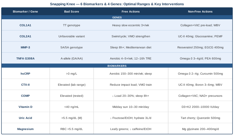
This article covers six biomarkers that are worth tracking if you are dealing with snapping knee syndrome, followed by four genetic variants with meaningful evidence for connective tissue and joint health. After that, you will find a synthesis of research — including work discussed extensively in clinical and science communication circles — that challenges some standard rehabilitation thinking, plus evidence-based complementary approaches with demonstrated benefit for knee soft-tissue conditions. No cure claims. Just better information pointing toward better decisions.
6 Biomarkers Worth Tracking for Snapping Knee Syndrome
Most people with snapping knee syndrome never have specific blood or urine markers assessed beyond a basic panel, if at all. Yet several biomarkers can reveal whether the mechanical snap is occurring against a backdrop of systemic inflammation, active cartilage breakdown, tissue stress, nutritional insufficiency, or impaired repair capacity. Knowing which of these is true for you changes the intervention entirely.
The six biomarkers below were selected for clinical relevance to knee soft-tissue conditions, practical accessibility, and evidence of actionability. Most can be ordered through standard labs or direct-access testing services; a few require specialist or functional medicine access.
1. High-Sensitivity C-Reactive Protein (hsCRP)
Why it matters: hsCRP is the most accessible marker of low-grade systemic inflammation currently in routine clinical use. In the context of snapping knee syndrome, an elevated hsCRP does not identify the mechanical snap itself — but it tells you whether the soft tissues surrounding the knee are operating inside a chronically inflamed environment. Tendons, bursae, synovial folds (plica), and periarticular connective tissue all become more reactive, less resilient, and slower to repair when systemic inflammatory signaling is elevated. Research on iliotibial band syndrome and patellar tendinopathy — two of the most common contributors to lateral and anterior knee snapping — consistently finds elevated inflammatory cytokines in symptomatic tissue. hsCRP reflects the systemic side of that picture and is the first place to look.
How to measure it: A standard blood draw. Specify high-sensitivity CRP — not standard CRP — since the high-sensitivity assay detects low-grade inflammation in the range most relevant for musculoskeletal tissue health. Cost: $15–$50 depending on bundling. Optimal target: below 0.5 mg/L. Borderline concern: 1–3 mg/L. Significant concern: above 3 mg/L.
If the score is elevated — the plan without supplements: The most consistent non-pharmacological interventions for reducing hsCRP involve structured aerobic exercise at moderate intensity (150–300 minutes per week), sleep quality improvement targeting 7–9 hours with consistent timing, and meaningful reduction of ultra-processed foods, refined carbohydrates, and excess dietary omega-6 linoleic acid. For individuals with a symptomatic knee, low-impact aerobic modalities — swimming, cycling, rowing — achieve cardiovascular benefit without adding mechanical stress to the inflamed joint. Time-restricted eating (12–16 hour overnight fasting window) also consistently reduces inflammatory markers in randomized trials, making it a practical dietary lever without requiring caloric restriction.
If the score is elevated — the plan with supplements or equipment: Omega-3 fatty acids (EPA + DHA): 2–4 g/day of combined EPA and DHA is among the most evidence-dense interventions for systemic hsCRP reduction. Mechanism: competitive displacement of arachidonic acid from membrane phospholipids, reducing downstream pro-inflammatory eicosanoid synthesis. Frequency: continuous; effects become measurable at 6–8 weeks. Cycling: no established need for breaks. Side effects: mild anticoagulation at doses above 3 g/day — relevant if taking blood thinners; fishy reflux mitigated by enteric-coated formulations or triglyceride-form fish oil.
Curcumin with piperine: 500–1000 mg curcumin combined with 5–10 mg piperine, taken with a fat-containing meal. A 2017 meta-analysis of randomized controlled trials found significant hsCRP reduction from curcumin supplementation. Frequency: once daily with food. Cycling: 8–12 weeks on, assess response, then decide on continuation. Side effects: rare GI upset; avoid high doses with gallbladder disease.
Red light / photobiomodulation (PBM): 630–850 nm devices applied over the knee and lower extremity for 10–20 minutes daily have been studied for local anti-inflammatory tissue effects, including reductions in TNF-α and IL-1β in periarticular soft tissue. Home LED panel devices in the therapeutic wavelength range cost $150–$600 and represent a practical, side-effect-free adjunct.
2. CTX-II (C-Terminal Cross-Linked Telopeptide of Type II Collagen)
Why it matters: CTX-II is currently the most validated urine biomarker of articular cartilage degradation available outside of research settings. Type II collagen is the structural backbone of hyaline cartilage, and when that cartilage is under abnormal mechanical load or enzymatic breakdown, CTX-II fragments are released and detectable in urine. Snapping knee syndrome is frequently associated with plica irritation, subtle meniscal stress, altered joint kinematics, or early patellofemoral changes — all of which can accelerate cartilage wear over time even before it is visible on imaging. Elevated CTX-II does not necessarily indicate MRI-visible cartilage damage, but it does signal a catabolic tilt in the joint environment that is worth addressing proactively, before damage accumulates.
How to measure it: Second morning void urine, corrected for creatinine. Not routinely available through standard primary care labs; requires a specialty or direct-access laboratory. Cost: $80–$200. Reference ranges vary by age, sex, and menopausal status. Trend over successive measurements — taken at consistent time points, at least 24 hours after significant loading — matters more than a single reading.
If the score is elevated — the plan without supplements: Load management is the most direct cartilage-protective intervention. Reducing high-impact repetitive loading — running on hard surfaces, deep barbell squats, high-volume stair climbing — decreases mechanical stress at the cartilage surface. Replacing some impact loading with aquatic exercise or cycling maintains cardiovascular and muscular fitness while dramatically reducing joint contact forces. Simultaneously, progressive strengthening of the quadriceps (particularly the VMO) and hip external rotators improves joint alignment and distributes load more evenly across the cartilage surface, which is as important as reducing total load.
If the score is elevated — the plan with supplements or equipment: Undenatured type II collagen (UC-II), 40 mg/day: UC-II targets cartilage-specific collagen via oral tolerization through the gut-associated lymphoid tissue, modulating the immune response to cartilage antigens. Randomized trials have shown reductions in joint discomfort and biomarkers of cartilage stress. Frequency: once daily, fasted if possible. Cycling: 90-day blocks with re-measurement of CTX-II to assess tissue response. Side effects: minimal; occasional mild GI tolerance issues.
Boron (3–6 mg/day): Boron has early human evidence for reducing urinary CTX-II, with proposed mechanisms involving modulation of calcium and magnesium metabolism and sex hormone bioavailability, both of which influence cartilage maintenance. Frequency: continuous. Side effects: safe at these doses; avoid supplementing above 10 mg/day.
Whole-body vibration (WBV) therapy: Low-frequency vibration protocols (25–45 Hz, 10–15 minutes, 3×/week) have shown cartilage-protective signals in controlled trials on knee OA populations, with proposed mechanisms including stimulation of chondrocyte anabolic activity and improved subchondral bone quality. Equipment cost: $300–$1,500 for platforms with appropriate frequency ranges.
3. COMP (Cartilage Oligomeric Matrix Protein)
Why it matters: COMP is a non-collagenous pentameric protein found in cartilage, tendon, and ligament. It is released into serum under mechanical stress or inflammatory conditions affecting joint and periarticular tissues. Serum COMP rises acutely after heavy loading and returns to baseline with adequate rest — making it a useful real-time indicator of mechanical demand on joint tissues. In individuals with snapping knee syndrome where a tendon or cartilage structure is repetitively irritated, chronically elevated COMP suggests that tissue is not recovering between loading sessions. Research has associated elevated COMP with early knee osteoarthritis, patellar tendinopathy, and a range of soft-tissue overuse conditions relevant to knee snapping.
How to measure it: Serum COMP via blood draw. Available through specialty labs and some sports medicine or functional medicine practices. Cost: $100–$200. Timing is critical: measure in a rested state, at least 24 hours after significant lower-body loading. Intra-individual trends over 3–6 month intervals are more informative than single readings.
If the score is elevated — the plan without supplements: Structured recovery management is the primary intervention. Reducing total weekly knee-loading volume by 20–30% during the assessment period, increasing sleep duration toward 8+ hours to maximize growth hormone-mediated tissue repair, and replacing complete rest with active recovery — walking, gentle hip mobility work, swimming — maintains tissue adaptability while allowing COMP to normalize. Compression therapy (graded compression sleeves or intermittent pneumatic compression devices used post-exercise) has supporting evidence for reducing post-exercise joint swelling markers in knee conditions.
If the score is elevated — the plan with supplements or equipment: Hydrolyzed collagen peptides + Vitamin C before loading: 10–15 g of hydrolyzed collagen combined with 50 mg Vitamin C, consumed 30–60 minutes before a loading session, was shown in a randomized crossover study (Shaw et al., 2017) to significantly increase collagen synthesis markers in the bloodstream during the post-exercise anabolic window. This intervention times substrate delivery to the period of peak collagen synthesis activity. Frequency: before each loading session. Cycling: continuous with loading program. Side effects: minimal at these doses.
NAD+ precursors (NMN or NR, 250–500 mg/day): Early mechanistic and human studies suggest that raising NAD+ levels supports mitochondrial function in cartilage and connective tissue cells and reduces NF-κB inflammatory signaling. Evidence is still emerging for joint applications specifically. Cycling: 12 weeks on, 4 weeks off, is a commonly used protocol. Side effects: generally mild; occasional GI effects; NMN is typically better tolerated.
Compression sleeve with post-activity elevation: Consistent use of a graduated knee compression sleeve (20–30 mmHg) during and after loading sessions reduces intra-articular pressure fluctuation and post-exercise synovial fluid volume shifts, supporting faster normalization of circulating COMP between sessions.
4. 25-Hydroxyvitamin D (25-OH Vitamin D)
Why it matters: Vitamin D deficiency impairs musculoskeletal function at multiple levels simultaneously: reduced muscle strength and contractile efficiency, impaired calcium signaling in tendons, increased production of pro-inflammatory cytokines including IL-6 and TNF-α, and decreased proteoglycan synthesis in cartilage matrix. For snapping knee syndrome, low Vitamin D is a modifiable factor that directly undermines connective tissue resilience, slows recovery from repetitive mechanical irritation, and amplifies the inflammatory response to tissue stress. Multiple systematic reviews have confirmed consistent associations between Vitamin D insufficiency and musculoskeletal pain, tendon pathology, and slower recovery from joint conditions. Evidence-focused clinicians including Peter Attia typically target a functional optimal of 40–60 ng/mL, well above the clinical deficiency threshold.
How to measure it: Standard blood draw. Universally available at any lab. Cost: $30–$80. Optimal functional range: 40–60 ng/mL. Insufficiency: 20–29 ng/mL. Deficiency: below 20 ng/mL. Potential toxicity zone: above 100 ng/mL (rare without aggressive supplementation).
If the score is low — the plan without supplements: Midday sun exposure (10–30 minutes on the arms and legs without sunscreen, between 10am and 2pm) remains the most efficient natural source of Vitamin D synthesis, though effectiveness depends heavily on latitude, skin tone, and season. Dietary contributions from fatty fish (salmon, mackerel), egg yolks, and liver are modest and rarely sufficient to correct established insufficiency on their own. In northern latitudes during winter, supplementation is practically unavoidable to reach the functional optimal range.
If the score is low — the plan with supplements or equipment: Vitamin D3 with Vitamin K2 (MK-7 form): For insufficiency (20–29 ng/mL): 2,000–4,000 IU D3 daily combined with 100–200 mcg MK-7 K2 to direct calcium to bone rather than soft tissue. For deficiency (below 20 ng/mL): 5,000–10,000 IU D3 for 8–12 weeks before retesting. Frequency: daily with a fat-containing meal (Vitamin D is fat-soluble). Cycling: continuous once the optimal range is reached; taper to maintenance dose (2,000–3,000 IU). Side effects: toxicity is possible above 10,000 IU chronically; retest at 3 months to calibrate dose. Always co-supplement with K2 when using doses above 2,000 IU.
Importantly, magnesium is required for several steps in Vitamin D activation and conversion. If magnesium is depleted — which is common — supplemental D3 may fail to raise serum levels effectively, making biomarker #6 below directly relevant here.
5. Serum Uric Acid
Why it matters: The association between uric acid and joints is typically reduced to gout in clinical conversations. But emerging research demonstrates that hyperuricemia — uric acid elevated well below the crystal deposition threshold — promotes low-grade joint and periarticular soft-tissue inflammation through activation of the NLRP3 inflammasome, driving production of IL-1β and IL-18. These inflammatory signals sensitize tendons and synovial tissue, amplify the pain response to mechanical snapping, and impair tissue repair. For individuals who notice that their snapping knee feels distinctly worse after high-protein meals, alcohol, excess fructose, or dehydration, uric acid is a plausible contributing mechanism worth quantifying.
How to measure it: Standard blood draw. Cost: $15–$40. Functional optimal (not just clinical gout threshold): 3.5–5.5 mg/dL for men, 2.5–4.5 mg/dL for women. Clinicians targeting metabolic health and joint longevity increasingly treat the lower end of these ranges as optimal rather than simply avoiding the clinical gout threshold.
If the score is elevated — the plan without supplements: Reduce high-fructose corn syrup and fruit juice (fructose is the primary dietary driver of uric acid synthesis via hepatic fructokinase activity, independent of purine metabolism). Reduce alcohol, particularly beer and spirits. Maintain hydration at 2.5–3.5 L of water daily to support renal uric acid excretion. Moderate rather than eliminate red meat and shellfish. Importantly, structured aerobic exercise consistently improves uric acid metabolism; the effect is largely mediated by improved renal clearance and reduced insulin resistance, both of which enhance urate excretion.
If the score is elevated — the plan with supplements or equipment: Tart cherry extract (concentrated capsules, equivalent to 480 ml of tart cherry juice): Tart cherry anthocyanins have consistent evidence for reducing serum uric acid through xanthine oxidase inhibition and improved urate excretion. Prefer concentrated capsule forms over juice to avoid high sugar content. Frequency: daily. Cycling: continuous; re-measure at 8 weeks. Side effects: minimal; ensure extract is standardized for anthocyanin content.
Quercetin (500–1000 mg/day): Quercetin inhibits xanthine oxidase — the same enzyme targeted by the pharmaceutical allopurinol — reducing uric acid synthesis. Evidence is preliminary but mechanistically coherent. Frequency: once or twice daily with food. Cycling: 8 weeks on, 4 off. Side effects: occasional headache; take with food.
Vitamin C (500 mg/day): A modest but consistent uricosuric effect from Vitamin C supplementation has been observed in randomized trials, with a meta-analysis finding an average reduction of approximately 0.35 mg/dL per 500 mg daily dose. This pairs well with the Vitamin D protocol above given the collagen cross-linking benefits at the same dose.
6. Serum Magnesium and RBC Magnesium
Why it matters: Magnesium participates in over 300 enzymatic reactions, including those governing muscle contraction and relaxation, tendon elasticity, anti-inflammatory cytokine regulation, and collagen cross-linking. Magnesium deficiency — affecting an estimated 45–50% of adults in developed countries despite serum levels appearing normal — is associated with increased muscle tone, tendon stiffness, joint sensitivity, reduced resilience to mechanical loading, and a blunted anabolic response to exercise. For snapping knee syndrome, inadequate magnesium can elevate resting muscle tension in the quadriceps, IT band complex, and hip musculature, mechanically tightening the structures that snap. It also amplifies inflammatory signaling, impairs sleep quality (reducing GH-mediated overnight tissue repair), and blunts Vitamin D activation. It is often the overlooked link when other interventions are not performing as expected.
How to measure it: Serum magnesium is available on any standard metabolic panel but is a poor indicator of total body magnesium status — only approximately 1% of total body magnesium is extracellular, meaning serum levels remain normal until intracellular stores are significantly depleted. RBC magnesium (red blood cell magnesium) reflects intracellular status far more accurately and is the preferred test for assessing functional magnesium sufficiency. Cost: $40–$100. Optimal serum range: 2.0–2.5 mg/dL. Optimal RBC magnesium range: 5.5–6.5 mg/dL.
If the score is low — the plan without supplements: Dietary sources with meaningful magnesium content include dark leafy greens (particularly spinach and Swiss chard), pumpkin seeds, almonds, dark chocolate above 70% cacao, and legumes. Reducing caffeine and alcohol — both of which significantly increase renal magnesium excretion — helps preserve dietary magnesium. Many people consume adequate dietary magnesium but lose it through stress, alcohol, and high coffee intake, making lifestyle modification as important as dietary optimization.
If the score is low — the plan with supplements or equipment: Magnesium glycinate or magnesium malate, 200–400 mg elemental magnesium per day: Both forms have superior bioavailability compared to magnesium oxide (which is poorly absorbed and primarily acts as a laxative). Glycinate is favored for its calming effect and sleep support; malate is preferred when energy metabolism is a concern. Frequency: once daily in the evening to leverage parasympathetic effects and improve sleep quality, which compounds the tissue repair benefit. Cycling: continuous. Side effects: loose stools at higher doses — titrate up from 200 mg over 2–3 weeks. Retest RBC magnesium at 8–12 weeks.
Transdermal magnesium oil (applied to thighs and calves): Evidence for transdermal absorption is moderate; some practitioners use it as an adjunct when GI tolerance limits oral dosing or as a direct soft-tissue intervention for tight quadriceps and hamstrings. Cost: $15–$30/month. Practical for targeted local application before sleep or after training sessions.
Tracking these six biomarkers does not require a specialist referral. A direct-access blood draw can cover hsCRP, Vitamin D, uric acid, and serum magnesium for under $150 through services like LabCorp or your GP. CTX-II and COMP require more targeted orders, accessible through functional medicine practitioners or sports medicine physicians. What matters is not a single snapshot but a trend across 3–6 month intervals as you implement changes. That feedback loop is something generic protocols never provide.
The Genetic Side: 4 Key Variants That Shape Your Knee Tissue
Biomarkers reflect what is happening in your biology right now. Genetics reveals the structural and inflammatory tendencies your tissue was built with — and where it may need sustained, specific support. Genetic variants individually have modest effect sizes and are always modified by training history, nutrition, and environment. But understanding your tendencies helps you calibrate expectations, prioritize interventions, and recognize why the same approach that works well for someone else may underperform for you.
The four variants below are the most studied in connective tissue, cartilage biology, and joint inflammatory response. Most are accessible through consumer genetic platforms (23andMe, AncestryDNA) combined with third-party analysis tools, or through clinical genetic panels offered by functional medicine practitioners.
COL1A1 — The Tendon Architecture Gene
What it does: COL1A1 encodes the alpha-1 chain of type I collagen — the dominant structural protein in tendons, ligaments, joint capsules, and periarticular connective tissue. The Sp1 binding site polymorphism (rs1800012) has been studied extensively in sports medicine populations. Carriers of the TT genotype tend to produce tendons that are structurally stiffer and less mechanically compliant — meaning they tolerate load more poorly at the extremes of range, are more susceptible to repetitive irritation, and recover more slowly from microtrauma. For snapping knee syndrome driven by iliotibial band, biceps femoris, or popliteus tendon involvement, this variant represents a meaningful biological risk factor.
What the evidence says: A meta-analysis published in the British Journal of Sports Medicine confirmed significant associations between COL1A1 Sp1 variants and soft-tissue injury risk across multiple athletic populations. Effect sizes are modest, as is typical for complex trait genetics, but the association is directionally consistent across cohorts.
If the gene variant is unfavorable — the plan without supplements: Progressive tendon loading through heavy slow resistance (HSR) training is the primary and most evidence-supported epigenetic lever for improving tendon quality regardless of genetic starting point. The protocol: 3–4 sets of slow eccentric-concentric movements (3 seconds down, 3 seconds up) using exercises that load the relevant knee structures — eccentric step-downs, leg press, single-leg Romanian deadlifts — performed 3 times per week with progressive load over 12 weeks. The mechanical stimulus drives collagen fiber realignment and cross-link maturation, effectively improving the tendon's functional properties even when the architectural tendency is toward stiffness.
Eccentric loading also deserves specific attention. Eccentric muscle contractions (lengthening under load) produce the highest tendon strain and are the most potent stimulus for collagen remodeling. For knee tendon involvement, eccentric single-leg decline squats (8–15 reps, 3 sets, twice weekly) represent the most rigorously studied eccentric protocol in knee tendon research.
If the gene variant is unfavorable — the plan with supplements or equipment: Hydrolyzed collagen + Vitamin C (10–15 g + 50 mg, 30–60 minutes before loading): As detailed in the COMP biomarker section, this pre-loading protocol specifically upregulates collagen synthesis in tendons during the post-exercise anabolic window. For COL1A1 TT carriers, this represents a direct substrate support strategy for a gene affecting collagen structural quality. Frequency: before each loading session, 4–5 times per week. Cycling: continuous with loading program.
Whole-body vibration training (25–40 Hz, 10–15 minutes, 3×/week): Vibration platform use during static and dynamic knee exercises has been studied for promoting tendon collagen remodeling and improving musculotendinous mechanical properties. Protocols typically run in 8–12 week blocks. Equipment cost: $300–$1,500 for appropriate-quality platforms.
COL2A1 — The Cartilage Blueprint Gene
What it does: COL2A1 encodes the alpha-1 chain of type II collagen — the primary structural protein of articular cartilage. Variations in this gene influence cartilage thickness, proteoglycan retention capacity, and long-term joint resilience under repetitive load. While rare high-penetrance mutations in COL2A1 cause severe skeletal dysplasias, common polymorphisms influence more subtle differences in cartilage matrix organization that compound progressively over decades of mechanical use. Snapping knee syndrome that involves plica thickening, meniscal stress, or subtle patellofemoral loading abnormalities is particularly relevant here, as cartilage quality directly influences how the joint responds to the repetitive mechanical events associated with snapping.
What the evidence says: Research in early-onset knee OA populations has linked COL2A1 variants with altered cartilage matrix organization and increased susceptibility to degradation under load. Human evidence remains largely associative at this stage; mechanistic evidence from cell and animal models is more developed. This is a field where genetic research is still maturing.
If the gene variant is unfavorable — the plan without supplements: Preserving cartilage integrity when COL2A1 function may be suboptimal requires careful management of the load-to-recovery ratio. Swimming and cycling are cartilage-friendly because they provide muscular and cardiovascular training load without the impact peaks that drive cartilage compression-decompression cycles at their highest intensity. Progressive quadriceps and hip external rotator strengthening remains the single most evidence-supported intervention for reducing cartilage contact stress at the patellofemoral and tibiofemoral joints — not by reducing loading but by improving load distribution. Unloaded deep flexion (full-squat positions without body weight) practiced daily as a mobility drill also maintains cartilage health by facilitating synovial fluid circulation through the joint surfaces.
If the gene variant is unfavorable — the plan with supplements or equipment: Undenatured type II collagen (UC-II, 40 mg/day): The oral tolerization mechanism of UC-II is specifically relevant to cartilage-targeted intervention — it targets the immune response to type II collagen fragments in the joint, which is the same collagen encoded by COL2A1. Cycling: 90-day blocks with measurement of CTX-II to assess response. Side effects: minimal.
Glucosamine sulfate (1500 mg/day) + Chondroitin sulfate (1200 mg/day): Evidence is genuinely mixed in the broader OA literature, but a subset of well-designed trials shows structural benefit — particularly in individuals with elevated cartilage turnover markers at baseline. Best approached as a 6–12 month monitored trial rather than indefinite use. Side effects: minimal; occasional GI discomfort; glucosamine may affect blood glucose in insulin-resistant individuals.
Pulsed electromagnetic field (PEMF) therapy: PEMF has early clinical trial evidence for stimulating chondrocyte proliferation and reducing cartilage degradation markers in knee OA populations. Proposed mechanism: electromagnetic field-mediated activation of adenosine receptors and anti-inflammatory signaling in cartilage cells. Home devices range $200–$800; typical protocols involve 30 minutes daily for 12-week cycles.
MMP-3 (Matrix Metalloproteinase-3) — The Tissue Remodeling Regulator
What it does: MMP-3 (stromelysin-1) is an enzyme that degrades components of the extracellular matrix including collagen, proteoglycans, and fibronectin in cartilage and synovial tissue. Its expression is controlled partly by a common promoter polymorphism (5A/6A, rs3025058). Homozygous 5A carriers produce significantly more MMP-3 activity, which, when combined with inflammatory triggers, accelerates breakdown of the cartilage matrix and soft-tissue collagen structures around the joint. For snapping knee syndrome, this variant is relevant because plica thickening, synovial inflammation, and accelerated collagen turnover in tendons are all driven in part by MMP-3 activity. It also means that stressors that other people's joints shrug off — heavy loading without adequate recovery, poor sleep, metabolic inflammation — produce more tissue-level damage in 5A/5A carriers.
What the evidence says: MMP-3 variants have been associated with knee OA progression, tendon matrix disorganization, and differential soft-tissue responses to exercise loading across multiple cohort studies. The variant also interacts meaningfully with inflammatory markers — when both TNF-α and MMP-3 activity are elevated simultaneously, the degradation effect on joint tissue is amplified beyond what either produces alone.
If the gene variant is unfavorable — the plan without supplements: Avoiding the primary environmental activators of MMP-3 expression matters as much as any loading strategy. Chronic psychological stress (via elevated cortisol), excessive alcohol, smoking, and hyperglycemia all upregulate MMP-3 gene expression epigenetically, meaning lifestyle factors can either amplify or suppress the variant's impact. Sleep quality is a particularly direct modulator — sustained sleep under 6 hours is associated with elevated MMP activity in joint tissue, making consistent deep sleep a frontline structural intervention. The Mediterranean dietary pattern has specific anti-MMP-3 evidence through its effects on NF-κB signaling, the primary transcriptional driver of MMP-3 upregulation.
If the gene variant is unfavorable — the plan with supplements or equipment: Resveratrol (250–500 mg/day, trans-resveratrol form): Trans-resveratrol inhibits NF-κB activation and downregulates MMP-3 expression in cartilage tissue in both cell studies and some human trials with OA populations. Cycling: 8–12 weeks on, 4 weeks off. Side effects: generally well tolerated; potential interaction with blood thinners at high doses.
EGCG (epigallocatechin-3-gallate from green tea extract, 400–800 mg/day): EGCG suppresses MMP-3 upregulation via NF-κB inhibition in multiple studies, with cartilage-protective effects in human cell studies and some OA-related research. Frequency: once or twice daily, with food to reduce GI effects. Cycling: continuous. Side effects: mild caffeine effect; take with food; avoid on empty stomach.
Sub-antimicrobial doxycycline (20 mg twice daily, prescription only): Doxycycline at sub-antibiotic doses is a direct MMP inhibitor that has clinical trial data for slowing cartilage loss in OA populations. This is a physician-supervised intervention relevant only when documented progressive joint involvement is established and conservative measures are insufficient.
TNFA G308A — The Inflammatory Amplifier
What it does: The TNF-α G308A polymorphism (rs1800629) is one of the most extensively studied inflammatory genetics variants in the entire human disease literature. The A allele — present in GA heterozygotes and AA homozygotes — is associated with higher baseline TNF-α transcription in response to mechanical, metabolic, and immunological triggers. For snapping knee syndrome, elevated TNF-α amplifies the inflammatory response to repetitive tendon and synovial irritation, slows soft-tissue repair rate, lowers the pain threshold for mechanical stimulation, and sensitizes nociceptors around the joint. In practical terms: TNFA A-allele carriers may experience significantly more pain and tissue reactivity from the same degree of mechanical snapping as non-carriers, and may recover more slowly between loading sessions. This is not a reason for pessimism — it is a reason for precision.
What the evidence says: TNF-α G308A has been associated with greater severity of tendinopathy, faster OA progression, higher perioperative inflammatory responses, and altered pain sensitivity in multiple human genetic cohort studies. It also interacts with the MMP-3 variant above — when both are present in their high-expression forms, the combined effect on tissue degradation under inflammatory conditions is more than additive.
If the gene variant is unfavorable — the plan without supplements: Anti-inflammatory lifestyle fundamentals matter more for TNFA A-allele carriers than for most people, because the same behavioral inputs produce larger inflammatory outputs. The evidence-based levers: Mediterranean dietary pattern (consistent meta-analytic support for TNF-α reduction through omega-3 intake, polyphenol activity, and reduced saturated fat); structured moderate aerobic exercise 4–5 days per week (chronic downregulation of TNF-α expression via IL-10 and PGC-1α-mediated pathways); time-restricted eating with a 12–16 hour overnight fast; elimination of smoking (which directly increases TNF-α promoter activity); and sleep quality targeting 8 hours (sleep deprivation is one of the strongest acute TNF-α inducers known). These are not secondary considerations for this genotype — they are the frontline intervention.
If the gene variant is unfavorable — the plan with supplements or equipment: Omega-3 (EPA + DHA, 3–4 g/day): Competes with arachidonic acid at the enzymatic level, reducing the substrate available for TNF-α-driven eicosanoid synthesis. This is the most consistently evidence-supported anti-TNF-α supplement available without prescription. Frequency: continuous. Cycling: none needed. Side effects: mild anticoagulation above 3 g/day.
Palmitoylethanolamide (PEA), 600–1200 mg/day: PEA is an endogenous lipid mediator that activates PPAR-α receptors, reducing mast cell and macrophage activity and directly downregulating TNF-α and IL-1β release in periarticular tissue. It has a specific and growing body of human clinical trial evidence for joint and soft-tissue pain with an excellent safety profile. Frequency: twice daily. Cycling: 8–12 week blocks; re-evaluate response. Side effects: very well tolerated; no significant drug interactions reported.
Low-dose naltrexone (LDN, 1.5–4.5 mg at night, prescription required): LDN modulates microglial and peripheral immune cell inflammatory activity, with a growing body of evidence for tendon, joint, and soft-tissue pain conditions. Mechanism involves transient opioid receptor blockade driving endorphin upregulation and separate Toll-like receptor 4 antagonism that reduces TNF-α signaling. This is a physician-supervised intervention for cases where inflammatory-driven pain remains high despite lifestyle and supplement optimization.
The genetic picture rarely operates in isolation. COL1A1 variants have greater impact when Vitamin D is low; MMP-3 activity escalates more when uric acid is elevated; TNFA variants create compounding damage when hsCRP is chronically high. Reading genetics and biomarkers together — not separately — is where the genuinely actionable signal emerges.
10 Things the Research on Connective Tissue Loading Gets Right That Clinical Practice Often Ignores
Some of the most practically useful research on connective tissue repair over the past decade has come from laboratories studying the mechanics of collagen synthesis under load — work that has been synthesized and communicated extensively in clinical and science education circles, including discussions with researchers like Keith Baar (University of California, Davis), whose laboratory has produced foundational findings on collagen metabolism and tendon adaptation. These findings challenge several assumptions embedded in standard physical therapy and sports medicine practice.
1. Collagen Synthesis Has a Narrow Anabolic Window After Loading
Collagen synthesis in tendons and ligaments peaks approximately 6 hours after a mechanical loading stimulus and returns to baseline by 24 hours. The critical implication: consuming substrate — hydrolyzed collagen with Vitamin C — precisely 30–60 minutes before loading, not after, ensures peak availability during this post-loading synthetic window. This timing effect has been demonstrated in human randomized trials. Post-workout collagen supplementation misses the window entirely.2. Static Stretching Does Not Structurally Remodel Tendons
Passive elongation of a tendon through static stretching does not produce the mechanical strain required to drive collagen fiber turnover and cross-link maturation. Only loaded elongation — tissue placed under meaningful tension while being lengthened — produces the mechanical stimulus for adaptation. For the knee, this makes slow eccentric loading far more relevant than prolonged passive stretching, however much the latter feels productive.3. Heat Accelerates Collagen Gel Stiffness After Loading
Laboratory work from the Baar group demonstrated that brief heat application (10–15 minutes at 40–42°C) to connective tissue following loading increased collagen gel stiffness and cross-linking in vitro. The practical translation: sauna exposure or heated pad application over the knee after loading sessions may accelerate the maturation of newly synthesized collagen, potentially improving the structural benefit of each session beyond what exercise alone provides.4. Icing After Exercise Blunts Connective Tissue Adaptation
Ice applied immediately post-exercise reduces reactive oxygen species and prostaglandin signaling — the same signals that drive connective tissue adaptation. For acute traumatic injury management, this trade-off may be acceptable. For routine exercise recovery where the goal is tissue adaptation and remodeling, avoiding icing in the 2–3 hours post-loading may preserve the anabolic signaling cascade that the session was intended to generate.5. Tendons Need 36–48 Hours to Recover Between Sessions
Unlike skeletal muscle, which can recover its contractile properties within 24 hours after moderate loading, tendons require 36–48 hours for structural remodeling to occur between sessions. Consecutive-day loading of the same tendinous structure may produce a net catabolic effect rather than adaptation, particularly in individuals with compromised genetic or nutritional support for collagen synthesis. Knee-specific loading sessions should be separated by at least 48 hours for tissue quality to accumulate.6. Isometric Contractions Reduce Pain Without Suppressing Adaptation
Sustained isometric quadriceps contractions — wall sits, isometric leg press holds at 60–90 degrees of knee flexion — produce reliable short-term reductions in patellar and periarticular knee pain for 20–45 minutes post-exercise, mediated by cortical inhibition of pain pathways. Crucially, unlike NSAIDs, this analgesic effect does not suppress prostaglandin-mediated tissue adaptation. Used as a warm-up before loading sessions, isometrics allow symptom-managed exercise without pharmacological interference with healing.7. Heavy Slow Eccentric Loading Is the Most Evidence-Supported Tendon Stimulus
Across multiple randomized trials and meta-analyses, heavy slow eccentric-concentric loading (3 seconds eccentric, 3 seconds concentric, with meaningful load progression over 8–12 weeks) represents the most consistently effective protocol for tendon remodeling in clinical populations. For knee tendons, single-leg eccentric decline squats, leg press with controlled descent, and slow step-downs have the strongest supporting data. Volume should begin conservatively and increase by approximately 10% per week to avoid exceeding the tendon's remodeling capacity.8. Sleep Is the Primary Anabolic Stimulus for Connective Tissue Repair
Growth hormone and IGF-1 — the dominant hormonal signals driving tendon and cartilage repair — are secreted predominantly during slow-wave sleep in the first half of the night. Consistently sleeping below 7.5 hours interrupts this repair cascade as effectively as under-loading. Optimizing sleep quantity and quality (consistent timing, cool and dark room, minimal alcohol, reduced evening light exposure) is a connective tissue intervention in its own right, not merely a lifestyle recommendation.9. Vitamin C Is Rate-Limiting for Collagen Cross-Linking
Prolyl hydroxylase and lysyl hydroxylase — the enzymes responsible for adding the hydroxyproline and hydroxylysine residues required for stable collagen cross-linking — are both Vitamin C-dependent. Without adequate circulating Vitamin C (dietary plus supplemental intake above 200 mg/day), newly synthesized collagen chains are structurally weaker and degrade more rapidly. This makes consistent Vitamin C intake a precondition for the collagen synthesis benefits of loading, not merely an adjunct.10. The Musculotendinous Junction Is the Point of Highest Strain Concentration
Biomechanical studies consistently show that during eccentric loading, peak strain concentrates at the junction between muscle belly and tendon — not in the mid-tendon, as often assumed. For snapping knee conditions involving the iliotibial band complex, biceps femoris, or quadriceps tendon, controlling the rate of load progression at this junction — not just managing absolute load — is the most important variable in preventing setbacks during rehabilitation. Increasing load by more than 10% per week exceeds the adaptive rate at this junction in most individuals.Complementary Approaches With Clinical Evidence for Knee Soft-Tissue Conditions
The following modalities have meaningful human evidence for knee soft-tissue conditions and serve as practical adjuncts to the biomarker and genetic optimization strategies above. Each is presented with specific evidence and a realistic protocol.
Low-Level Laser Therapy and Photobiomodulation
Low-level laser therapy (LLLT) — also called photobiomodulation (PBM) — uses red and near-infrared light (630–1000 nm) to penetrate tissue and stimulate mitochondrial cytochrome c oxidase activity, reduce local inflammatory cytokine production (TNF-α, IL-1β), and accelerate repair processes in tendons, ligaments, and periarticular soft tissue. For snapping knee syndrome, its primary relevance is in addressing the inflammatory and repair-impaired tissue microenvironment around the knee without systemic effects, drug interactions, or recovery interference.
A Cochrane-reviewed systematic review published in Brosseau et al. (2009) found significant short-term pain reduction from LLLT for knee osteoarthritis and periarticular soft-tissue conditions versus sham. Separate randomized trials in patellar tendinopathy populations have shown accelerated collagen synthesis markers and pain score improvements with 3×/week PBM over 8 weeks. The evidence is most consistent for wavelengths of 810–850 nm with doses in the 4–8 J/cm² range for superficial knee structures.
Practically: Class 3B or 4 laser devices (or consumer-grade 630–850 nm LED panels with sufficient power density) applied directly over the lateral, medial, or anterior knee for 10–20 minutes per session, 3–5 times per week. Home LED panels in the therapeutic range cost $150–$600. For deeper tissue penetration (popliteal tendon, posterior knee), class 4 laser treatment through a sports medicine clinic or physiotherapy practice with laser certification is more effective than home LED panels. Duration: minimum 8–12 week trial. No significant side effects; avoid direct eye exposure to the beam.
Massage Therapy and Soft-Tissue Mobilization
Manual therapy to the soft tissues surrounding the knee — including the iliotibial band, quadriceps, hamstrings, calves, and hip external rotators — addresses muscular tension and fascial restrictions that directly alter joint mechanics and contribute to snapping by increasing tension on the structures that catch. By improving tissue compliance and reducing passive mechanical tension along the snapping structure, targeted massage can reduce symptom frequency and improve the quality of subsequent loading sessions.
A randomized controlled trial in Manual Therapy demonstrated significant improvements in knee pain and function from soft-tissue massage combined with exercise compared to exercise alone over an 8-week protocol. Myofascial release techniques targeting the IT band, lateral hip rotators (piriformis, gemelli), and tensor fasciae latae are particularly relevant for lateral knee snapping presentations where IT band tension is the mechanical driver.
Practically: Two sessions per week of 45–60 minutes focusing on hip, thigh, and knee musculature during the active rehabilitation phase, tapering to once weekly as symptoms resolve. For self-care between sessions: foam rolling the IT band (10–15 slow passes per side before loading sessions), hip external rotators, and quadriceps has supporting evidence as a preparatory tool for reducing passive tissue tension. Caution: deep transverse friction massage applied directly over an acutely inflamed tendon insertion should be avoided until the acute phase resolves, as it can increase local tissue irritation during the reactive phase.
Biofeedback and Neuromuscular Retraining
EMG-based biofeedback devices attached to the quadriceps (particularly the VMO), hamstrings, or hip external rotators provide real-time feedback on muscle activation patterns during movement. For snapping knee syndrome where abnormal or asymmetric muscle firing sequences allow a structure to move off its anatomical track, biofeedback retraining addresses a neuromuscular root cause that strengthening exercises alone cannot reliably correct — since strengthening without biofeedback does not guarantee correct activation sequencing.
A controlled study in the Journal of Orthopaedic and Sports Physical Therapy demonstrated that EMG biofeedback-assisted quadriceps training produced significantly superior VMO activation symmetry and pain reduction compared to standard resistance exercise in patellofemoral pain patients — a finding directly relevant to anterior and lateral knee snapping presentations. Gait retraining using real-time feedback has also been applied successfully to iliotibial band syndrome, with kinematic corrections at the hip and knee producing symptom reductions at 1-year follow-up in prospective studies.
Practically: EMG biofeedback is available through physical therapy clinics, typically delivered across 6–12 sessions. For home retraining after initial clinic-based learning, consumer wearable EMG systems allow independent practice with appropriate target settings established by a sports physiotherapist. The clinical goal is not simply strengthening but ensuring the correct muscles activate in the correct sequence and at the correct magnitude during the specific movement pattern that provokes snapping — which requires objective feedback, not assumption.
Yoga
Yoga combines controlled loaded stretching, body-weight resistance, proprioceptive challenge, and structured breathwork — making it one of the few exercise modalities that simultaneously addresses the muscular tightness, connective tissue loading, and neuromuscular control deficits commonly contributing to snapping knee syndrome. Hip-opening sequences (pigeon pose, lizard, figure-four) directly target hip external rotator and IT band tension; standing balance poses load the knee in a controlled single-leg context that closely simulates functional movement demands.
A randomized trial published in Annals of Internal Medicine (2015) found that yoga was non-inferior to physical therapy for chronic musculoskeletal knee pain, with improvements in pain, function, and movement confidence sustained at a 12-month follow-up. The hip-focused component of yoga is particularly relevant for lateral knee snapping presentations where IT band tension and hip external rotator weakness are the biomechanical drivers.
Practically: 2–3 sessions per week of 45–60 minutes, integrating hip mobility, single-leg balance work, and slow eccentric lower-body loading. Yin yoga — which uses passive holds of 2–5 minutes in hip-opening and knee-loading positions — provides connective tissue-specific loading through sustained elongation that more dynamic yoga styles do not. Avoid deep full-weight knee flexion in the early phase if it provokes snapping or discomfort; build range of motion progressively under qualified guidance rather than pushing through tissue resistance.
Tai Chi
Tai chi provides slow, coordinated, closed-chain loading of the knee through controlled weight-shifting, hip rotation, and sustained single-leg stance — the exact movement quality that most benefits tendon remodeling without adding the impact loading that frequently aggravates snapping knee presentations. Its simultaneous training of proprioception, neuromuscular timing, and slow eccentric control addresses the mechanical precision deficit that allows snapping structures to cycle off-track under dynamic load.
A well-powered randomized trial published in Annals of Internal Medicine (2016) found that 12 weeks of tai chi produced outcomes equivalent or superior to physical therapy for knee osteoarthritis — including pain scores, stiffness ratings, physical function, and quality of life measures — with benefits maintained at 52-week follow-up. Proprioceptive training specifically, which tai chi delivers systematically across every session, is among the strongest predictors of improved knee mechanics and reduced symptom recurrence in knee soft-tissue conditions.
Practically: Two to three sessions per week of 45–60 minutes, preferably with a certified instructor familiar with Yang-style tai chi (the style most studied in clinical trials). The first 4 weeks involve learning the movement forms and will feel more like skill acquisition than therapeutic exercise. The therapeutic effect accumulates over 8–12 weeks of consistent practice. Tai chi pairs effectively with both biofeedback retraining and the eccentric loading protocols described above when scheduled on alternating days — the tai chi sessions providing neuromuscular coordination work, the loading sessions providing the structural collagen stimulus.
Conclusion
Snapping knee syndrome sits at the intersection of biomechanics, connective tissue biology, inflammation, and individual genetic variance. The six biomarkers covered here — hsCRP, CTX-II, COMP, Vitamin D, uric acid, and magnesium — provide a window into the biological environment your knee operates in. The four genetic variants — COL1A1, COL2A1, MMP-3, and TNFA — reveal structural and inflammatory tendencies that shaped that environment long before the snap became a problem. Neither picture is complete without the other, but together they offer a degree of precision that generic rehabilitation protocols simply do not reach.
The most useful next step is rarely the most ambitious one. Request a basic panel — hsCRP, Vitamin D, uric acid, and serum magnesium — from your GP or a direct-access laboratory. If you have consumer genetic data, run it through a reputable third-party analysis tool. Bring those findings to a sports medicine physician, physiotherapist, or functional medicine clinician who can interpret them in the context of your mechanics, loading history, and recovery patterns. The information will not eliminate the snap overnight — but it will help you stop guessing and start making decisions grounded in what your own biology is actually doing.
Musculoskeletal: Joint Conditions Tendon & Ligament Conditions Sports Injuries
Autoimmune: Inflammatory Conditions Connective Tissue Conditions