This article contains AI generated content.
· UpdatedSpinal Stenosis Genes and Biomarkers: 6 Genes And 6 Biomarkers To Track
Introduction
Living with spinal stenosis often means navigating a frustrating combination of pain, limited mobility, and vague explanations. You stop mid-walk because your legs go heavy and numb. You shift position constantly because no chair, bed, or posture feels right for long. The answers you receive are often structurally accurate but practically useless: "your canal is narrowed," "try physical therapy," "avoid heavy lifting." What rarely gets addressed is why it happened to you specifically, and what that means for how to actually slow it down.
Spinal stenosis is not a single-cause condition. It develops through a combination of mechanical wear, inflammatory dysregulation, cartilage breakdown, and genetic predispositions that differ substantially from person to person. Two people with identical MRI findings can have completely different symptom profiles, different rates of progression, and different responses to the same treatment. That's not a mystery — it's a signal that the underlying biology differs, and that generic protocols have clear limits.
This article takes a more targeted approach. Blood-based biomarkers can reveal whether chronic inflammation, matrix degradation, or abnormal bone remodeling is the dominant driver in your case right now. Genetic variants can explain why your connective tissue may be structurally vulnerable, or why your inflammatory response is wired toward excess — and point toward interventions that actually address those mechanisms. Neither biomarkers nor genetics are a complete answer, but together they offer something far more useful than a blanket diagnosis.
Better information leads to better decisions. If your hsCRP is elevated and your Vitamin D is low, your path looks different than if your COMP is elevated or your bone turnover is off-balance. This article covers six actionable blood biomarkers with specific tracking and intervention protocols, followed by six gene variants worth knowing about, a deep dive into the most clinically rigorous spine health framework available, and a review of complementary approaches backed by real human evidence.
6 Biomarkers That Reveal What's Really Happening in Your Spine
Spinal stenosis is rarely purely a structural problem. The narrowing of the canal happens over time, driven by inflammatory signals, tissue degradation enzymes, bone remodeling imbalances, and hormonal deficits. Blood biomarkers can reveal which of these processes is most active right now — and that matters, because interventions that target the actual driver of your problem are far more effective than interventions aimed at symptoms alone.
The six biomarkers below are among the most clinically informative for understanding the biological landscape of spinal stenosis. Some cost under $30; others require specialty panels. Each one points toward a specific mechanism and, importantly, toward a specific response strategy.
1. hsCRP — The Inflammation Signal
High-sensitivity C-reactive protein (hsCRP) is produced by the liver in response to systemic inflammation. It is one of the most widely studied inflammatory markers in musculoskeletal research and has been consistently associated with chronic low back pain, disc degeneration, nerve root sensitization, and ligamentum flavum hypertrophy — the thickening of the spinal ligament that directly narrows the canal in many stenosis cases.
In spinal stenosis, chronic low-grade inflammation does more than cause pain. It accelerates osteophyte formation, promotes connective tissue thickening, and increases pain sensitivity through central sensitization. An elevated hsCRP — even mildly elevated, between 1.0 and 3.0 mg/L — may indicate that active inflammation is a primary driver of your symptoms rather than fixed structural damage alone. Research consistently links elevated inflammatory markers to worse functional outcomes and more severe pain in lumbar spinal stenosis.
How to measure it
hsCRP is a standard blood test available at any clinical lab. Most primary care physicians can order it, and it is often included in cardiovascular risk panels. Cost: $10–$40 out of pocket, frequently covered by insurance. Optimal range (per Peter Attia and most functional medicine frameworks): below 0.5 mg/L. Borderline: 0.5–1.0 mg/L. Elevated: 1.0–3.0 mg/L. High risk: above 3.0 mg/L. For stenosis tracking, aim to measure it fasting, away from acute illness or vigorous exercise (both transiently raise hsCRP).
If the score is bad, the plan without supplements
The most evidence-backed lifestyle intervention for hsCRP is dietary. An anti-inflammatory eating pattern — reducing ultra-processed foods, refined sugars, seed oils, and trans fats while increasing omega-3-rich fish (sardines, mackerel, salmon), leafy greens, olive oil, and polyphenol-rich foods — has been shown to lower hsCRP by 20–40% over 8–12 weeks in multiple controlled trials. Regular moderate exercise also reduces resting hsCRP independently of weight change: 30 minutes of walking daily can produce measurable reductions within 6 weeks. Sleep quality matters directly: consistently under 6 hours of sleep raises hsCRP. Eliminating smoking and reducing alcohol to one or fewer drinks per day are non-negotiable for anyone with chronically elevated levels.
If the score is bad, the plan with supplements or equipment
Omega-3 fatty acids (EPA + DHA, 2–4g daily) are the most evidence-supported supplement for hsCRP reduction — a 2012 meta-analysis in Atherosclerosis confirmed significant reductions at this dose range. Curcumin with piperine (500–1000mg curcumin + 5–10mg piperine/day) has shown consistent hsCRP reduction in multiple randomized trials for inflammatory conditions. Magnesium glycinate (300–400mg nightly) reduces inflammatory tone, particularly in the roughly 50% of adults who are deficient. Cycling: omega-3 is well tolerated year-round; curcumin can be cycled 8 weeks on, 2 weeks off. Side effects: high-dose omega-3 may mildly affect platelet function — flag this if you are on anticoagulants. Equipment option: a red light therapy device (660–850nm) applied to the lumbar spine for 10–15 minutes daily has emerging evidence for reducing local inflammatory markers and is a reasonable adjunct at moderate cost ($100–$400 for a quality panel).
2. Vitamin D (25-OH-D) — Bone, Nerve, and Inflammatory Regulation
25-hydroxyvitamin D is the standard serum measure of Vitamin D status, and it functions more like a hormone than a vitamin. It regulates calcium absorption, bone mineral density, immune signaling, and — critically for spinal stenosis patients — nerve function and myelin integrity. When spinal stenosis involves nerve root compression, the health of the nerve itself affects both symptom severity and recovery potential.
Vitamin D deficiency is extremely common in people with chronic back pain. A level below 20 ng/mL promotes secondary hyperparathyroidism, which accelerates bone resorption and may worsen vertebral instability over time. Low Vitamin D also enhances pro-inflammatory cytokine production, creating a feedback loop that worsens both tissue degradation and pain. Studies have found that low Vitamin D correlates with more severe stenosis symptoms and slower recovery following spinal interventions. Peter Attia consistently emphasizes Vitamin D as one of the highest-leverage, most under-optimized biomarkers in general health tracking.
How to measure it
A 25-OH-D blood test is standard and widely available. Cost: $30–$60 out of pocket; often covered when ordered for deficiency screening. Optimal range per most functional medicine practitioners including Peter Attia: 40–60 ng/mL (100–150 nmol/L). Below 30 ng/mL is insufficient. Below 20 ng/mL is deficient. Above 100 ng/mL may indicate toxicity, especially if supplementing. Retest every 3–6 months while actively adjusting levels.
If the score is bad, the plan without supplements
Midday sun exposure — 15–30 minutes on arms and legs without sunscreen, 4–5 days per week — is the most natural way to raise Vitamin D. This is effective for lighter-skinned individuals in sunny climates but insufficient at latitudes above 35° during winter months. Dietary sources (fatty fish, egg yolks, fortified dairy) provide modest support. Regular outdoor walking achieves two goals at once: sun exposure plus the anti-inflammatory benefits of movement.
If the score is bad, the plan with supplements or equipment
Vitamin D3 (cholecalciferol, 2000–5000 IU/day) is the standard recommendation for deficiency correction. Always pair with Vitamin K2 MK-7 (100–200mcg/day) to direct calcium into bone rather than soft tissue. If levels are severely low (below 20 ng/mL), a loading protocol of 10,000 IU/day for 8–12 weeks under physician oversight may be warranted before dropping to maintenance. Side effects: Vitamin D toxicity is rare but possible at sustained very high doses — monitor serum calcium if supplementing above 5,000 IU long-term. Equipment alternative for those with limited sun access: a UVB phototherapy lamp (311nm narrowband) used 5–10 minutes per session, 3× per week, produces meaningful Vitamin D synthesis in clinical trials.
3. MMP-3 (Matrix Metalloproteinase-3) — The Tissue Breakdown Marker
MMP-3 is an enzyme that breaks down extracellular matrix components including collagen, aggrecan, and fibronectin. When elevated in serum, it is a sign that active cartilage and connective tissue degradation is occurring — relevant not only for disc health but for the facet joint cartilage and spinal ligaments directly involved in stenosis.
MMP-3 elevation is documented in inflammatory arthritis, disc herniation, and progressive degenerative joint disease. In spinal stenosis specifically, persistently high MMP-3 suggests the degradative process is still active rather than stabilized — which matters, because it means the window to slow progression remains open. Multiple studies have documented elevated serum MMP-3 as a marker of progressive disc and facet joint degeneration.
How to measure it
MMP-3 serum levels are available through major specialty labs (Quest Diagnostics, LabCorp). Less commonly ordered than hsCRP but fully accessible without specialist referral in most countries. Cost: $50–$120 out of pocket. Normal range: generally below 59.7 ng/mL in women and below 121.0 ng/mL in men, though lab-specific reference ranges vary. Rheumatology panels often include MMP-3. It is best interpreted alongside hsCRP and COMP for a full picture.
If the score is bad, the plan without supplements
Reducing mechanical stress on degrading tissues is the most direct free intervention. This means avoiding repeated spinal flexion under load, prolonged axial compression without breaks, and impact loading activities. Replacing these with aquatic exercise (pool walking, water aerobics) allows movement with significantly reduced joint loading while maintaining circulation to damaged tissue. An anti-inflammatory dietary pattern reduces the signaling environment that upregulates MMP-3. Intermittent fasting (16:8) has preliminary evidence for reducing inflammatory enzyme activity through autophagy. Body weight normalization in overweight individuals significantly reduces MMP-3 and related markers over 12–24 weeks.
If the score is bad, the plan with supplements or equipment
Boswellia serrata extract (AKBA fraction, 100–200mg of 30% AKBA daily) is the most evidence-supported natural MMP inhibitor with clinical trial data in joint disease conditions. Green tea catechins (EGCG, 400–600mg/day) have demonstrated MMP inhibition in both laboratory studies and some human trials for arthritis. Hydrolyzed collagen peptides (10–15g/day) may support matrix integrity while modulating catabolic enzyme signaling. Cycling: Boswellia 8 weeks on, 4 weeks off to prevent adaptation; EGCG continuous at moderate doses. Important: do not combine high-dose EGCG (above 800mg/day) with anticoagulants or hepatotoxic medications without physician input. Equipment: photobiomodulation (red/near-infrared light) at 830nm, applied locally, has early evidence for reducing MMP activity and inflammatory enzyme signaling in joint tissue.
4. IL-6 — The Cytokine Behind Pain Amplification
Interleukin-6 (IL-6) is a pro-inflammatory cytokine produced by immune cells, fat tissue, and injured muscle. It sits at the center of the inflammatory cascade and plays a direct role in pain sensitization at the spinal cord level. Chronically elevated IL-6 is associated with central sensitization — a state in which the nervous system amplifies pain signals beyond what the physical tissue damage alone would generate.
For people with spinal stenosis, this distinction is practically significant. If your IL-6 is chronically high, part of your pain experience may be driven not by active nerve compression but by a hyperactive inflammatory-pain response. Addressing that mechanism — even without any change in the structural narrowing — can meaningfully reduce symptom burden. Research has linked elevated IL-6 to central sensitization and amplified pain processing in chronic lumbar conditions.
How to measure it
IL-6 is measured through a serum cytokine panel, available at major labs but less commonly ordered in routine care. Cost: $50–$150 out of pocket. Normal fasting range: below 5–7 pg/mL (lab-dependent). Because IL-6 fluctuates significantly with recent infection, acute stress, and exercise, it must be measured during a stable period while fasting and at rest. Peter Attia discusses IL-6 in the context of chronic disease biological age assessment.
If the score is bad, the plan without supplements
Sleep quality optimization is among the highest-leverage free interventions for IL-6. Consistently under 6 hours of sleep, or fragmented sleep, drives IL-6 production significantly. Establishing 7–8 hours with a consistent schedule produces measurable anti-inflammatory effects within weeks. Visceral fat reduction is the other major lever — adipose tissue, especially abdominal fat, is a major IL-6 producer. A moderate caloric deficit (300–500 calories/day) combined with resistance training reduces adipose-derived inflammation more effectively than aerobic training alone. Cold exposure progression (working up to 2–3 minute cold showers) has some evidence for modulating IL-6 signaling through sympathetic activation.
If the score is bad, the plan with supplements or equipment
Melatonin (0.5–3mg, 30 minutes before bed) has demonstrated IL-6 suppression in randomized trials beyond its role as a sleep aid. Resveratrol (250–500mg/day taken with a fatty meal for absorption) inhibits NF-kB, the primary transcription driver of IL-6 production. Palmitoylethanolamide (PEA) at 600mg twice daily has been studied for both chronic pain and neuroinflammation with promising results for reducing cytokine load and pain sensitization. Cycling: use melatonin at the lowest effective dose; resveratrol 8 weeks on, 2 weeks off. Equipment: far-infrared sauna (30 minutes, 3× per week) has demonstrated IL-6 modulation in small trials of people with chronic inflammatory conditions and may serve as a useful adjunct.
5. Bone Turnover Markers (CTX and P1NP) — The Remodeling Balance
CTX (C-terminal telopeptide of type I collagen) reflects the rate of bone resorption — how fast bone is being broken down. P1NP (procollagen type I N-terminal propeptide) reflects bone formation. Together they reveal whether bone turnover is balanced or skewed toward net loss. These markers are directly relevant to vertebral body integrity, osteophyte formation patterns, and long-term fracture risk in the stenotic spine.
In spinal stenosis, dysregulated bone turnover plays a dual role. Elevated CTX (excess resorption) weakens vertebral bodies and can worsen mechanical instability over time. Abnormal P1NP may indicate that the irregular bone growth forming osteophytes — the bony outgrowths that narrow the canal — is proceeding without adequate quality control. Understanding which end of the balance is off-kilter informs a very different intervention strategy. Bone remodeling dysregulation has been documented as a contributing feature of accelerated vertebral and facet joint degeneration.
How to measure it
Both CTX and P1NP require a fasting morning blood draw before exercise — CTX is highly sensitive to eating and physical activity, which dramatically alter results. Available at major labs. Cost: $50–$100 per marker. Optimal targets for adults: CTX below 300 pg/mL (premenopausal women and men under 60), P1NP 15–74 mcg/L. Bone specialists (endocrinologists, rheumatologists) routinely order these; a functional medicine physician can also order them. Tracking both together gives the most actionable picture.
If the score is bad, the plan without supplements
Resistance training is the most powerful free intervention for bone turnover optimization. Weight-bearing exercises adapted for stenosis — goblet squats, hip hinges from a neutral spine, resistance band work — stimulate osteoblast activity and increase P1NP within 12 weeks of consistent training (3 days/week). Protein intake must be adequate: falling below 1g/kg body weight significantly impairs P1NP generation. Target 1.2–1.6g/kg daily. Eliminating excess alcohol (which accelerates bone resorption) and excessive caffeine intake (above 4 cups/day) are additional free measures with documented effects on bone turnover.
If the score is bad, the plan with supplements or equipment
Vitamin D3 + K2 (as described above) is foundational for improving the CTX/P1NP ratio. Calcium from food or supplements (500mg/day supplemental maximum) supports bone matrix when dietary intake is consistently below 800mg/day — but excess calcium supplementation above need carries potential cardiovascular risk and no added bone benefit. Creatine monohydrate (5g/day), paired with resistance training, has shown improvements in bone turnover markers and lean mass in older adults in randomized controlled trials. Equipment: whole-body vibration platforms (10 minutes, 3× per week at 25–40Hz) have demonstrated improvements in bone density and turnover markers in postmenopausal women in controlled studies — a lower-impact option when exercise tolerance is limited.
6. COMP (Cartilage Oligomeric Matrix Protein) — Cartilage Degradation in Real Time
COMP is a glycoprotein found in articular cartilage, tendons, and intervertebral discs. When cartilage is actively degrading, COMP fragments are released into the bloodstream — making serum COMP a real-time signal of ongoing tissue breakdown. It is well-validated in osteoarthritis research, and its relevance to facet joint degeneration and disc breakdown in spinal stenosis is increasingly recognized in the literature.
The key clinical value of COMP is its dynamic nature: elevated COMP indicates that cartilage is currently degrading, not just that it was damaged in the past. In younger or middle-aged adults developing stenosis, elevated COMP may represent an early warning — the process is active and potentially modifiable. In older adults already symptomatic, high COMP may predict faster functional decline if not addressed. COMP has been validated as a sensitive early biomarker of cartilage degradation in joint disease and is increasingly studied in spinal conditions.
How to measure it
COMP is measured via serum testing through specialty labs including Quest Diagnostics and LabCorp, typically as part of a musculoskeletal or rheumatology panel. Cost: $80–$150 out of pocket. Normal range: generally below 12 U/L, though reference ranges vary by lab. Best interpreted alongside MMP-3 and hsCRP — together these three markers paint a complete picture of the tissue degradation environment.
If the score is bad, the plan without supplements
Joint load reduction through activity substitution — not rest — is the most direct free approach. Replace high-impact, axially-loaded activities (running on hard surfaces, heavy loaded spinal flexion) with lower-impact alternatives (cycling, swimming, water walking). Intermittent fasting (16:8) activates autophagy, the cellular cleaning process that clears damaged matrix components and may reduce ongoing COMP release. Maintaining a healthy body weight is the single most impactful long-term strategy for joint preservation: every kilogram of body weight adds approximately 3–4kg of force across lumbar facet joints during daily activities.
If the score is bad, the plan with supplements or equipment
Undenatured type II collagen (UC-II, 40mg/day) — specifically the undenatured form, which works through an oral tolerance immune mechanism rather than as a substrate — has the strongest trial evidence for reducing cartilage degradation markers including COMP in joint disease. Glucosamine sulfate (1500mg/day) and chondroitin sulfate (1200mg/day) show mixed overall evidence but consistent benefit in cartilage-degradation subgroups, making them reasonable additions when COMP is elevated. Cycling: UC-II requires at least 90 days of continuous use before assessing response. Equipment: photobiomodulation (low-level laser at 830nm) applied to the lumbar spine, 10–15 minutes per session, 3× per week, has early but meaningful evidence for reducing joint degradation markers and improving pain in degenerative joint conditions.
With these six biomarkers, you have a working biological map of which mechanisms are most active in your case. The next layer — genetic variants — explains why your biology may be predisposed to these patterns in the first place.
What Your Genes Say About Spinal Stenosis Risk
Genetic variants don't determine fate, but they do shift the playing field. Understanding which variants you carry can explain why standard advice produces limited results for some people, and why the same lifestyle choices lead to very different spinal outcomes across individuals. The six genes below are among the most consistently implicated in disc degeneration, connective tissue vulnerability, and inflammatory dysregulation relevant to spinal stenosis.
Gene testing is accessible through direct-to-consumer services (23andMe, AncestryDNA) — raw data can be uploaded to interpretation tools like Genetic Genie or FoundMyFitness — or through clinical panels via services like Dante Labs or Genomind. Gary Brecka, who has worked extensively with genetic-guided supplementation protocols, emphasizes that raw genetic data combined with bloodwork produces a far more actionable picture than either alone. Always interpret genetic data in the context of biomarker results and clinical findings.
COL1A1 — Structural Collagen Vulnerability
COL1A1 encodes the alpha-1 chain of type I collagen, the primary structural protein of intervertebral discs, vertebral endplates, bone matrix, and spinal ligaments. The Sp1 binding site polymorphism (rs1800012) is the most studied variant: carriers of the risk "s" allele produce less collagen and yield structurally weaker connective tissue overall. Research has repeatedly documented that Sp1 "s" allele carriers have higher rates of disc degeneration and osteoporotic fracture. The COL1A1 Sp1 variant is among the best-replicated genetic associations with reduced bone mineral density and accelerated disc degeneration.
If the gene is bad, the plan without supplements
Focus on collagen-stimulating mechanical loading as the primary intervention. Resistance training with controlled eccentric loading — slow lowering phase of 3–4 seconds — is the most effective stimulus for collagen synthesis in tendons, ligaments, and disc tissue. Prioritize hip-dominant movements that load the spine in its neutral position. Consistently avoid repeated flexion of the lumbar spine under load: each cycle adds disproportionate micro-damage to already-weaker discs in COL1A1 risk carriers. High-protein diet (1.4–1.6g/kg body weight daily) provides amino acid building blocks needed for collagen repair.
If the score is bad, the plan with supplements or equipment
Vitamin C (500–1000mg/day) is an essential and often overlooked cofactor for collagen synthesis — without adequate Vitamin C, prolyl hydroxylase cannot form stable collagen triple helices. Hydrolyzed collagen peptides (10–15g/day) taken 30–60 minutes before resistance exercise have the strongest evidence for stimulating type I collagen production in tendons and bone — a timing-dependent effect shown in randomized trials. Adding lysine (500mg/day) is a simple cofactor addition if dietary protein is suboptimal. Cycling: collagen peptides and Vitamin C can be taken year-round without issues. Side effects are minimal; Vitamin C above 2g/day may cause GI discomfort in sensitive individuals.
MMP3 — The 5A/6A Variant and Accelerated Matrix Breakdown
The MMP3 5A/6A promoter polymorphism (rs3025058) is one of the most replicated genetic risk factors for disc degeneration. The 5A allele drives significantly higher MMP-3 enzyme production — meaning 5A carriers have a genetically elevated baseline rate of extracellular matrix breakdown in disc and joint tissue. Studies show that 5A homozygotes have faster disc degeneration progression and higher risk for disc herniation and subsequent canal narrowing. The MMP3 5A variant is one of the most replicated genetic associations with lumbar disc degeneration and herniation risk.
If the gene is bad, the plan without supplements
Since the 5A variant cannot be changed, the non-supplement strategy centers on reducing the upstream inflammatory signals that further upregulate MMP-3 activity. Avoid sustained spinal compression: take movement breaks every 45–60 minutes during seated work. An anti-inflammatory dietary pattern reduces the NF-kB signaling that drives MMP-3 transcription. Intermittent fasting may downregulate MMP-3 activity through reduced inflammatory tone. Managing body weight is particularly important for 5A carriers because excess adipose tissue amplifies the inflammatory signal that the variant is already genetically inclined to overproduce.
If the score is bad, the plan with supplements or equipment
Intervention strategy mirrors the MMP-3 biomarker section. Boswellia serrata (100–200mg AKBA/day) and EGCG from green tea (400–600mg/day) are the two most evidence-supported natural MMP-3 inhibitors. Curcumin with piperine (500mg + 5mg) inhibits NF-kB-driven MMP-3 transcription. Cycling: Boswellia 8 weeks on, 4 weeks off; EGCG continuous at moderate doses. Do not combine high-dose EGCG with anticoagulants. Note: if serum MMP-3 is also elevated, the combination of the genetic and biomarker findings confirms this as a high-priority intervention target.
VDR — Vitamin D Receptor Variants
VDR polymorphisms — particularly FokI (rs2228570), BsmI (rs1544410), and TaqI (rs731236) — affect how efficiently Vitamin D signals are translated into cellular response. Risk alleles reduce receptor sensitivity: identical blood levels of Vitamin D produce less biological effect in risk-allele carriers. The downstream consequences include reduced bone mineral density, impaired anti-inflammatory response to Vitamin D, and increased susceptibility to inflammatory conditions affecting spinal tissue. Gary Brecka specifically identifies VDR variants as a key reason many people remain symptomatic despite adequate-looking Vitamin D levels. VDR variants have been associated with lower bone mineral density, impaired inflammatory resolution, and increased susceptibility to chronic musculoskeletal pain.
If the gene is bad, the plan without supplements
Since VDR variants reduce receptor signaling efficiency, the non-supplement strategy focuses on maximizing natural Vitamin D production and activation. Extended midday sun exposure within safe limits, combined with adequate dietary magnesium (dark chocolate, almonds, legumes, seeds), is the foundation — magnesium activates the enzymes that convert Vitamin D to its active form, and deficiency blocks activation regardless of VDR status. Weight-bearing exercise stimulates VDR expression in bone tissue, partially compensating for reduced receptor sensitivity through exercise-driven receptor upregulation.
If the score is bad, the plan with supplements or equipment
VDR risk-allele carriers need higher Vitamin D3 doses to achieve the same biological effect as non-carriers. Rather than the standard 2000 IU/day, many practitioners target 4000–8000 IU/day for known VDR risk carriers, aiming for serum levels at the higher end of optimal (50–70 ng/mL). Magnesium glycinate (300–400mg/day) is a non-negotiable cofactor. Vitamin K2 MK-7 (200mcg/day) ensures calcium direction. Retest at 3 months to calibrate dose. In cases of severe conversion impairment, a physician may consider calcitriol (the biologically active Vitamin D form), though this requires monitoring due to hypercalcemia risk. Note: if 25-OH-D serum levels appear adequate but symptoms persist, VDR variant testing may explain the disconnect.
ADAMTS5 — Aggrecan Degradation and Disc Hydration
ADAMTS5 encodes an aggrecanase enzyme that cleaves aggrecan — the primary proteoglycan responsible for the water-retaining capacity of intervertebral discs. When ADAMTS5 is overexpressed or carries risk variants, aggrecan breaks down faster, discs lose hydration and height more rapidly, and the cushioning capacity of the spine diminishes. This contributes directly to facet joint overloading and progressive canal narrowing. ADAMTS5 has been identified as a primary driver of aggrecan degradation in disc degeneration and is considered a therapeutic target in spine research.
If the gene is bad, the plan without supplements
Hydration discipline matters more for ADAMTS5 risk carriers than for most: intervertebral discs rehydrate during sleep and during periods of unloaded rest. Consistent daily water intake (2.5–3L for most adults), combined with extended horizontal rest (adequate sleep duration), allows disc matrix to recover fluid lost during daily loading. Avoiding sustained spinal compression without adequate breaks — particularly prolonged seated work — is critical. Anti-inflammatory dietary patterns reduce the cytokine environment that upregulates ADAMTS5 expression.
If the score is bad, the plan with supplements or equipment
Undenatured type II collagen (UC-II, 40mg/day) works through an oral tolerance mechanism to reduce aggrecan-cleaving activity. Chondroitin sulfate (1200mg/day) may provide substrate competition that limits ADAMTS5 activity on native aggrecan. Resveratrol (250–500mg/day) has shown ADAMTS5 inhibitory effects in multiple cell-based studies, with human trial evidence emerging. Cycling: UC-II for continuous 90-day blocks with 4-week breaks to assess response. Equipment: intermittent mechanical spinal traction (spinal decompression therapy) creates negative intradiscal pressure that draws fluid and nutrients back into dehydrated discs. This has clinical trial support for pain and function improvement in spinal stenosis and is particularly logical for ADAMTS5 carriers whose discs lose hydration faster.
IL6 Gene — The Inflammatory Baseline Setter
The IL6 -174 G/C promoter polymorphism (rs1800795) determines baseline IL-6 production capacity. Carriers of the G allele — particularly GG homozygotes — produce more IL-6 both at rest and in response to inflammatory triggers compared to CC carriers. In spinal stenosis, this means genetically elevated IL-6 signaling accelerates ligamentum flavum hypertrophy, promotes central pain sensitization, and may drive faster inflammatory progression of facet joint degeneration. The IL-6 -174 G allele is associated with higher baseline inflammatory output and increased risk for chronic inflammatory musculoskeletal conditions.
If the gene is bad, the plan without supplements
GG carriers benefit most from interventions that directly suppress the upstream drivers of IL-6 transcription. Visceral fat reduction is the highest-leverage single intervention: adipose tissue, especially abdominal fat, is a major IL-6 producer. Sleep optimization (7–9 hours, consistent schedule, sleep hygiene practices) powerfully suppresses resting IL-6. Resistance training, though it acutely raises IL-6, chronically reduces resting IL-6 through muscle's anti-inflammatory adaptations (myokine production). Reducing chronic psychological stress — which drives cortisol and consequently IL-6 — through whichever sustainable method suits the individual is a meaningful additional lever.
If the score is bad, the plan with supplements or equipment
Intervention strategy mirrors the IL-6 serum biomarker plan: omega-3 (EPA/DHA, 2–4g/day), curcumin with piperine, and PEA (600mg twice daily) are the three most evidence-supported options for reducing IL-6 driven inflammation. Melatonin (0.5–3mg) offers dual benefit for GG carriers: IL-6 suppression plus sleep quality improvement, addressing two root causes simultaneously. Equipment: far-infrared sauna (3× per week, 30 minutes per session) has demonstrated resting IL-6 modulation in small studies of chronic inflammatory conditions. For GG homozygotes with elevated serum IL-6, combining the genetic and biomarker findings creates the clearest case for a consistent anti-inflammatory protocol.
ACAN — Aggrecan Core Protein and Early Disc Vulnerability
ACAN encodes aggrecan, the key proteoglycan responsible for disc compressive resilience. It contains a variable number tandem repeat (VNTR) in exon 12, and shorter VNTR alleles produce functionally less effective aggrecan molecules — weaker water retention, less compressive resistance, earlier structural compromise. Population studies have found that shorter VNTR alleles predict earlier onset and more severe disc degeneration, which may partly explain why spinal stenosis develops decades earlier in some individuals. Shorter ACAN VNTR repeats have been linked to earlier onset and more severe intervertebral disc degeneration in population studies.
If the gene is bad, the plan without supplements
ACAN risk carriers should prioritize disc preservation through intelligent loading: alternating loaded periods with genuine unloaded recovery, optimizing sleep position (side-lying with a pillow between the knees maintains lumbar alignment and decompresses facet joints during the night's rehydration period), and strictly avoiding sustained hyperflexion of the lumbar spine. Dietary protein at 1.2–1.6g/kg body weight supports matrix synthesis; adequate hydration (2.5L daily) maintains disc turgor pressure between loaded periods.
If the score is bad, the plan with supplements or equipment
The same stack as ADAMTS5 applies here: UC-II (40mg/day) and chondroitin sulfate (1200mg/day) to support aggrecan integrity. Oral hyaluronic acid (80–200mg/day of low-molecular-weight form) has emerging evidence for supporting joint matrix hydration from within. Cycling: UC-II for 90-day continuous blocks. Equipment: mechanical spinal decompression therapy (intermittent traction, 20–30 minutes/session, 2–3× per week over 6–8 weeks) is the most directly relevant equipment-based intervention for ACAN carriers, as it physically restores disc height and negative intradiscal pressure, temporarily reversing the hydration deficit that faster aggrecan degradation creates.
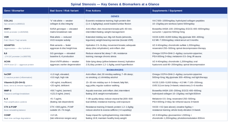
The table below summarizes both the genetic and biomarker findings covered in this article, along with their key action steps at a glance.
The Spine Principles That Could Change How You Manage This Condition
The Huberman Lab podcast episode featuring Dr. Stuart McGill — Professor Emeritus of Spine Biomechanics at the University of Waterloo and author of Low Back Disorders and Back Mechanic — is arguably the most clinically dense, study-grounded conversation on spine health available in public media. McGill has published over 240 peer-reviewed studies on lumbar spine mechanics, and his work has directly influenced clinical protocols for spinal stenosis, disc herniation, and chronic low back pain worldwide. The following ten insights from his work apply directly and practically to life with spinal stenosis.
1. Your Spine Has a Damage Budget — And Flexion Depletes It Fastest
McGill's foundational concept is that discs have a finite tolerance for repeated stress cycles. Each flexion cycle under load — bending forward to pick something up, rounding during a sit-up, slumping while seated — chips away at that budget. For people with structural stenosis, where disc and facet integrity is already compromised, this budget is smaller. Protecting it requires recognizing that common movements many people think are neutral or beneficial (yoga forward folds, crunches, toe-touches) can be among the most damaging if performed repeatedly.
2. Walking Is Medicine for Spinal Stenosis — With One Modification
McGill distinguishes walking from most other exercises as uniquely beneficial for spinal stenosis because it loads the spine in a rhythmic, decompressive pattern while also promoting neurological recovery. The specific modification he recommends: a slight forward lean from the hips (not rounding the back, but inclining the torso slightly forward as if walking into a mild wind). This posteriorly tilts the pelvis, opens the posterior spinal canal, and creates more space for compressed neural tissue. Many patients with neurogenic claudication find they can walk further with this adjustment.
3. Spine Hygiene Before Strengthening — The Sequence Matters
One of McGill's most important clinical observations is that strengthening a painful, poorly moving spine accelerates damage rather than reducing it. The correct sequence is: first find pain-free movement patterns (the "spine hygiene" phase), then build endurance in those patterns, then add strength. Skipping to the strengthening phase because someone has been told they need a "stronger core" is a common error that worsens many stenosis cases.
4. The Big Three Exercises for Spinal Stenosis
McGill's three foundational exercises — the curl-up, the side bridge, and the bird dog — are specifically designed to create spinal stiffness (stability) without spinal flexion. Unlike crunches or sit-ups, the curl-up maintains a neutral lumbar spine while activating the rectus abdominis. The side bridge and bird dog similarly create anti-rotation and anti-extension stability. These three exercises alone, performed daily in appropriate volume, have been shown to reduce pain and improve function in chronic low back conditions including stenosis.
5. Core Stability Is Not the Same as Core Strength
McGill draws a critical distinction: the goal for a stenotic spine is stiffness and stability, not maximal muscular strength. A stiff, well-controlled spine transfers load efficiently and protects compressed neural structures. Heavy core strength training — high-load deadlifts, maximal kettlebell swings — can increase spinal compression forces beyond what a narrowed canal can tolerate. The question is not "how strong is your core?" but "how well does it prevent unwanted movement under load?"
6. Hip Mobility Deficits Force the Lumbar Spine to Move More
When hips lack mobility — a common consequence of prolonged sitting and aging — the lumbar spine compensates by moving through greater range during activities like walking, bending, and rising from a chair. Every degree of additional lumbar motion in a stenotic spine means more stress on already-compromised structures. McGill's clinical work consistently identifies hip mobility restoration as a high-leverage intervention that reduces lumbar load without any spinal exercise at all.
7. Sleep Position Is a Clinical Intervention
McGill treats sleep position as a genuine therapeutic variable, not a comfort afterthought. For spinal stenosis, he recommends [BOLD]side-lying with a pillow between the knees[/TITLE] or fetal position to maintain lumbar decompression during the 6–8 hours of spinal rest. Prone sleeping increases lumbar extension and compresses posterior neural structures. Supine sleeping with a pillow under the knees allows a moderate amount of decompression. Getting this right may provide several hours of passive therapeutic benefit every night.
8. Neural Flossing — Moving the Nerve Rather Than Stretching It
McGill distinguishes between neural mobilization (flossing) and neural tension stretching. For compressed nerves in spinal stenosis, sustained stretching of already-compromised neural tissue can worsen symptoms. Neural flossing — gentle, rhythmic alternating movements that slide the nerve through its sheath without sustained tension — promotes circulation and healing without adding stress. This is a practical protocol that many physiotherapists trained in McGill's approach can teach in a single session.
9. Intra-Abdominal Pressure and Breathing Patterns Protect the Spine
Properly timed breathing — inhaling to create moderate intra-abdominal pressure before a challenging movement — creates a hydraulic cylinder effect that takes load off the spinal column. McGill's research quantifies the protective force this generates. The implication: learning to breathe and brace correctly before any loaded movement (rising from a chair, picking up objects, getting in or out of a car) reduces spinal stress on every rep of every movement throughout the day.
10. Progressive Loading Timing — Starting Too Heavy or Too Early Sets You Back
McGill's clinical data consistently shows that patients who begin loaded rehabilitation too aggressively — before pain-free movement patterns are established — recover more slowly than patients who begin lighter and progress gradually. His framework is start below perceived tolerance, maintain pain-free movement as the primary criterion, and increase load only when 3 consecutive sessions at the current level are pain-free. This patience-first approach consistently outperforms the "push through it" model for stenosis rehabilitation.
Evidence-Based Complementary Approaches for Spinal Stenosis
The following modalities have meaningful human clinical evidence relevant to spinal stenosis — specifically for pain management, functional improvement, or nervous system regulation. These are not alternatives to medical care or the strategies above; they work best as integrated additions to a comprehensive approach.
Yoga
Yoga combines controlled movement, postural training, breath work, and body awareness in a way that addresses several of the mechanisms relevant to spinal stenosis simultaneously. Extension-focused yoga protocols open the posterior spinal canal, and mindful movement practice develops the proprioceptive awareness that protects a vulnerable spine during daily activities. Evidence for yoga in spinal stenosis has grown substantially in recent years, with specific protocols designed for this population showing clear benefits.
A randomized controlled trial published in Spine found that a yoga program tailored for lumbar spinal stenosis significantly improved walking capacity, pain, and disability scores compared to standard care over 6 months. The most supportive yoga styles for stenosis are Iyengar yoga and chair yoga — both prioritize alignment, use props liberally, and avoid aggressive spinal flexion. Randomized trial evidence supports tailored yoga programs for improving function and reducing pain in lumbar spinal stenosis.
To apply this realistically: seek an instructor experienced with spinal conditions or chronic back pain populations. Avoid Bikram (hot yoga) and Vinyasa styles that involve rapid forward folds or unsupported spinal flexion. A home practice of 3×15-minute sessions per week, focusing on cat-cow, supported bridge, and extended child's pose variations, is a reasonable and safe starting point for most stenosis patients. Always communicate your diagnosis to the instructor before the first session.
Tai Chi
Tai chi is a slow, flowing movement practice that builds postural stability, balance, hip mobility, and body awareness — all of which are directly relevant to functional decline in spinal stenosis. The neurogenic claudication characteristic of advanced stenosis often reduces walking confidence and increases fall risk; tai chi has particularly strong evidence for improving balance and reducing fall frequency in older adults.
A 2016 meta-analysis published in PLoS ONE reviewed 18 randomized controlled trials and found that tai chi significantly reduced chronic low back pain intensity and disability, with effects comparable to conventional physiotherapy in several trials. Meta-analyses confirm tai chi's effectiveness for chronic low back pain intensity and functional disability in older adults. Condition-specific evidence for stenosis is more limited but emerging.
For practical application: community tai chi classes designed for older adults or beginners are widely available. The Sun style is the most accessible for people with limited mobility and includes a characteristic step-close-step pattern that reduces demands on balance. Two to three 45-minute sessions per week is the most commonly studied frequency. Tai chi is extremely low-risk for most stenosis patients — one of its advantages is that it can be practiced even when pain limits more vigorous exercise.
Mindfulness-Based Stress Reduction (MBSR)
MBSR is an 8-week structured program developed by Jon Kabat-Zinn that combines body scan meditation, sitting meditation, and mindful movement to alter the relationship between the nervous system and pain signals. It is particularly relevant for stenosis patients because chronic spinal pain frequently involves central sensitization — a state where the nervous system amplifies pain signals — and MBSR directly targets this mechanism through neuroplasticity-driven retraining of pain processing.
A landmark JAMA Internal Medicine study (2016, PMID: 26903761) compared MBSR, cognitive behavioral therapy, and usual care for chronic low back pain in 342 participants. Both MBSR and CBT significantly outperformed usual care for pain and functional improvement at 26 and 52 weeks. While not specific to stenosis, the central sensitization mechanism it addresses is directly applicable. This large randomized trial established MBSR as an evidence-based intervention for chronic low back pain with effects sustained at one year.
MBSR programs are available in-person through hospital systems, community health centers, and online (the Palouse Mindfulness program offers a free, structured 8-week course). Realistic application means committing to 8 weeks of 30–45 minute daily practice — the evidence is specifically tied to the full program duration, not sporadic sessions. Side effects are minimal; some individuals experience temporary emotional discomfort as body awareness increases. For stenosis patients with high psychological distress around their condition, this is among the highest-value additions to a physical management plan.
Low-Level Laser Therapy (Photobiomodulation)
Low-level laser therapy (LLLT), also called photobiomodulation, uses specific wavelengths of red and near-infrared light (typically 630–1000nm) to penetrate tissue and stimulate cellular energy production, reduce local inflammation, and modulate pain signaling. In the context of spinal stenosis, it has the most direct relevance for reducing local inflammatory processes in the posterior spinal tissues and for supporting nerve tissue health in areas of compression.
A 2015 Cochrane systematic review of low-level laser therapy for non-specific low back pain found moderate evidence for short-term pain relief and improvement in disability, with effects particularly pronounced when applied to specific lumbar levels. Randomized trials and systematic reviews support LLLT for short-term pain and function improvement in lumbar conditions. Condition-specific evidence for stenosis remains limited but is growing, and the mechanism is plausible given LLLT's documented effects on inflammation and nerve healing.
For practical application: professional LLLT sessions are available through physiotherapy practices and chiropractic offices (10–15 sessions over 3–5 weeks is a typical initial course). Home devices in the 100–200mW range at 830nm are available for $200–$500 and allow ongoing self-treatment. Apply to the lumbar region for 10–15 minutes per session, 3–5 times per week. LLLT is safe, non-invasive, and well-tolerated — there are no serious adverse effects when used at therapeutic (non-ablative) power levels. It works best as an adjunct to active rehabilitation rather than as a standalone treatment.
Massage Therapy
Massage therapy addresses several of the secondary contributors to stenosis symptom burden: paraspinal muscle hypertonicity, fascial restrictions, and the psychological stress that both exacerbates pain sensitivity and raises inflammatory markers. While massage does not address the structural narrowing itself, it can meaningfully reduce the muscular guarding and referred pain patterns that amplify functional limitation — and its effects on the nervous system extend beyond the local tissue.
A 2014 Annals of Internal Medicine systematic review found that massage therapy provided significant short-term relief for chronic non-specific low back pain, with effects comparable to acupuncture and exercise for pain intensity at short-term follow-up. Systematic reviews confirm massage's short-term effectiveness for chronic low back pain intensity and function. Direct evidence for stenosis specifically is limited; most studies address chronic lumbar pain broadly.
For realistic application: deep tissue massage and myofascial release targeting the paraspinals, gluteal muscles, and hip flexors are the most relevant techniques for stenosis patients. A qualified therapist should be informed of the diagnosis — direct high-pressure work over the stenotic spinal levels should be avoided. A frequency of once per week for 4–6 weeks is appropriate for an initial course, with reassessment afterward. Monthly maintenance sessions may be sufficient once the muscular tension baseline has been reduced. Cost varies widely ($60–$120 per session); some insurance plans cover massage under physical therapy or therapeutic massage codes.
Conclusion
Spinal stenosis is not a single event — it is a biological process with identifiable drivers, and those drivers vary from person to person. Tracking six key biomarkers (hsCRP, Vitamin D, MMP-3, IL-6, bone turnover markers, and COMP) gives you a real-time biological picture of which mechanisms are most active. Understanding six relevant gene variants (COL1A1, MMP3, VDR, ADAMTS5, IL6, and ACAN) explains why those mechanisms may be disproportionately active in the first place. Together, these two layers of information move the conversation from "you have narrowing" to "here is specifically what is driving it and what to do about it."
The next smart step is not to implement everything at once — it is to start with what is measurable. Order an hsCRP and a Vitamin D test at your next blood draw. Consider requesting MMP-3 and bone turnover markers if your physician is receptive. If you have genetic data from 23andMe or AncestryDNA, upload the raw file to a free interpretation tool and look specifically for the variants covered here. Bring that information to a conversation with a physician, functional medicine practitioner, or physiatrist who is willing to interpret it in the context of your clinical picture. Better data, applied intelligently, leads to better decisions — and that is the most reliable path forward that exists.
Musculoskeletal: Bone Conditions Joint Conditions Spine Conditions
Neurological: Nerve Conditions
Autoimmune: Inflammatory Conditions Connective Tissue Conditions