This article contains AI generated content.
· UpdatedSpinocerebellar Ataxia: 7 Genes and 6 Biomarkers to Track
Introduction
Living with spinocerebellar ataxia places you in a frustrating paradox. The genetic cause is often precisely known — a specific gene, a specific repeat count, sometimes the exact size of the expansion — yet the clinical conversation frequently ends at "degenerative condition, supportive care only." That gap between molecular precision and practical guidance is real, and this article is an attempt to close some of it.
The science on SCA has moved considerably faster over the last decade than most neurology appointments reflect. Protein clearance pathways — autophagy, the ubiquitin-proteasome system — are now understood well enough that meaningful modulation is within reach using tools available today. A recently identified gene, FGF14, explains thousands of cases previously labeled idiopathic late-onset cerebellar ataxia, and a pharmacological treatment exists that appears specifically effective for this subtype. Biomarkers like neurofilament light chain can tell you in real time how much ongoing axonal damage is happening, regardless of genetic subtype. None of this is noise — it is actionable biology.
Generic neurological advice — exercise, sleep, reduce inflammation — is not wrong, but it is too blunt for a condition this specific. SCA3 driven by ataxin-3 aggregation has a different mechanistic profile than SCA6 driven by calcium channel dysfunction, and both differ from the intronic RFC1 expansion now known to cause CANVAS syndrome in thousands of undiagnosed patients. Understanding what your specific gene actually does at the cellular level is where meaningful management begins.
This article works through two parallel frameworks. The first goes gene by gene through seven major SCA genetic variants, explaining the molecular mechanism and what the evidence supports for compensation — without supplements, and with them — including dosing, frequency, cycling, and side effects. The second covers six biomarkers that function as real-time readouts of neurological and systemic health, with measurement guides and corrective plans. Beyond these, a book summary presents neuroplasticity findings that challenge the idea cerebellar degeneration is simply linear and irreversible, and four evidence-supported complementary approaches round out a genuinely complete picture.
Summary
This article examines seven of the most clinically significant SCA-associated genes — ATXN1, ATXN2, ATXN3, CACNA1A, ATXN7, FGF14, and RFC1 — explaining what each mutation does at the molecular level and what the current evidence supports for compensation, with protocols, dosing, cycling, and side effect notes for each. For every gene, you will find both a supplement-free plan and a targeted supplementation or equipment-based plan.
The biomarker section then covers six measurable indicators: neurofilament light chain, BDNF, IGF-1, CoQ10, homocysteine, and fasting insulin with HbA1c — each with a cost estimate, an optimal range, and a corrective protocol. Beyond genetics and biomarkers, you will find a summary of The Brain's Way of Healing by Norman Doidge, which presents neuroplasticity evidence with direct implications for cerebellar disease, plus four evidence-supported complementary modalities with published human data in balance disorders and ataxia. The underlying premise: knowing what your genes and biomarkers are telling you leads to better decisions than any generic protocol.
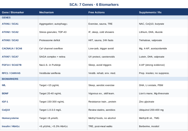
The Genetic Architecture of Spinocerebellar Ataxia: 7 Genes That Shape the Disease
Spinocerebellar ataxia is not one disease. It is a family of over forty genetically distinct conditions that share the same primary target — the cerebellum — but diverge sharply in molecular mechanism, cellular vulnerability, progression rate, and response to intervention. That divergence is precisely why genetic subtyping matters for management and not just for diagnosis.
Most common SCA subtypes are caused by CAG trinucleotide repeat expansions in coding regions of their respective genes. When the repeat count crosses a threshold, the resulting protein acquires toxic new properties: it misfolds, aggregates, and disrupts normal cellular machinery through gain-of-function toxicity. But the downstream pathways differ meaningfully. Some subtypes primarily impair protein quality control; others disrupt transcription; still others alter ion channel function. Two of the seven genes covered here — FGF14 and RFC1 — do not follow the CAG expansion model at all, which explains why they went unrecognized for decades.
ATXN1 — SCA1: When Purkinje Cell Survival Depends on Protein Clearance
ATXN1 encodes ataxin-1, a nuclear protein involved in transcriptional regulation and RNA metabolism. A normal allele carries fewer than 39 CAG repeats; expansions at or above 39 cause SCA1, a cerebellar ataxia that progresses to include dysarthria, dysphagia, and pyramidal signs. Repeat length inversely correlates with age of onset — longer expansions mean earlier onset, sometimes in the thirties.
The pathogenic mechanism centers on Serine 776 phosphorylation. When phosphorylated, mutant ataxin-1 forms stable nuclear inclusions and abnormally engages the CIC (Capicua) transcriptional repressor complex, disrupting the gene regulatory networks that Purkinje cells depend on for survival. Autophagy — the cellular self-cleaning system — is the primary route for clearing these inclusions. Anything that upregulates autophagy without introducing additional Purkinje cell stress is a legitimate target.
If the gene is bad, the plan without supplements
Balance-demanding aerobic exercise at moderate-to-vigorous intensity (cycling on varied terrain, swimming, dance) for thirty to forty-five minutes, five times per week upregulates BDNF and drives autophagy through AMPK and FOXO signaling pathways — both of which promote clearance of ataxin-1 inclusions. Flat treadmill walking is less effective than balance-challenging modalities. Sauna exposure at 80–90°C for fifteen to twenty minutes, three to four times per week, activates heat shock proteins HSP70 and HSP90, which function as molecular chaperones and reduce protein aggregation; avoid if cardiovascular conditions are present without physician clearance. Time-restricted eating within a ten-hour window (fourteen hours overnight fasting minimum) suppresses mTOR and triggers autophagy without caloric restriction; contraindicated in diabetic patients on insulin or sulfonylureas without medication adjustment.
If the gene score is bad, the plan with supplements or equipment
NAC (N-acetylcysteine) at 600–1200mg/day supports glutathione synthesis and reduces the oxidative stress generated by ataxin-1 aggregates; take with food; mild GI effects are the main side effect; no cycling required at standard doses. Ubiquinol (CoQ10) at 200–400mg/day with a fatty meal supports mitochondrial function impaired by ATXN1 pathology; reassess plasma levels after eight to twelve weeks. Sodium butyrate — available through dietary sources (ghee, resistant starch) or at 300–600mg supplemental form — provides HDAC inhibition that counteracts the transcriptional dysregulation caused by CIC complex disruption; reasonable safety profile. For patients engaged with a neurologist, mTOR inhibitor strategies (including rapamycin discussion) are increasingly considered in geroscience-oriented practice given the central role of mTOR in ataxin-1 clearance; not an over-the-counter option.
ATXN2 — SCA2: RNA Metabolism, Stress Granules, and the ALS Connection
ATXN2 encodes ataxin-2, a cytoplasmic protein critical for RNA metabolism and mRNA stability. CAG expansions above 35 repeats cause SCA2; expansions in the intermediate range (27–33 repeats) significantly increase risk of ALS through the interaction of ataxin-2 with TDP-43. SCA2 is notably prevalent in Cuban populations and is clinically characterized by slow saccades — a distinctive eye movement finding — in addition to progressive cerebellar ataxia.
The mechanism involves ataxin-2's role in stress granule dynamics. Under cellular stress, ataxin-2 promotes formation of RNA-protein condensates; expanded ataxin-2 causes these granules to persist abnormally, sequestering TDP-43 and disrupting normal RNA processing. This stress granule pathology is directly amplified by cellular stressors including oxidative damage, glucose dysregulation, and chronic sleep disruption — making lifestyle inputs unusually relevant for this specific variant.
If the gene is bad, the plan without supplements
Intermittent fasting combined with low-carbohydrate eating reduces glucose variability and oxidative stress — both of which promote pathological stress granule formation in ATXN2 pathology. Consistent sleep of seven to nine hours at fixed timing reduces cortisol-driven cellular stress, which is relevant because sustained cortisol elevation disrupts TDP-43 homeostasis in ways directly relevant to this variant. Cold exposure at 10–15°C for two to three minutes daily (cold shower or immersion) activates cold shock proteins including RBM3, which counteract stress granule persistence and support mRNA stability; approach gradually; avoid in cardiovascular disease without clearance.
If the gene score is bad, the plan with supplements or equipment
Lithium at low therapeutic doses (150–300mg lithium carbonate, physician-prescribed and monitored) was specifically investigated in SCA2 in a published randomized controlled trial (Lei et al., Movement Disorders, 2016) showing modest but measurable functional benefit. Lithium inhibits GSK-3β, reduces tau phosphorylation, and promotes autophagy. Requires regular serum level monitoring and kidney function assessment; narrow therapeutic window. DHA omega-3 at 2–3g/day reduces TDP-43 mislocalization in cellular models and attenuates neuroinflammation; safe long-term; no cycling needed; take with food. Riluzole (FDA-approved for ALS, also studied in SCA) reduces cerebellar excitotoxicity through sodium channel modulation; requires prescription and liver function monitoring.
ATXN3 — SCA3/Machado-Joseph Disease: The Proteasome Problem
ATXN3 encodes the most globally common SCA subtype, particularly prevalent in individuals with Portuguese, Brazilian, or Azorean ancestry. Ataxin-3 normally functions as a deubiquitinase — an enzyme that removes ubiquitin tags from proteins to allow their recycling through the proteasome. When the CAG repeat exceeds approximately 60 repeats (pathogenic range 55–87), ataxin-3 misfolds and forms nuclear and cytoplasmic inclusions while simultaneously losing its deubiquitinase activity. This creates a double hit: toxic aggregates accumulate while the protein quality control system that should clear them is disabled.
SCA3 has broad clinical features including cerebellar ataxia, parkinsonism in some families, peripheral neuropathy, and prominent muscle cramps and spasticity. This variability reflects the breadth of systems affected by ubiquitin pathway dysfunction.
If the gene is bad, the plan without supplements
High-intensity interval training (HIIT) increases proteasome activity and autophagy flux more potently than steady-state exercise — a specific advantage for the proteasome deficit at the core of SCA3 pathology. A protocol of four to six rounds of thirty seconds at near-maximal effort followed by ninety-second recovery, three times per week, upregulates the ubiquitin-proteasome pathway measurably. Sauna exposure (80–90°C, twenty minutes, three to four times per week) activates HSP70 and HSP40 chaperones that assist in ataxin-3 refolding and solubilization. Periodic prolonged fasting (twenty-four to thirty-six hour fasts once monthly) maximally activates autophagy through mTOR suppression; contraindicated in individuals with hypoglycemia tendency, eating disorder history, or active hepatic disease.
If the gene score is bad, the plan with supplements or equipment
Trehalose, a disaccharide that activates autophagy through TFEB (Transcription Factor EB), has been specifically studied in SCA3 animal models showing reduced ataxin-3 aggregates and improved motor function. Human trial data are limited but preliminary results are promising. Dose: 3–5g/day dissolved in water; generally safe; watch for blood glucose elevation in diabetics; no established cycling protocol. Valproate (an HDAC inhibitor) has published clinical trial data in SCA3 (Aizawa et al., Movement Disorders) showing slowed progression over twelve months; requires prescription and liver function monitoring. NAC at 600mg twice daily reduces oxidative damage from ataxin-3 aggregates; safe for most adults; monitor blood pressure at high doses. Resveratrol at 500mg/day activates SIRT1, which deacetylates ataxin-3 in ways that reduce its aggregation propensity; take with a fat-containing meal; cycle eight weeks on, two weeks off; mild GI effects possible.
CACNA1A — SCA6: When Calcium Channels Misbehave in Purkinje Cells
CACNA1A encodes the pore-forming subunit of P/Q-type voltage-gated calcium channels, which are expressed at extremely high density in cerebellar Purkinje cells. Unlike most other SCA genes, the pathogenic CAG expansion in CACNA1A is remarkably small — only 20–33 repeats causes SCA6 — making it easy to overlook without careful attention. SCA6 is notable for late onset (typically the forties through seventies) and comparatively slow progression.
The same gene causes three distinct conditions depending on mutation type: point mutations cause familial hemiplegic migraine type 1, other mutations cause episodic ataxia type 2, and the small CAG expansion causes SCA6. The unifying thread is calcium channel dysfunction in Purkinje cells, leading to inappropriate calcium influx, mitochondrial calcium overload, and eventual Purkinje cell death.
If the gene is bad, the plan without supplements
Reducing neuronal excitotoxicity is the primary target. A ketogenic or low-carbohydrate diet (50–100g carbohydrates/day) stabilizes neuronal membrane potentials and reduces the frequency of depolarization-induced calcium overload in Purkinje cells; this level is achievable long-term without strict ketosis. Trigger avoidance is specifically relevant: alcohol, fever, physical overexertion, and sleep deprivation each destabilize CACNA1A channel gating and can precipitate episodic ataxia episodes. Sleep timing consistency — same bedtime and wake time, seven to nine hours — reduces cortisol fluctuations that aggravate channel instability.
If the gene score is bad, the plan with supplements or equipment
Magnesium glycinate or malate at 300–400mg/day directly modulates calcium channel activity by occupying calcium channel binding sites, reducing inappropriate influx; safe and well-tolerated; loose stool at high doses; no cycling needed. 4-Aminopyridine (4-AP) in extended-release form has published evidence for episodic ataxia type 2 and has been used in SCA6 with an episodic component; requires physician prescription and QTc cardiac monitoring. Acetazolamide (a carbonic anhydrase inhibitor) is the standard treatment for the episodic component of CACNA1A-related disorders; prescription only; monitor for kidney stones with long-term use. Memantine (NMDA receptor antagonist) has been proposed for SCA6 based on its calcium-dampening mechanism and has a favorable safety profile; limited human evidence but reasonable theoretical support.
ATXN7 — SCA7: The Subtype That Threatens Both Cerebellum and Retina
ATXN7 encodes ataxin-7, a component of the SAGA transcriptional coactivator complex — a multi-protein assembly critical for regulating gene expression through histone acetylation. When the CAG repeat in ATXN7 exceeds 37 repeats, mutant ataxin-7 disrupts SAGA function, leading to widespread transcriptional dysregulation in cerebellar and retinal cells.
What distinguishes SCA7 from all other subtypes is the consistent involvement of the retina. Cone-rod dystrophy — progressive degeneration of photoreceptors affecting color vision first, then visual acuity — occurs in essentially all SCA7 patients. In severe cases, significant visual impairment or blindness can precede or accompany cerebellar ataxia. This makes SCA7 a condition requiring active ophthalmological management alongside neurological care, and it opens a specific set of interventions targeting retinal health.
If the gene is bad, the plan without supplements
The retinal component demands specific protective behaviors. UV and high-energy visible (blue) light avoidance is directly protective: quality UV-blocking sunglasses with amber-tinted lenses outdoors, and blue light filtering for screen use indoors. A high-carotenoid diet — kale, spinach, egg yolks, orange and red vegetables — provides lutein and zeaxanthin that accumulate in the macula and filter damaging wavelengths. Smoking cessation and elimination of passive smoke exposure reduces retinal oxidative stress significantly. For the cerebellar component, balance-integrated aerobic exercise (dance, swimming, yoga) four to five times per week remains the non-negotiable baseline.
If the gene score is bad, the plan with supplements or equipment
Lutein (10mg) and zeaxanthin (2mg) daily is supported by strong evidence for macular and retinal health; absorption is enhanced with fat in the same meal; safe long-term; reassess with ophthalmologist every six to twelve months. DHA omega-3 at 2g/day is the primary structural fatty acid of photoreceptor outer segments and supports retinal membrane integrity; take with meals; no cycling needed. HDAC inhibitor research for ATXN7 is active given the SAGA complex disruption — valproate has preclinical support and an evolving evidence base; this is an area to watch for clinical trials. Acetyl-L-carnitine at 500–1000mg/day supports mitochondrial energy production in photoreceptors and may slow cone-rod deterioration; generally safe; consult physician if on anticoagulants.
FGF14 — GAA-FGF14 Ataxia: The Diagnosis Hidden in Plain Sight for Decades
FGF14 — Fibroblast Growth Factor 14 — breaks the typical SCA mold entirely. It does not involve a CAG repeat expansion. The disease it causes, GAA-FGF14 ataxia (also called SCA27B), was only formally characterized in 2022. Yet preliminary data suggest it is one of the most common causes of late-onset cerebellar ataxia, explaining a substantial proportion of cases previously labeled idiopathic. This means thousands of people have been sitting with an undiagnosed condition for which an effective pharmacological treatment exists.
The mutation is an intronic GAA pentanucleotide repeat expansion in FGF14. FGF14 protein normally modulates voltage-gated sodium channels — specifically Nav1.6 — in cerebellar Purkinje cells. When FGF14 function is reduced by the repeat expansion, Nav1.6 channel availability at the Purkinje cell axon initial segment decreases, impairing the cell's ability to fire reliably. The clinical result is typically late-onset cerebellar ataxia with episodic features, often appearing in the fifth or sixth decade.
If the gene is bad, the plan without supplements
Trigger management for the episodic component is immediately practical: fever, alcohol, and physical overexertion reliably worsen symptoms in GAA-FGF14 ataxia by further reducing sodium channel availability. Strict sleep hygiene is particularly relevant for this variant because sleep deprivation directly reduces Nav1.6 expression in Purkinje cells. Avoiding sodium channel-suppressing medications — certain anticonvulsants (carbamazepine, phenytoin), local anesthetics, and some antiarrhythmics — is critical; discuss every new medication with a neurologist who knows your diagnosis. Vestibular and balance-specific physiotherapy emphasizing gaze stabilization exercises (not just generic balance training) should begin early.
If the gene score is bad, the plan with supplements or equipment
4-Aminopyridine (4-AP) has shown remarkable documented efficacy specifically in GAA-FGF14 ataxia. It works by blocking voltage-gated potassium channels, which compensates for the reduced sodium channel drive in Purkinje cells. Published case series (Cen et al., 2023, Brain) documented substantial functional improvements in multiple GAA-FGF14 patients treated with 4-AP, including improvements in the Scale for the Assessment and Rating of Ataxia (SARA) score. Extended-release 4-AP (Ampyra/Fampyra, prescription) and immediate-release 4-AP are both used; dosing requires physician supervision due to dose-dependent seizure risk; QTc monitoring is required. This is one of the few SCA subtypes where pharmacological treatment may produce the most dramatic improvement — identifying the FGF14 repeat expansion and starting 4-AP is a clinical priority.
RFC1 — CANVAS Syndrome: A Hidden Epidemic of Misdiagnosed Ataxia
RFC1 (Replication Factor C Subunit 1) carries a biallelic intronic AAGGG pentanucleotide repeat expansion that causes CANVAS syndrome — Cerebellar Ataxia, Neuropathy, and Vestibular Areflexia Syndrome. Formally described by Cortese et al. in Brain (2019), this condition accounts for a significant proportion of previously "idiopathic" sporadic late-onset ataxia worldwide. The autosomal recessive inheritance and the intronic repeat location mean it is typically missed by standard genetic panels and even whole exome sequencing without specific repeat-primed PCR testing.
The vestibular component is often the most functionally disabling. Both vestibular organs are typically non-functional (bilateral vestibular areflexia), meaning the patient cannot stabilize gaze during head movement. This produces oscillopsia, severe imbalance in the dark, and an inability to maintain posture without visual or proprioceptive input. Peripheral neuropathy adds further instability. The RFC1 mechanism involves DNA repair pathway disruption and possibly RNA toxicity from the intronic repeat, though the exact downstream pathway continues to be characterized.
If the gene is bad, the plan without supplements
Specialized vestibular rehabilitation — not generic balance training — is the cornerstone of management. Standard physiotherapy misses the most critical element: gaze stabilization exercises that force the vestibulo-ocular reflex to compensate through visual and cervical inputs (VSR exercises). A trained vestibular physiotherapist is not optional for this subtype. Vibration platform training (whole-body vibration at 20–40 Hz, ten to fifteen minutes, three to four times per week) provides proprioceptive input that partially compensates for absent vestibular function. Environmental modification — improving lighting in all rooms, removing fall hazards, using contrasting floor colors — is essential rather than optional; with bilateral vestibular loss, falls in the dark are near-certain without adaptation. Nordic walking poles during all outdoor movement reduce fall risk substantially and enable continued physical activity.
If the gene score is bad, the plan with supplements or equipment
There is currently no established pharmacological disease-modifying treatment for RFC1/CANVAS — gene therapy research is active. For symptomatic management, vestibular suppressants (meclizine, diazepam) should generally be avoided in CANVAS: suppressing the already-absent vestibular signal paradoxically impairs the compensatory neuroplasticity the central vestibular system needs to develop. This is a critical distinction some clinicians miss, and flagging it explicitly with your neurology team is worthwhile. Proprioceptive insoles with enhanced textural feedback (available through specialist physiotherapy) and balance exergaming systems (commercial balance board programs) provide structured proprioceptive training. Emerging vestibular implant research for bilateral vestibular loss represents a legitimate medium-term horizon to watch.
Six Biomarkers That Reveal What Your Nervous System Is Doing Right Now
Genetic subtyping tells you what your nervous system is working against. Biomarkers tell you how the battle is going in real time. These six measurements are accessible, interpretable, and — most importantly — modifiable. They do not replace genetic diagnosis or clinical neurological assessment, but they fill in a picture that genetic testing alone cannot provide.
Monitoring these markers over time also gives you feedback on whether your interventions are working. Quarterly or biannual biomarker tracking functions as a feedback loop that clinical visits every six months, focused primarily on SARA scale scoring, cannot adequately provide. Think of them as real-time readouts for the systems most relevant to cerebellar health.
Neurofilament Light Chain (NfL) — The Neurodegeneration Speedometer
NfL is a structural protein found inside neuronal axons. When axons are damaged — by disease, neuroinflammation, or mechanical injury — NfL leaks into the cerebrospinal fluid and subsequently into the bloodstream. It does not distinguish between SCA subtypes, but it does tell you how much ongoing axonal damage is occurring right now. It is one of the most informative single measurements available for tracking neurological disease activity.
The EUROSCA consortium has published extensively showing that serum NfL correlates with ataxia severity scale scores and predicts progression rate in SCA1, SCA2, and SCA3. Research published in Neurology established that NfL rises significantly before clinical worsening becomes detectable, making it a true early-warning marker — view related publications on PubMed. Trending NfL over time is more clinically useful than a single snapshot.
How to measure it
Serum NfL via Simoa (Single Molecule Array) technology; cost $120–200 USD through specialty labs. Not yet universally available through primary care but increasingly accessible. Optimal range: generally below 10–15 pg/mL for adults under sixty, though age-adjusted values are more informative. Re-measure every six to twelve months for trend data.
If the score is bad, the plan without supplements
Maximize glymphatic clearance: the brain's waste-clearance network operates primarily during slow-wave sleep, removing neurotoxic proteins including damaged structural proteins. Prioritize seven to nine hours of sleep, sleeping in a lateral (side) position (shown to improve glymphatic flow), and eliminating alcohol (which disrupts slow-wave sleep and impairs glymphatic function by up to forty percent). Aerobic exercise at 60–70% maximum heart rate, thirty to forty-five minutes, four to five times per week has been shown to reduce serum NfL in multiple sclerosis patients — the mechanism likely involves exercise-driven autophagy reducing ongoing axonal damage. Eliminating chronic neuroinflammation sources (ultra-processed foods, trans fats, chronic alcohol) removes avoidable contributors to axonal stress.
If the score is bad, the plan with supplements or equipment
DHA omega-3 (2–3g/day) reduces neuroinflammation and supports axonal membrane integrity; NfL has been shown to decrease with omega-3 supplementation in some neuroinflammatory conditions; take with meals; no cycling needed. Lithium orotate (5mg/day), a low-dose neuroprotective form distinct from psychiatric lithium dosing, has evidence for reducing axonal injury markers and protecting neuronal integrity; inform your physician even at low doses given lithium's narrow therapeutic window. Near-infrared photobiomodulation (PBM) using scalp-applied devices delivering 810–850nm light (such as Vielight or similar clinical units) is an emerging approach for reducing neuroinflammation; twenty-minute sessions three to five times per week; limited but growing clinical evidence in neurodegenerative conditions.
BDNF — The Growth Factor Your Purkinje Cells Depend On
Brain-derived neurotrophic factor is essential for Purkinje cell survival, growth, and synaptic maintenance — the very neurons that degenerate in most SCA subtypes. BDNF binds TrkB receptors on Purkinje cells and activates PI3K/Akt/mTOR survival signaling. While serum BDNF (mostly platelet-derived) does not perfectly mirror brain BDNF, population-level evidence consistently links lower serum BDNF to worse neurological outcomes, and the interventions that raise serum BDNF are the same ones shown to raise cerebellar BDNF in preclinical studies.
How to measure it
Serum BDNF via ELISA; cost $60–120 through specialty labs. A morning fasting sample is preferred for reproducibility. Reference values vary by lab, with many reporting 20–40 ng/mL as a typical adult range. Note: vigorous exercise the prior day significantly elevates values — standardize timing and activity around measurements.
If the score is bad, the plan without supplements
Vigorous aerobic exercise remains the single most potent known BDNF stimulus. Multiple published studies confirm that thirty-plus minutes at elevated heart rate (above 60–70% maximum) acutely raises BDNF two to threefold. Chronic regular exercise raises baseline. Motor skill acquisition — learning new physical skills like a musical instrument, complex dance choreography, or juggling — drives cerebellar-specific BDNF upregulation through learning-dependent synaptic plasticity; this is qualitatively different from repetitive practice. Morning bright light exposure (ten to twenty minutes of natural sunlight within thirty minutes of waking) activates serotonergic pathways that support BDNF synthesis throughout the day.
If the score is bad, the plan with supplements or equipment
Lion's mane mushroom (Hericium erinaceus) at 500–1500mg/day of standardized extract produces hericenones and erinacines that stimulate nerve growth factor and may upregulate BDNF; a 2009 Japanese randomized controlled trial showed cognitive improvement in older adults; start at 500mg and increase gradually; generally safe with occasional mild GI effects. Magnesium L-threonate at 1.5–2g/day (providing approximately 140–150mg elemental magnesium) is the form with the strongest preclinical evidence for crossing the blood-brain barrier; allow a three-month trial before reassessment; mild GI effects possible. Rhodiola rosea at 400–600mg/day standardized to 3% rosavins has adaptogenic effects with some BDNF-relevant evidence; cycle eight weeks on, two to three weeks off; avoid in individuals with bipolar disorder.
IGF-1 — The Overlooked Cerebellar Trophic Factor
Insulin-like growth factor 1 plays a unique and underappreciated role in cerebellar maintenance. Unlike BDNF, IGF-1 crosses the blood-brain barrier efficiently from circulation, directly supporting Purkinje cell dendrite complexity and synaptic density. Research from Torres-Aleman's group showed that systemic IGF-1 administration reduced cerebellar atrophy and improved motor performance in SCA1 mouse models. Low IGF-1 (below 130 ng/mL in adults) is consistently associated with faster neurodegenerative progression across multiple conditions, making it a relevant optimization target.
How to measure it
Serum IGF-1 with IGFBP-3; cost $40–80; widely available at standard labs. Optimal adult range: 150–300 ng/mL (age-adjusted). Levels below 100 ng/mL are consistently associated with faster neurodegeneration. Measure fasting in the morning; values are affected by recent illness, prolonged fasting, and recent alcohol intake.
If the score is bad, the plan without supplements
Progressive resistance training (three to four sessions per week, compound movements, progressive overload) is the most potent endogenous IGF-1 stimulus. Adequate protein intake (1.6–2.2g per kg of bodyweight daily, emphasizing leucine-rich complete protein sources — meat, fish, eggs, dairy) is necessary for IGF-1 axis function; chronic low protein independently suppresses IGF-1 regardless of training. Deep sleep optimization is critical: the largest IGF-1 pulses occur during slow-wave sleep stages; disrupted sleep architecture substantially reduces daily IGF-1 output.
If the score is bad, the plan with supplements or equipment
Zinc glycinate or zinc bisglycinate at 15–30mg/day is required for IGF-1 axis function; deficiency consistently suppresses IGF-1 production; take with food; balance with copper at a 10:1 zinc-to-copper ratio; reassess at three months. Avoid chronic caloric restriction below basal metabolic rate — this is among the most powerful suppressors of IGF-1 and is surprisingly common in individuals managing fatigue and mobility challenges. If IGF-1 remains consistently below 100 ng/mL despite optimization, endocrinology consultation to assess the growth hormone axis is warranted.
CoQ10 (Ubiquinol) — Mitochondrial Currency for High-Demand Neurons
The cerebellum's energy demands are extreme. Purkinje cells fire at rates of 50–200 Hz and maintain massive dendritic arborizations requiring continuous ATP. CoQ10 is the electron shuttle in the mitochondrial respiratory chain; without adequate CoQ10, ATP synthesis stalls and reactive oxygen species accumulate. Mitochondrial dysfunction has been documented in ataxin-1, ataxin-2, and ataxin-3 pathology specifically, making CoQ10 status directly relevant to SCA biology rather than a generic wellness metric.
How to measure it
Plasma CoQ10 (total coenzyme Q10); cost $70–150 through specialty labs. Optimal plasma range: 1.0–3.0 mg/L. Most unsupplemented adults fall between 0.4–0.8 mg/L. Statin users routinely fall below 0.4 mg/L. Measure fasting for consistency.
If the score is bad, the plan without supplements
Statin review with physician is the first step — statins inhibit the mevalonate pathway that synthesizes CoQ10 endogenously, and for someone with SCA and a genetic predisposition to mitochondrial dysfunction, the risk-benefit calculation deserves explicit discussion. Aerobic exercise upregulates mitochondrial biogenesis and endogenous CoQ10 synthesis. CoQ10-rich foods (sardines, organ meats, beef, peanuts) contribute modestly to plasma levels.
If the score is bad, the plan with supplements or equipment
Ubiquinol (reduced CoQ10) at 200–400mg/day is superior to ubiquinone in bioavailability, especially in older individuals and those with fat absorption challenges; take with a fat-containing meal; no cycling required; mild GI effects at high doses; reassess plasma levels after eight to twelve weeks. PQQ (Pyrroloquinoline Quinone) at 20mg/day supports mitochondrial biogenesis — the formation of new mitochondria, not just improved function of existing ones — and complements CoQ10 effectively; safe long-term.
Homocysteine — The Methylation Marker with Direct Neurotoxic Consequences
Elevated homocysteine above 10 μmol/L is directly neurotoxic. It induces oxidative stress in neurons, promotes NMDA receptor-mediated excitotoxicity, disrupts DNA repair, and is independently associated with accelerated brain atrophy on MRI. For someone with SCA already experiencing neurodegeneration, elevated homocysteine functions as an avoidable additional burden — it is one of the most addressable contributors to neurological decline.
Homocysteine elevation is most commonly driven by MTHFR polymorphisms — particularly the C677T variant — which reduce the enzyme responsible for converting folate to the active methylated form needed for methionine metabolism. Gary Brecka has extensively discussed how MTHFR variants affect methylation capacity and why methylated B vitamins are the appropriate response. Ali Torkamani's work on genomics reinforces that common variants like MTHFR have disproportionate consequences when environmental factors amplify them, particularly chronic stress and poor dietary methyl donor intake.
How to measure it
Serum homocysteine fasting; cost $20–50; available at any standard lab. Optimal: below 8 μmol/L. Above 12 μmol/L is a concern; above 15 μmol/L warrants active intervention. Add MTHFR genotyping ($50–100, one-time test) for mechanistic context on whether the driver is genetic.
If the score is bad, the plan without supplements
Dietary methyl donors: leafy greens (folate), eggs (choline and betaine), beets (betaine), and liver (B12, folate, B6) directly reduce homocysteine through the methylation cycle. Eliminate alcohol entirely — alcohol is among the most potent dietary suppressors of B vitamins and directly raises homocysteine; this single step can reduce levels substantially. Support kidney function — kidneys clear homocysteine from the bloodstream; chronic dehydration and kidney disease raise levels independently of methylation.
If the score is bad, the plan with supplements or equipment
Methylated B vitamins are the targeted response: methylfolate (400–1000mcg/day) + methylcobalamin (1000mcg/day) + P5P/pyridoxal-5-phosphate (25–50mg/day). This combination is specifically appropriate for anyone with MTHFR C677T or A1298C variants who cannot efficiently convert standard folic acid or cyanocobalamin. Avoid high-dose non-methylated folic acid in MTHFR-positive individuals. Re-test homocysteine at three months; target below 8 μmol/L. TMG (trimethylglycine/betaine) at 1–3g/day serves as an alternative methylation donor for those who cannot tolerate methyl B vitamins; start at a low dose as some individuals with methylation sensitivity experience mood effects; no cycling required but recheck every six months.
Fasting Insulin and HbA1c — Metabolic Health as Neurological Insurance
Insulin resistance is not just a metabolic disease risk factor. The brain is highly insulin-sensitive — insulin signaling modulates synaptic plasticity, BDNF expression, and mitochondrial function in neurons. Elevated fasting insulin above 10 μIU/mL and HbA1c above 5.4% are associated with accelerated cerebral and cerebellar atrophy, independent of diabetes diagnosis. Peter Attia has described insulin resistance as the metabolic contribution to neurological deterioration — preventable and modifiable, but requiring measurement to catch in its early stages, since most clinicians do not order fasting insulin until overt diabetes is already established.
How to measure it
Fasting insulin ($15–30) + HbA1c ($20–40) + fasting glucose ($10–20); any standard lab; order all three together. Optimal targets: fasting insulin below 6 μIU/mL, HbA1c below 5.3%, fasting glucose below 90 mg/dL. Calculate HOMA-IR (fasting insulin × fasting glucose / 405) — below 1.0 indicates excellent insulin sensitivity. Most standard labs flag insulin only above 15–20 μIU/mL, which misses the relevant range for neurological protection.
If the score is bad, the plan without supplements
Time-restricted eating within a ten to twelve hour window (adjusting to eight hours if HOMA-IR exceeds 2.5) reduces insulin secretion frequency and improves receptor sensitivity without caloric restriction. Post-meal walking of ten to fifteen minutes after each meal reduces postprandial glucose spikes by 20–30% through non-insulin-mediated glucose uptake in active muscles. Resistance training three times per week improves skeletal muscle glucose uptake independent of insulin pathways. Eliminating refined carbohydrates and ultra-processed foods is the foundational dietary step.
If the score is bad, the plan with supplements or equipment
Berberine at 500mg three times daily with meals has published RCT evidence showing insulin sensitivity improvement comparable in magnitude to metformin; cycle eight weeks on, two weeks off to prevent gut microbiome disruption; GI effects (diarrhea, cramping) are common initially and improve with dose titration. Myo-inositol at 4g/day improves insulin receptor signaling through second-messenger pathways; safe; mild GI effects; no cycling required. Metformin (prescription only) activates AMPK — the cellular energy sensor that also promotes autophagy and neuroprotection — and epidemiological data links metformin use to reduced neurodegeneration rates in several conditions; discuss with physician, particularly given the overlap with autophagy mechanisms relevant in SCA.
What The Brain's Way of Healing Gets Right About Cerebellar Neuroplasticity
Norman Doidge's 2015 book The Brain's Way of Healing is not specifically about spinocerebellar ataxia, but it contains a body of evidence and clinical case material that is directly relevant to anyone navigating a cerebellar condition. The book challenges the clinical assumption that neurological degeneration proceeds on a fixed trajectory unaffected by input, and it presents convergent evidence from multiple research programs showing that movement, sensation, and attention can drive neuroplastic changes even in disease contexts previously considered refractory.
What follows are the ten most clinically impactful concepts from the book for anyone approaching cerebellar disease.
The Cerebellum Has Far More Plasticity Than Classical Neurology Assumed
For most of the twentieth century, the cerebellum was considered a largely fixed structure — a precise biological clock that could be damaged but not rewired. Contemporary neuroscience has overturned this. The cerebellum contains roughly half of the brain's neurons and exhibits forms of synaptic plasticity (long-term depression at parallel fiber-Purkinje cell synapses) that are directly activity-dependent. Doidge documents multiple cases of patients with significant cerebellar and brainstem compromise achieving functional recovery beyond what structural damage would predict, driven by targeted sensory and motor input.
Motor Learning Is Error-Driven — and That Changes Everything About Practice
The cerebellum learns by computing the difference between expected and actual movement outcomes and updating internal models accordingly. This means that error-based practice — attempting movements that are genuinely challenging — drives cerebellar learning, while repetitive practice of already-mastered movements does not. For SCA patients, this reframes the goal of exercise: the target is not to repeat movements you can already do comfortably, but to consistently work at the edge of current capability with adequate rest between attempts. Boring repetition is less valuable than novel difficulty.
The Feldenkrais Method: Slow, Attentive Movement as Neural Input
The Feldenkrais Method — a movement education approach developed by physicist Moshe Feldenkrais — features slow, varied, attention-directed movement sequences specifically designed to drive cortical remapping. Doidge presents evidence that this approach activates neuroplastic mechanisms more effectively in some patients than conventional rehabilitation, because the slow pace allows sensory feedback to be fully processed and integrated. For cerebellar conditions involving proprioceptive integration deficits, the method's emphasis on sensory attention during movement (rather than force or speed) is mechanistically appropriate. Practitioner-led sessions are available internationally.
The PoNS Device: Tongue Stimulation Reaching the Cerebellum
The Portable Neuromodulation Stimulator (PoNS) is a device that delivers gentle electrical stimulation to the tongue, which is richly innervated and projects through the trigeminal and facial nerves to the brainstem and cerebellum. Clinical trials in multiple sclerosis-related balance disorders and traumatic brain injury have shown improved balance and coordination with PoNS-combined physical therapy. While SCA-specific data is limited, the brainstem/cerebellar projection pathway is directly relevant, and the device has received Health Canada and FDA clearance for specific indications. Doidge covers the research origin and early clinical findings in detail.
Disuse Accelerates Functional Decline Beyond the Disease Itself
One of Doidge's most important clinical observations is that neurological patients who restrict activity to avoid symptoms often experience a secondary functional decline driven by disuse — independent of disease progression. Restriction-based adaptation is physiologically costly in the nervous system: underused circuits downregulate, and the territory they occupy is progressively ceded to other functions. For SCA patients, this means that the conservative management of avoiding challenging movements to prevent falls can accelerate overall decline. Supervised challenging practice with appropriate safety measures is physiologically preferable to restriction.
Neuroinflammation as Both Symptom and Cause
Doidge synthesizes evidence showing that chronic low-grade neuroinflammation is not merely a downstream symptom of neurological disease but an active driver of its progression. Microglial activation, cytokine signaling, and blood-brain barrier compromise each contribute to ongoing neuronal loss. Addressing neuroinflammation — through diet, sleep, exercise, and targeted supplementation — is therefore therapeutic, not simply supportive. This framing supports the biomarker monitoring approach described earlier and reinforces the value of tracking inflammatory indicators.
Sound and the Cerebellum: Auditory-Motor Integration as a Therapeutic Target
The cerebellum receives substantial projections from auditory pathways and is directly involved in temporal processing and rhythm. Doidge covers auditory-motor integration as a therapeutic pathway — specifically, how rhythmic auditory input can drive cerebellar timing circuits in ways that support motor rehabilitation. This mechanistic basis underlies the evidence for rhythmic auditory stimulation (RAS) in gait and coordination disorders, covered in more detail in the complementary approaches section below.
Sleep as the Primary Window for Neurological Consolidation
Every learning and repair process described in the book converges on sleep as its primary consolidation window. During slow-wave sleep, motor memory consolidation in the cerebellum occurs, synaptic pruning optimizes circuits, and the glymphatic system removes neurotoxic proteins. Doidge argues — with supporting research — that poor sleep quality is one of the most reversible and consequential factors in neurological disease trajectory. Sleep architecture optimization is not a peripheral concern but a central one.
Social Engagement and Environmental Enrichment Drive Trophic Factor Production
Environmental enrichment — complex, stimulating environments with social engagement, novel challenges, and varied sensory input — is the most potent inducer of BDNF and other trophic factors in animal models. Doidge translates this to human terms: social isolation and environmental monotony are neurologically harmful in ways that go beyond psychological wellbeing. Deliberately maintaining social complexity, varied environments, and intellectually and physically challenging activities has measurable trophic consequences.
The Rate of Functional Decline Is Partly Determined by Neuroplastic Reserve
Perhaps the book's most practically important message: two individuals with the same genetic mutation and the same repeat count can have meaningfully different functional trajectories based on their neuroplastic reserve — the accumulated capacity of the nervous system to respond to challenge with adaptation. Neuroplastic reserve is built over a lifetime through cognitive engagement, physical challenge, and sensory richness, and it can be actively maintained and partially rebuilt. This reframes the question from "what is happening to me?" to "what can I actively do to maintain the nervous system's capacity to respond?"
Evidence-Based Complementary Approaches for Cerebellar Ataxia
The following modalities are selected for having meaningful human clinical evidence in balance disorders, cerebellar dysfunction, or closely related neurological conditions. They are not alternatives to genetic management or biomarker monitoring — they are additions to it. Each brings a specific mechanism that complements the physiological targets already described.
Tai Chi
Tai chi is a Chinese movement practice involving slow, continuous, weight-shifting movements that require sustained balance, proprioceptive attention, and lower extremity control. For cerebellar ataxia, its primary relevance lies in its combination of balance challenge, dual-task training (cognitive and motor), and controlled movement at the edge of stability. Unlike most exercise modalities, tai chi is inherently practiced at the limits of balance capacity, which aligns with the error-driven learning model described above.
A randomized controlled trial by Li et al. (New England Journal of Medicine, 2012) in Parkinson's disease patients showed tai chi reduced falls by forty-seven percent compared to resistance training and stretching, with significantly improved functional reach and gait speed. For cerebellar ataxia specifically, a pilot study published in The Cerebellum found improved SARA scores and balance confidence in SCA patients following a structured tai chi program. Evidence is promising but based on smaller studies; it is not a replacement for formal physiotherapy but a complement to it.
A practical protocol: twenty to thirty minutes of guided tai chi, three to four times per week, using instructor-led sessions (in-person or video-based) rather than self-taught practice. For fall risk, beginners should practice near a wall or with a chair for support until stability improves. Progress is typically measurable within six to eight weeks of consistent practice.
Balance Biofeedback Training
Biofeedback for balance disorders involves providing real-time sensory feedback about body position — typically through visual displays showing center-of-pressure sway, or through auditory or vibrotactile signals — to help the nervous system recalibrate postural control. For cerebellar ataxia, where internal feedback mechanisms are impaired, external augmented feedback can partially substitute for the degraded proprioceptive and cerebellar error-correction signals.
Multiple published studies have examined balance biofeedback in ataxia and related cerebellar conditions. Baram and Miller (2007, Multiple Sclerosis Journal) published a randomized trial showing significant improvements in gait parameters with visual feedback during walking in patients with cerebellar dysfunction. A systematic review in Gait and Posture identified consistent benefit of biofeedback training across balance disorders including cerebellar ataxia. Wii balance board-based training has been validated as a low-cost accessible platform in small clinical trials.
Practical application: commercial force plate biofeedback systems are available in balance rehabilitation clinics; Nintendo Balance Board-based programs (Wii Fit) provide an affordable home option. Start with two to three ten-minute sessions per week under supervised conditions before transitioning to home practice. Sessions should be challenging — targeting sway reduction at the edge of comfortable balance — rather than simply repeating stable positions.
Music Therapy and Rhythmic Auditory Stimulation
Rhythmic Auditory Stimulation (RAS) is a specific music therapy technique developed by Michael Thaut that uses precisely timed rhythmic auditory cues to drive motor timing and gait coordination. The mechanism exploits the tight coupling between auditory-temporal processing and cerebellar motor timing circuits. The cerebellum uses rhythmic input as a timing scaffold for movement sequencing, which makes auditory rhythmic entrainment a direct therapeutic target in cerebellar conditions affecting timing.
Thaut and colleagues have published extensively on RAS in gait disorders. A meta-analysis of RAS in neurological gait disorders (NeuroRehabilitation and Neural Repair) showed significant improvements in gait velocity, cadence, and stride symmetry across Parkinson's disease, stroke, and cerebellar conditions. For cerebellar ataxia specifically, the data are more limited but mechanistically compelling — the auditory-motor timing pathway bypasses some of the degraded cerebellar timing signals with an external rhythmic reference.
A practical protocol: walking to music with a fixed, clearly audible beat that matches slightly above your comfortable walking cadence (typically 100–120 bpm); thirty minutes, four to five times per week. For ataxic gait, metronome apps (such as ProMetronome) can be more precise than music with variable tempos. Sessions should begin seated or in a supported environment to allow adjustment to the rhythmic cue before attempting challenging surfaces.
Yoga
Yoga's relevance for cerebellar ataxia lies in its systematic cultivation of proprioceptive awareness, controlled one-leg balance, trunk stability, and breathing regulation. Unlike aerobic exercise, yoga emphasizes the quality of attention during movement — sensing subtle weight shifts, correcting alignment in real time — which creates the error-based sensory learning relevant to cerebellar rehabilitation.
A randomized trial by Schmid et al. examining yoga in multiple sclerosis (a condition with significant cerebellar involvement) published in Clinical Rehabilitation showed improvements in balance and mobility compared to waiting-list control, with maintained effects at six months. For SCA specifically, a pilot study in BMC Complementary Medicine and Therapies examining yoga in spinocerebellar ataxia patients documented improvements in functional mobility and patient-reported balance confidence. The evidence base is smaller than for tai chi but growing.
For practical application: modified yoga adapted for neurological conditions (sometimes called therapeutic yoga or chair yoga) is important for patients with significant gait instability — standard yoga classes may present fall risk. A thirty-minute session two to three times per week, working with a teacher experienced in neurological conditions or following a video protocol specifically adapted for balance disorders, is a reasonable starting protocol. Poses emphasizing single-leg balance (tree pose with wall support), trunk rotation, and controlled slow transitions are most mechanistically relevant.
Conclusion
Spinocerebellar ataxia is genetically driven, but the trajectory of its progression is not entirely fixed. The molecular mechanisms underlying each SCA subtype — whether protein aggregation, calcium channel dysfunction, RNA metabolism disruption, or DNA repair failure — are increasingly well understood, and each opens specific points of intervention. The recently identified GAA-FGF14 ataxia subtype, with its responsive pharmacological treatment, is a concrete reminder that precision in diagnosis translates directly into clinical opportunity.
The six biomarkers covered here function as a real-time monitoring layer — not a replacement for clinical assessment, but a way of tracking disease activity, trophic support, and metabolic health between appointments. Tracking NfL, BDNF, IGF-1, CoQ10, homocysteine, and metabolic markers over time provides feedback that clinical visits alone cannot offer. And the complementary approaches, book-derived neuroplasticity principles, and lifestyle strategies described throughout this article build on — rather than replace — that molecular foundation.
The most useful next step is rarely everything at once. Identifying your genetic subtype, ordering the two or three most relevant biomarkers, and implementing one or two evidence-supported interventions from the framework that matches your situation is more sustainable than wholesale lifestyle overhaul. Better information does not solve everything, but it changes the conversation — from passive management of an inevitable decline to active engagement with a biology that is more modifiable than most clinical conversations currently reflect. Discuss the specifics with a neurologist familiar with your subtype, and bring the biomarker data when you do.
Neurological: Brain Conditions Nerve Conditions Movement Disorders
Eye: Retinal Conditions
Ear, Nose & Throat: Hearing & Balance Conditions