This article contains AI generated content.
· UpdatedSpondyloenchondromatosis - 5 Genes And 6 Biomarkers To Track
Introduction
Living with spondyloenchondromatosis means navigating a condition that most physicians have never treated, most labs have never specifically tested for, and most search results reduce to a handful of case reports. If you have received this diagnosis, or suspect it in yourself or a family member, you already know that finding practical, honest information is harder than it should be.
Generic skeletal health advice, aimed at osteoporosis or common arthritis, does not map well onto a rare enchondromatosis affecting the spine and long bones simultaneously. The cartilage abnormalities at the center of this condition are driven by molecular events that differ meaningfully from ordinary bone loss, and treating them the same way leads to wasted effort and missed opportunities.
This article takes a different approach. It focuses on the specific genes and molecular pathways that research in enchondromatosis has identified, and the handful of measurable biomarkers that can tell you how active bone and cartilage turnover currently is in your body. Neither of these angles is a cure, and this article does not pretend otherwise. But better information genuinely changes what questions you can ask a specialist, what monitoring is worth requesting, and what lifestyle decisions are grounded in evidence rather than guesswork.
What follows is organized around two main strategies: first, a deep look at the five most relevant genes and epigenetic factors tied to enchondromatosis-spectrum disorders, with practical plans for each; and second, a focused guide to six biomarkers worth tracking over time. After that, you will find a summary of a relevant research-rich resource, and a review of the complementary modalities with the strongest clinical evidence for musculoskeletal and bone health.
Summary
Spondyloenchondromatosis is a rare skeletal dysplasia characterized by enchondromas distributed through the vertebrae and long bones. The molecular drivers behind it overlap significantly with better-studied enchondromatosis syndromes, and five genes stand out: IDH1/IDH2, COL2A1, IHH, PTPN11, and SOX9. For each of these, there are concrete implications, monitoring strategies, and lifestyle or supplemental interventions that can shift the trajectory. On the biomarker side, six measurements, from bone-specific alkaline phosphatase to IGF-1 and 25-OH vitamin D, can reveal whether bone and cartilage metabolism is running out of balance right now, not just years from now when imaging changes become obvious. The article also covers a relevant Andrew Huberman episode packed with actionable bone health science, and five evidence-supported complementary modalities including low-level laser therapy and mindfulness-based stress reduction, both of which have clinical data behind them for skeletal and pain outcomes. If you are trying to find the most useful next step for a condition this rare, this article is written for exactly that goal.
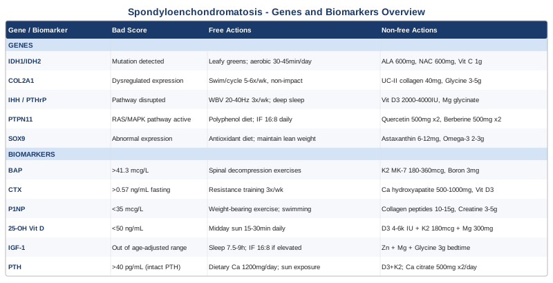
5 Genes and Epigenetic Factors That Shape Spondyloenchondromatosis
Spondyloenchondromatosis sits within the broader enchondromatosis spectrum, a family of disorders in which benign cartilage tumors, enchondromas, accumulate in skeletal sites where they do not belong. The spinal involvement that defines the spondylo- prefix makes this variant particularly complex because vertebral enchondromas add neurological risk and postural consequence that limb-only forms do not carry. The five genes below are the ones most consistently implicated by current research in enchondromatosis and closely related skeletal dysplasias. For each, the current state of evidence is noted honestly, because strong human data and early mouse-model data require different levels of confidence.
Gene 1: IDH1 and IDH2 — The Metabolic Disruption at the Core
IDH1 and IDH2 encode isocitrate dehydrogenase enzymes, which are central to the citric acid cycle and to the regulation of cellular differentiation. Somatic gain-of-function mutations in these genes, particularly the R132H substitution in IDH1 and the R172K substitution in IDH2, produce an oncometabolite called 2-hydroxyglutarate (2-HG). This molecule competitively inhibits alpha-ketoglutarate-dependent dioxygenases, a class of enzymes that includes the TET family of DNA demethylases and Jumonji-domain histone demethylases. The result is widespread hypermethylation of DNA and histones, which blocks normal chondrocyte differentiation.
This mechanism was first clearly described in Ollier disease and Maffucci syndrome, the two most studied enchondromatosis conditions, with somatic IDH mutations found in roughly 87 percent of Ollier disease enchondromas according to a landmark 2011 study published in Nature Genetics. Evidence for spondyloenchondromatosis specifically is more limited and largely extrapolated from the broader enchondromatosis literature, but the mechanistic overlap is strong enough that IDH status is clinically relevant to test.
If the gene score is concerning — the plan without supplements
If IDH1 or IDH2 mutations are found in tissue or identified through liquid biopsy, the primary non-supplemental approach is epigenetic diet strategy: maximizing dietary methyl donors and alpha-ketoglutarate precursors to partially counter the hypermethylation environment. This means prioritizing leafy greens, cruciferous vegetables, eggs, and legumes at every meal. Reducing refined sugar is also important because glycolytic flux feeds IDH-mutant tumor metabolism preferentially. Movement, specifically low-impact daily aerobic activity for 30 to 45 minutes, improves mitochondrial efficiency and reduces the substrate availability for 2-HG production. No cycling needed for these lifestyle measures; maintain consistently.
If the gene score is concerning — the plan with supplements
Alpha-lipoic acid (600 mg daily, with food) acts as a mitochondrial cofactor and may support the alpha-ketoglutarate pathway. NAC (N-acetylcysteine) at 600 to 1200 mg per day supports glutathione recycling, which is depleted in high-2-HG environments. Vitamin C at 1000 mg per day is a required cofactor for TET enzymes and collagen synthesis. Cycling: consider 5 days on, 2 days off for high-dose vitamin C to avoid oxalate buildup. Side effects to monitor: gastrointestinal sensitivity with NAC; always take with meals.
Gene 2: COL2A1 — The Structural Backbone of Cartilage
COL2A1 encodes type II collagen, the dominant structural protein of hyaline cartilage and the nucleus pulposus of intervertebral discs. Mutations in this gene are the primary driver of a wide family of skeletal dysplasias including achondrogenesis type II, Stickler syndrome, and spondyloepiphyseal dysplasia. In enchondromatosis-spectrum disorders, COL2A1 variants are not always the causative mutation, but COL2A1 expression is frequently dysregulated downstream of IDH mutations and IHH pathway disruptions, making it a key secondary target.
When COL2A1 function is impaired, chondrocytes produce structurally abnormal collagen fibrils that accumulate in cartilage matrix rather than assembling into ordered networks. This creates the disorganized enchondroma structure characteristic of spondyloenchondromatosis. The GeneReviews entry on type II collagenopathies provides a detailed clinical framework, though it addresses the broader family rather than spondyloenchondromatosis specifically.
If the gene score is concerning — the plan without supplements
Mechanical loading of cartilage through consistent but non-impact movement is the most evidence-supported way to stimulate healthy collagen synthesis without aggravating existing enchondromas. Swimming, cycling, and aquatic exercise provide joint compression without shear stress. Frequency: 5 to 6 days per week, 30 to 60 minutes. Avoiding prolonged immobility is equally important because cartilage receives nutrients through compression and release cycles, not blood vessels. Spinal decompression postures, such as gentle inversion or hanging from a bar for 30 to 60 seconds, may support disc cartilage health without loading the spine axially.
If the gene score is concerning — the plan with supplements
Undenatured type II collagen (UC-II) at 40 mg per day has human clinical trial evidence for reducing joint degradation markers, including a 2009 randomized controlled trial showing significant improvement over placebo and glucosamine/chondroitin combinations. Vitamin C is essential for prolyl hydroxylase activity in collagen synthesis. Glycine at 3 to 5 grams per day provides the dominant amino acid in collagen structure; it is generally very well tolerated. No strict cycling needed for these. Monitor for digestive changes with UC-II; start at lower dose if sensitive.
Gene 3: IHH and the PTHrP Signaling Axis
Indian Hedgehog (IHH) and its receptor-partner Patched 1 (PTCH1), along with parathyroid hormone-related protein (PTHrP / PTHLH), form one of the most critical signaling axes in growth plate biology. IHH produced by pre-hypertrophic chondrocytes signals back to resting zone chondrocytes to promote PTHrP expression, which in turn inhibits chondrocyte hypertrophy and keeps growth plates in a proliferating state. Disruption of this feedback loop allows premature or disorganized hypertrophy, which is a hallmark of enchondroma formation.
Research in multiple enchondromatosis models consistently shows dysregulated IHH signaling in lesional tissue. Loss-of-function variants in PTCH1 are associated with enchondroma development in sporadic cases. A 2006 study in Journal of Pathology confirmed IHH pathway aberrations in multiple enchondromatosis tumor samples. For spondyloenchondromatosis, this pathway is mechanistically plausible given the spinal growth plate involvement, though direct mutation screening data for this specific variant remains sparse.
If the gene score is concerning — the plan without supplements
IHH pathway regulation is closely tied to mechanical stimulation of chondrocytes. Therapeutic vibration, specifically whole-body vibration at low frequencies (20 to 40 Hz) for 10 to 20 minutes, 3 times per week, has been shown to modulate Hedgehog pathway gene expression in chondrocyte studies. This should be approached cautiously in patients with vertebral enchondromas; always consult the treating specialist before initiating vibration therapy. Adequate sleep, particularly slow-wave sleep, is also relevant because growth hormone pulsatility during this phase directly modulates IHH expression in growth zones.
If the gene score is concerning — the plan with supplements
Vitamin D3 at 2000 to 4000 IU per day modulates Hedgehog signaling through vitamin D receptor-dependent pathways, and its deficiency is well-documented to impair chondrocyte maturation. Magnesium glycinate at 300 to 400 mg per night supports chondrocyte calcium flux and enhances Hedgehog receptor sensitivity. Cycling: not strictly required for either. Side effects: excessive vitamin D supplementation can elevate serum calcium; monitor 25-OH-D and corrected calcium every 6 months when supplementing.
Gene 4: PTPN11 — The RAS Pathway Amplifier
PTPN11 encodes the SHP2 phosphatase, a central node in RAS/MAPK signaling. Gain-of-function mutations in PTPN11 cause Noonan syndrome, which includes skeletal abnormalities, and somatic PTPN11 mutations are increasingly recognized in sporadic skeletal tumor contexts. In enchondromatosis-spectrum research, RAS/MAPK pathway dysregulation has been identified in a subset of enchondromas, particularly those with more aggressive behavior or unusual distribution. The spinal distribution in spondyloenchondromatosis may reflect embryological differences in how vertebral chondrocytes respond to RAS pathway amplification.
RAS/MAPK pathway activity promotes chondrocyte proliferation and suppresses terminal differentiation, effectively keeping cells in a dysregulated proliferative state. This is mechanistically analogous to what IDH mutations achieve through epigenetic routes, and both pathways can operate simultaneously in the same tissue.
If the gene score is concerning — the plan without supplements
Dietary polyphenol loading is the best non-supplemental strategy for RAS/MAPK modulation, because quercetin, resveratrol, and curcumin all have documented RAS pathway inhibitory effects in cell studies. This means daily consumption of berries, green tea, turmeric, onions, and grape skins. Caloric restriction and intermittent fasting (16:8 daily) reduce insulin/IGF-1-driven RAS activation, which is relevant because both insulin and IGF-1 are upstream activators of the RAS/MAPK cascade.
If the gene score is concerning — the plan with supplements
Quercetin at 500 mg twice daily has documented RAS/ERK inhibitory effects. Berberine at 500 mg twice daily (always with food) reduces MAPK pathway activation and has a strong safety profile in adults. Curcumin with piperine (500 to 1000 mg curcumin, 5 to 10 mg piperine) has documented NF-kB and MAPK modulation effects. Cycling recommendation: use quercetin and berberine 5 days on, 2 days off. Berberine may lower blood glucose; monitor if diabetic or pre-diabetic. Avoid berberine if pregnant.
Gene 5: SOX9 — The Master Regulator of Chondrocyte Identity
SOX9 is often called the master transcription factor of chondrogenesis. It controls the differentiation of mesenchymal stem cells into chondrocytes, and it regulates the expression of COL2A1, COL11A2, and aggrecan, making it upstream of much of cartilage's structural identity. Haploinsufficiency of SOX9 causes campomelic dysplasia, a severe skeletal dysplasia, but partial or regulatory-region variants have been associated with more subtle phenotypes including enchondroma-like lesions in case series.
In enchondromatosis-spectrum tissue studies, SOX9 expression is consistently abnormal, often showing either overexpression in lesional chondrocytes that fail to mature or aberrant nuclear-cytoplasmic localization that impairs its transcriptional function. This makes SOX9 status a useful tissue marker but also a pathway target for intervention strategies aimed at normalizing chondrocyte differentiation.
If the gene score is concerning — the plan without supplements
SOX9 activity is highly sensitive to oxidative stress; high ROS environments cause post-translational modifications that impair its DNA binding. An antioxidant-rich whole-food diet with emphasis on colorful vegetables, olive oil, and minimal processed food reduces the oxidative load in cartilage tissue. Maintaining healthy body weight is particularly important because adipose tissue-derived cytokines (leptin, TNF-alpha, IL-6) directly suppress SOX9 expression in chondrocytes.
If the gene score is concerning — the plan with supplements
Astaxanthin at 6 to 12 mg per day is among the most potent lipid-soluble antioxidants known and has shown cartilage-protective effects in oxidative challenge models. Omega-3 fatty acids (EPA + DHA, 2 to 3 grams per day) reduce the pro-inflammatory cytokine environment that suppresses SOX9. Zinc at 15 to 30 mg per day is a required cofactor for SOX9 DNA binding activity. Cycling: omega-3 is maintained continuously; astaxanthin can be cycled 8 weeks on, 2 weeks off. Monitor for blood thinning effects with high-dose omega-3 if on anticoagulants.
6 Biomarkers to Track in Spondyloenchondromatosis
Genetic information tells you about predisposition and mechanism. Biomarkers tell you what is happening right now. For spondyloenchondromatosis, where imaging is the primary clinical monitoring tool but is costly and exposes the patient to radiation over time, blood and urine biomarkers offer a practical, repeatable window into bone and cartilage metabolic activity. The six below represent the most informative, clinically validated options available.
Biomarker 1: Bone-Specific Alkaline Phosphatase (BAP)
Bone-specific alkaline phosphatase is an enzyme produced exclusively by osteoblasts during bone matrix mineralization. Unlike total alkaline phosphatase, which includes liver and other tissue isoforms, BAP is a direct signal of osteoblast activity and bone formation rate.
Why it matters for this condition: enchondromas, when they are metabolically active, disrupt normal bone modeling in surrounding trabeculae. Elevated BAP signals that compensatory bone formation is occurring at an accelerated rate, which may indicate local mechanical stress from expanding enchondromas, particularly vertebral ones. Very low BAP can also be significant, suggesting inadequate bone formation capacity.
How to measure it
BAP is measured from a fasting morning blood draw. Cost range: $40 to $90 in most commercial labs. Optimal reference range: 15 to 41.3 mcg/L in adults (varies slightly by lab). It is distinct from total ALP on a standard metabolic panel; you must request it specifically.
If the score is elevated — the plan without supplements
Reduce mechanical overloading of the spine through activity modification and, if relevant, body weight reduction. Daily 10-minute spinal decompression exercises (supine knee-to-chest, child's pose variants) reduce axial load on vertebral enchondromas and may reduce compensatory osteoblast drive.
If the score is elevated — the plan with supplements or equipment
Vitamin K2 (MK-7) at 180 to 360 mcg per day directs osteoblast activity toward proper mineralization rather than dysregulated matrix deposition. Boron at 3 mg per day modulates osteoblast activity through sex hormone and vitamin D pathways. No cycling required; monitor calcium levels when combining K2 with D3.
Biomarker 2: CTX (C-Terminal Telopeptide of Type I Collagen)
CTX is a serum marker of osteoclast activity and bone resorption rate. It reflects how rapidly type I collagen in bone matrix is being broken down. Peter Attia consistently cites CTX alongside P1NP as the paired remodeling markers most worth tracking for understanding bone turnover dynamics.
In spondyloenchondromatosis, elevated CTX may indicate that enchondromas are causing local structural compromise that triggers osteoclast-driven remodeling, which paradoxically weakens the surrounding bone. Very low CTX after prolonged bisphosphonate use can indicate over-suppression of resorption, an important distinction.
How to measure it
Fasting morning blood draw (CTX levels drop significantly after eating; this is critical for accurate results). Cost: $50 to $120. Reference range: generally below 0.573 ng/mL for premenopausal women and below 0.704 ng/mL for men under 50; age-adjusted norms apply.
If the score is elevated — the plan without supplements
Resistance training is the most evidence-supported resorption reducer: 3 sessions per week of moderate resistance exercise consistently lowers CTX over 8 to 12 weeks. For spinal enchondromatosis patients, supervised physiotherapy-guided resistance training with spinal protection technique is essential before starting independently.
If the score is elevated — the plan with supplements or equipment
Calcium hydroxyapatite (rather than calcium carbonate) at 500 to 1000 mg per day, taken at night, reduces overnight CTX elevation. Vitamin D3 at therapeutic levels (target serum 25-OH-D of 50 to 80 ng/mL) reduces compensatory secondary hyperparathyroidism-driven resorption. Red bone light therapy (670 nm photobiomodulation, 10 minutes daily over relevant skeletal areas) has emerging evidence for reducing osteoclast activity.
Biomarker 3: P1NP (Procollagen Type 1 N-Terminal Propeptide)
P1NP is the formation-side counterpart to CTX, reflecting osteoblast production of new type I collagen. The ratio of P1NP to CTX gives a dynamic snapshot of whether bone remodeling is net-positive (formation exceeding resorption) or net-negative. Thomas Dayspring and Allan Sniderman have both emphasized paired remodeling markers as more informative than either alone.
For spondyloenchondromatosis, tracking P1NP over time alongside CTX provides an early warning system for deteriorating bone quality around enchondroma sites before structural changes are visible on imaging.
How to measure it
Standard morning blood draw, no fasting strictly required but recommended for consistency. Cost: $40 to $100. Target: P1NP above 35 mcg/L is generally indicative of adequate bone formation in adults; values below 20 mcg/L warrant investigation.
If the score is low — the plan without supplements
Weight-bearing exercise is the most reliable P1NP elevator. Walking, resistance bands, and body-weight exercises are sufficient stimuli. For vertebral enchondromatosis, axial loading must be approached with caution; swimming and cycling provide cardiovascular benefit without direct spinal compression, while upper and lower body resistance work increases P1NP without directly loading enchondroma sites.
If the score is low — the plan with supplements or equipment
Collagen peptides at 10 to 15 grams per day (taken 30 to 60 minutes before exercise, with vitamin C) consistently elevate P1NP in clinical studies by providing proline and glycine substrates for osteoblast collagen synthesis. Creatine monohydrate at 3 to 5 grams per day has a documented bone formation-stimulating effect in parallel with its muscular effects, supported by a 2007 randomized trial in Medicine & Science in Sports & Exercise. No cycling needed; take creatine daily.
Biomarker 4: 25-OH Vitamin D
25-hydroxyvitamin D is the most clinically relevant measure of vitamin D status. It functions as a steroid hormone precursor with receptors in virtually every tissue, including chondrocytes, osteoblasts, osteoclasts, and immune cells. Its role in enchondromatosis is multi-pathway: it regulates the IHH/PTHrP axis, controls calcium-phosphate homeostasis critical for mineralization, and modulates the inflammatory cytokine environment that affects SOX9 and COL2A1 expression.
Many patients with rare skeletal disorders have chronically low 25-OH-D without being aware, partly because routine metabolic panels do not always include it.
How to measure it
Standard blood draw, no fasting required. Cost: $30 to $80. Optimal target for skeletal and immune health: 50 to 80 ng/mL (125 to 200 nmol/L), as recommended by Peter Attia based on outcome data. Levels above 100 ng/mL warrant dose reduction.
If the score is low — the plan without supplements
Midday sun exposure on large skin surface area (arms, legs, back) for 15 to 30 minutes produces 10,000 to 20,000 IU of vitamin D3 in light-skinned adults. Frequency: daily when climate permits, with UV index of 3 or above. This is the most physiologically natural route and avoids the calcium-metabolism considerations of high-dose oral supplementation.
If the score is low — the plan with supplements or equipment
Vitamin D3 (cholecalciferol, not D2) at 4000 to 6000 IU per day, taken with vitamin K2 (MK-7, 180 to 360 mcg) and magnesium (300 to 400 mg), is the standard precision medicine approach. Retest 25-OH-D at 90 days and adjust dose to maintain the 50 to 80 ng/mL range. No cycling required; maintain year-round.
Biomarker 5: IGF-1 (Insulin-Like Growth Factor 1)
IGF-1 is the primary anabolic growth factor for skeletal tissue, produced mainly in the liver in response to growth hormone signaling. In children, it drives bone elongation through growth plate chondrocyte stimulation; in adults, it maintains bone density and cartilage matrix synthesis. In enchondromatosis-spectrum disorders, the relationship with IGF-1 is complex: excess IGF-1 signaling can promote dysregulated chondrocyte proliferation, while IGF-1 deficiency impairs the cartilage maintenance needed to keep existing enchondromas metabolically inactive.
How to measure it
Fasting morning blood draw (IGF-1 is less variable than growth hormone itself). Cost: $50 to $120. Age-specific reference ranges apply; for most adults, optimal range per Peter Attia is roughly the upper half of the age-adjusted normal range. Notably low or high values are both actionable.
If the score is out of range — the plan without supplements
Sleep optimization is the most powerful non-supplemental IGF-1 regulator: 80 percent of daily growth hormone secretion, which drives IGF-1 production, occurs during slow-wave sleep. Targeting 7.5 to 9 hours, maintaining a dark cool room, and avoiding alcohol within 3 hours of sleep collectively optimize GH pulsatility. Intermittent fasting can lower IGF-1 if it is pathologically elevated, while resistance training elevates it if low.
If the score is out of range — the plan with supplements or equipment
For low IGF-1: Zinc, magnesium, and vitamin D as a triad support GH receptor sensitivity. Glycine at 3 grams before bed enhances sleep quality and thus GH pulsatility. For elevated IGF-1 linked to excess body fat or high carbohydrate intake: a lower-glycemic diet combined with time-restricted eating (16:8) is the first-line approach. Do not attempt to suppress IGF-1 pharmacologically without specialist guidance.
Biomarker 6: PTH (Parathyroid Hormone)
PTH regulates calcium and phosphate homeostasis, and it is a direct modulator of osteoclast-driven bone resorption. In the context of spondyloenchondromatosis, PTH is relevant because the IHH/PTHrP signaling axis (discussed under Gene 3) uses a structurally similar peptide for growth plate regulation, and because secondary hyperparathyroidism from vitamin D deficiency or calcium dysregulation is very common in skeletal dysplasia patients and accelerates bone resorption around enchondroma sites.
How to measure it
Fasting blood draw. Cost: $40 to $90. Intact PTH reference range: 15 to 65 pg/mL; optimal for bone health is typically 20 to 40 pg/mL. Elevated PTH in the presence of normal calcium suggests secondary hyperparathyroidism, most commonly driven by vitamin D deficiency.
If the score is elevated — the plan without supplements
If elevated PTH is secondary to low calcium intake, increasing dietary calcium through dairy, leafy greens, almonds, and sardines to 1200 mg per day is the first step. If it is secondary to vitamin D deficiency, sun exposure (as above for biomarker 4) is the priority. Weight-bearing movement also has documented PTH-lowering effects through mechanical feedback to bone.
If the score is elevated — the plan with supplements or equipment
Correcting 25-OH-D to the 50-80 ng/mL range with D3/K2 supplementation is the most direct PTH normalizer in secondary hyperparathyroidism. If PTH remains elevated despite replete vitamin D, calcium citrate at 500 mg twice daily (with meals) provides the suppression signal PTH requires. Avoid calcium carbonate if digestive tolerance is an issue. Retest PTH and calcium together at 90 days.
What the Andrew Huberman Podcast Reveals About Bone and Cartilage Biology
The Huberman Lab episode titled "How to Build and Maintain Bone Strength & Density" (released in 2023) is one of the most research-dense public resources available for understanding bone biology in practical terms. While it does not address enchondromatosis specifically, it covers the mechanistic science that underpins most of the gene and biomarker strategies in this article. Here are the ten most impactful points it raises for anyone with a skeletal condition like spondyloenchondromatosis.
1. Bone Is a Living Tissue That Responds to Load
The episode opens by emphasizing that bone is not static mineral. Osteoblasts and osteoclasts are in constant conversation, and the primary signal that tips the balance toward formation is mechanical load. Even in a condition with pre-existing structural abnormalities, appropriate loading stimulates the healthy bone around enchondromas to maintain density and resistance to fracture.
2. Calcium Supplementation Is Less Critical Than Commonly Believed
Huberman reviews evidence suggesting that dietary calcium from whole foods is better handled physiologically than calcium supplements, and that supplement-driven calcium can deposit in soft tissues when vitamin D and K2 are not optimal. This is particularly relevant for spondyloenchondromatosis patients who may be advised by well-meaning physicians to supplement heavily.
3. Vitamin D Is Not Enough Alone
The episode stresses the D3/K2/magnesium triad repeatedly, noting that vitamin D cannot properly direct calcium to bone without K2 (which activates osteocalcin) and without adequate magnesium (which is required for vitamin D's activation enzyme). This triad recurs in nearly every biomarker plan above.
4. Resistance Training Has Systemic Bone Effects, Not Just Local Ones
One of the episode's most interesting findings is that loading a specific limb creates bone density increases in distant bones, suggesting systemic osteoblast-stimulating signals (likely IGF-1 and osteocalcin) are released during resistance exercise. This means upper-body resistance work benefits spinal bone even if spinal loading is restricted.
5. Collagen Peptides Before Exercise Are Specifically Timed for Reason
Huberman references studies showing that collagen peptides consumed 30 to 60 minutes before exercise produce higher cartilage collagen synthesis than the same supplements taken at rest. The mechanical stimulus amplifies the substrate utilization. This finding directly supports the P1NP optimization protocol described above.
6. Sleep Quality Determines Bone Remodeling Quality
The episode devotes significant time to growth hormone pulsatility during slow-wave sleep and its role in driving the nightly bone formation cycle. Poor sleep quality — not just duration — specifically impairs this pulse, raising CTX relative to P1NP and tilting remodeling toward resorption. Sleep hygiene is not optional for skeletal health.
7. Testosterone and Estrogen Both Protect Bone, in Both Sexes
Huberman discusses how both sex hormones suppress osteoclast activity, and how even modest declines (not just menopause or hypogonadism, but subclinical changes) accelerate bone resorption. This frames why tracking IGF-1 and managing lifestyle factors that support hormonal health (sleep, body composition, resistance training) matter for spondyloenchondromatosis patients.
8. Creatine Has Bone-Specific Effects Beyond Muscle
He reviews multiple studies showing creatine supplementation improves bone mineral density and P1NP independently of muscle mass gains. The mechanism appears to involve creatine's role as a phosphate buffer in osteoblast energy metabolism. Creatine monohydrate at 3 to 5 grams daily is supported by this evidence.
9. Fluoride, Iron Overload, and Cadmium Silently Impair Bone Quality
The episode discusses several underappreciated bone-degrading exposures. Fluoride in high concentrations displaces hydroxyapatite crystals. Iron overload (which can occur with genetic hemochromatosis or with excessive supplementation) increases oxidative damage to osteoblasts. Cadmium from smoking or food contamination mimics PTH and drives resorption. Testing for these exposures is reasonable in any patient with unexplained bone quality issues.
10. Omega-3s Have Dose-Dependent Anti-Resorptive Effects
The final key point is Huberman's synthesis of evidence showing that EPA and DHA at 2 to 3 grams per day consistently lower inflammatory cytokines that drive osteoclast activity, with dose-response data suggesting meaningful benefit starting at 2 grams. This is one of the best-supported supplement interventions for bone health with an excellent safety profile.
Complementary Approaches With Clinical Evidence
Moving from molecular strategies to body-level interventions, the modalities below have the strongest evidence for conditions involving bone, joint, or chronic musculoskeletal pain, which are the three domains most relevant to spondyloenchondromatosis. Evidence levels are noted honestly throughout.
Low-Level Laser Therapy / Photobiomodulation
Photobiomodulation (PBM) uses red and near-infrared light (typically 630 to 850 nm) applied to tissue to stimulate mitochondrial cytochrome c oxidase, increase cellular ATP production, reduce reactive oxygen species, and promote anti-inflammatory signaling. In musculoskeletal medicine, PBM has a substantial evidence base for joint pain, cartilage protection, and bone repair acceleration.
A 2009 systematic review and meta-analysis in The Lancet found PBM significantly reduced neck pain compared with sham in randomized trials, and separate studies have found enhanced osteoblast activity and accelerated fracture healing with near-infrared light in both animal models and some human studies. For enchondromatosis, direct evidence is absent, but the mechanisms relevant to cartilage oxidative stress, osteoblast support, and local inflammation reduction are applicable.
For practical application: an 810 nm or 850 nm device with at least 100 mW per cm² applied to painful or affected skeletal sites for 10 to 15 minutes per session, 4 to 5 times per week. Avoid direct application over actively growing lesions until you have discussed this with your specialist. Side effects are minimal, but avoid use over thyroid tissue and do not irradiate the eyes.
Mindfulness-Based Stress Reduction (MBSR)
MBSR is an 8-week structured program combining body scan meditation, sitting meditation, and gentle yoga developed by Jon Kabat-Zinn. Its relevance for spondyloenchondromatosis is primarily through pain modulation and cortisol reduction. Chronic pain from spinal enchondromas activates the hypothalamic-pituitary-adrenal axis, and sustained cortisol elevation is a documented driver of osteoclast activation and bone resorption, directly worsening the bone quality issues already present.
A 2016 meta-analysis in JAMA Internal Medicine confirmed MBSR's significant effect on chronic pain outcomes, with effect sizes comparable to standard pharmacological approaches. For rare skeletal conditions where pain management options are limited and opioid avoidance is a priority, MBSR offers meaningful benefit without drug interactions.
Realistic application: the standard 8-week MBSR program is available through university hospitals, mindfulness centers, and validated online platforms. 45 minutes of daily practice is the program standard, but studies show benefit at 20 to 30 minutes per day in adaptation-focused formats. The key is consistency over 8 to 12 weeks; effects compound rather than peak immediately.
Massage Therapy
For spondyloenchondromatosis involving the spine, therapeutic massage addresses paraspinal muscle tension that develops as postural compensation around structurally abnormal vertebrae. Chronic muscle guarding around affected spinal segments creates secondary myofascial pain, reduces local circulation, and impairs the movement-dependent cartilage nutrition described under COL2A1 above.
A 2015 Cochrane review on massage therapy for low back pain found moderate evidence for short-term pain and function improvement compared to passive controls. For spondyloenchondromatosis specifically, this evidence is extrapolated from low back pain populations, not enchondromatosis-specific trials.
Practical application: deep tissue massage of the paraspinal muscles and thoracic region, 45 to 60 minutes, every 2 to 4 weeks. The therapist must be informed of vertebral enchondromas; direct pressure over affected vertebral levels should be avoided or kept very gentle. Myofascial release techniques are preferable to high-pressure point-work over the spine itself.
Tai Chi
Tai chi combines slow, controlled movement sequences with diaphragmatic breathing and postural awareness. Its relevance for spondyloenchondromatosis comes from three directions: balance improvement reduces fracture risk from falls in patients with structurally vulnerable bone; its low-impact weight-bearing mechanics provide osteogenic stimulus without high axial load; and its meditative component reduces the pain-sensitization component of chronic skeletal discomfort.
A 2004 randomized controlled trial published in the British Journal of Sports Medicine found tai chi significantly improved bone mineral density and reduced fall frequency in older adults over 12 weeks. Separate studies in skeletal dysplasia-adjacent conditions (spondyloarthritis, osteoporosis) suggest functional mobility benefits.
Starting point: beginners' tai chi class (Yang style 24-form is the most studied) twice per week initially, building to 4 to 5 sessions per week of 30 to 45 minutes. Video-based programs are accessible when in-person classes are unavailable. Spinal extension movements should be modified based on vertebral enchondroma location; inform the instructor of the diagnosis.
Breathing-Based Therapies
Breathing-based therapies, including diaphragmatic breathing, Buteyko breathing, and resonance-frequency breathing, work through the vagus nerve to shift autonomic tone toward parasympathetic dominance. This has measurable downstream effects on inflammatory cytokine profiles (specifically reducing IL-6 and TNF-alpha), cortisol normalization, and pain threshold modulation, all of which are relevant to the inflammatory and stress-driven components of skeletal conditions.
A 2016 study in Frontiers in Human Neuroscience demonstrated that slow-paced breathing at 0.1 Hz (6 breaths per minute) significantly elevated heart rate variability and reduced salivary cortisol within a single session. For musculoskeletal conditions with a chronic pain and systemic inflammation component, resonance-frequency breathing (biofeedback-assisted pacing to personal resonance frequency, typically 4.5 to 7 breaths per minute) achieves the strongest and most consistent parasympathetic shift.
Practical protocol: 20 minutes per day of slow-paced breathing at 5 to 6 breaths per minute, using a free app such as Breathwrk or a dedicated HRV biofeedback device. No equipment is strictly required, but biofeedback accelerates skill acquisition. Build the habit daily before bed to also capture the sleep quality benefit; avoid forced hyperventilation-based techniques, which are contraindicated in skeletal and neurological vulnerability.
Conclusion
Spondyloenchondromatosis is a rare condition with limited dedicated research, but it exists within a well-studied molecular neighborhood. The IDH1/IDH2 metabolic pathway, COL2A1 structural integrity, IHH/PTHrP signaling, PTPN11 RAS amplification, and SOX9 differentiation control are all documented players in enchondromatosis biology, and each offers concrete leverage points through diet, movement, targeted supplementation, and lifestyle modification. The six biomarkers, BAP, CTX, P1NP, 25-OH-D, IGF-1, and PTH, form a practical monitoring panel that reveals the current metabolic state of bone and cartilage without waiting years for imaging changes.
None of these strategies replaces specialist oversight. Spondyloenchondromatosis requires imaging surveillance, orthopedic or neurosurgical consultation when spinal lesions carry neurological risk, and genetic counseling in familial cases. But within that framework, there is a meaningful amount that an informed patient can do to understand what their body is doing and shift conditions in a more favorable direction. The next smart step is to request a baseline panel of the six biomarkers at your next appointment, review any available genetic test results against the five genes above, and bring specific, evidence-based questions to your specialist rather than open-ended ones.
Musculoskeletal: Bone Conditions Joint Conditions Spine Conditions
Autoimmune: Connective Tissue Conditions