This article contains AI generated content.
· UpdatedSpontaneous Osteonecrosis of the Knee: 6 Genes and 7 Biomarkers to Track
Introduction
Spontaneous osteonecrosis of the knee (SONK) is a condition that tends to arrive without warning. One day you are walking, cycling, or climbing stairs without much thought, and then comes a sharp, localized pain — often in the medial femoral condyle — that does not resolve the way a muscle strain would. For most people who receive this diagnosis, the immediate questions are practical ones: why did this happen, what can be done, and is there a way to track whether things are getting worse or better?
What makes SONK frustrating is that the generic advice — rest, offload the joint, consider surgery if it progresses — leaves a great deal of biology unaddressed. The underlying mechanisms involve impaired blood supply to a small region of subchondral bone, but the reason that blood supply fails differs from person to person. Vascular fragility, clotting tendencies, vitamin D deficiency, and the way your body remodels bone under stress are all potentially involved, and each of those pathways has measurable markers.
This article takes a more targeted approach. Rather than stopping at symptoms and imaging, it looks at two converging angles: the biomarkers you can measure today that reveal what is happening inside your vascular and bone biology, and the genetic variants that may have set the stage long before your first MRI. Neither angle is a guarantee of reversal, but both offer something more actionable than waiting.
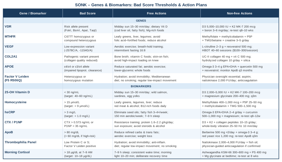
The goal here is not hope through vagueness — it is hope through better data. The biomarker section covers seven specific measurements, explains what each one reveals about SONK risk and progression, and outlines concrete steps for each. The genetics section covers six key gene variants worth understanding if you have access to a DNA report. Together, they offer a clearer map for navigating a condition that most people are told very little about.
7 Biomarkers That Matter in Spontaneous Osteonecrosis of the Knee
Tracking biomarkers is not about collecting numbers for their own sake. In SONK, the underlying biology — subchondral ischemia, impaired bone remodeling, low-grade inflammation, and microvascular stress — leaves traces in the blood that can be measured, interpreted, and acted on. The seven markers below are chosen because they reflect the mechanisms most directly implicated in SONK, and because interventions exist for most of them.
1. 25-OH Vitamin D
Why it matters: Vitamin D is not just a bone mineral supplement. Its receptor is expressed in osteoblasts, osteoclasts, endothelial cells, and immune cells. In SONK, vitamin D insufficiency compromises subchondral bone quality and impairs the vascular repair processes that could otherwise contain a small area of ischemia before it progresses to visible necrosis. Multiple studies link low 25-OH vitamin D to increased risk of bone stress injuries and impaired fracture healing — conditions mechanistically close to SONK.
How to measure it: A standard serum 25-hydroxyvitamin D test, available at any clinical lab. Cost typically ranges from $30 to $90 depending on provider and country. Optimal levels are debated, but most bone-focused clinicians (including those aligned with Peter Attia's approach) consider 40–60 ng/mL a functional target, not merely the lab reference range of 20 ng/mL.
If the score is bad (below 30 ng/mL) — plan without supplements: Direct sun exposure remains the most physiologically complete source of vitamin D, producing 10,000–20,000 IU per full-body session in midday sun. Prioritize this when geography and season allow. Wild-caught salmon, sardines, and egg yolks contribute modestly. Spending 15–30 minutes per day at solar noon with arms and legs exposed, without sunscreen, produces meaningful synthesis in most skin types during summer months.
If the score is bad — plan with supplements or equipment: Supplemental vitamin D3 (cholecalciferol) at 2,000–5,000 IU daily is a reasonable starting point for most adults with deficiency. Some clinicians use 10,000 IU daily for a 3-month correction phase, followed by re-testing. Vitamin D3 should always be paired with vitamin K2 (MK-7 form, 100–200 mcg/day) to direct calcium appropriately toward bone rather than vascular tissue. Magnesium (200–400 mg/day, glycinate form to minimize GI effects) is required as a cofactor for vitamin D activation. Re-test every 3 months during correction. Side effects at doses below 10,000 IU are uncommon when K2 and magnesium are co-administered; toxicity is primarily a risk above 40,000 IU/day sustained over months.
2. Homocysteine
Why it matters: Elevated homocysteine is a well-documented marker of endothelial dysfunction and small vessel disease. In the context of SONK, impaired microvascular perfusion of the subchondral bone is a central event — and homocysteine elevation is one of the more actionable contributors to that impairment. Homocysteine also directly inhibits osteoblast function and increases osteoclast activity, contributing to weakened subchondral architecture. Research published in the context of femoral head osteonecrosis has consistently identified elevated homocysteine as a risk factor, and the mechanism is applicable to knee osteonecrosis as well.
How to measure it: Serum homocysteine, ideally fasting. Cost ranges from $30 to $60. Functional target: below 9 µmol/L. Levels above 15 µmol/L are associated with meaningful cardiovascular and bone risk. Gary Brecka's work in applied genetics strongly emphasizes homocysteine reduction as a cornerstone of vascular and musculoskeletal longevity.
If the score is bad — plan without supplements: Dietary methionine restriction (reducing red meat, particularly processed forms) can modestly lower homocysteine. Increasing dietary folate through leafy greens, legumes, and liver is effective. Adequate protein intake with a food-first approach to B vitamins (eggs, meat, fish) supports methylation pathways. Reducing alcohol consumption has a measurable effect since alcohol depletes B6 and folate.
If the score is bad — plan with supplements or equipment: The methylation pathway — which converts homocysteine to methionine — requires folate (ideally methylfolate, not folic acid), B6 (pyridoxal-5-phosphate form), and B12 (methylcobalamin). A combined methylation support formula containing 400–1000 mcg methylfolate, 25–50 mg P5P, and 500–1000 mcg methylcobalamin, taken daily, reliably reduces homocysteine within 4–8 weeks in most people. Re-test at 8 weeks. Betaine (TMG, 500–1500 mg/day) provides an alternative methylation route (the BHMT pathway) and is particularly useful in those with MTHFR variants. No significant cycling required for these B vitamins at standard doses. Avoid high-dose isolated folic acid (non-methylated form) if MTHFR status is unknown.
3. High-Sensitivity CRP (hsCRP)
Why it matters: Low-grade systemic inflammation accelerates subchondral bone deterioration and impairs the angiogenic repair responses that healthy bone relies on. hsCRP is a sensitive marker of this background inflammatory state. While SONK is not classified as a purely inflammatory condition, elevations in hsCRP have been associated with worse outcomes in osteonecrosis in multiple studies, likely by accelerating cartilage and bone matrix degradation and impairing local healing.
How to measure it: Serum hsCRP, available at standard labs for $15 to $50. Functional target: below 1.0 mg/L. Values between 1 and 3 mg/L indicate moderate risk; above 3 mg/L suggests significant systemic inflammation requiring investigation.
If the score is bad — plan without supplements: Anti-inflammatory dietary patterns are evidence-based here. Eliminating seed oils (linoleic acid-heavy vegetable oils), reducing refined carbohydrates, and increasing omega-3-rich foods (fatty fish 3–4 times per week) typically reduce hsCRP within 6–8 weeks. Consistent aerobic exercise at moderate intensity (150 minutes per week) has a significant anti-inflammatory effect. Sleep optimization — targeting 7–9 hours with consistent timing — reduces baseline inflammatory tone substantially. Stress management (cortisol reduction) is also relevant since chronic cortisol elevation drives inflammatory cytokine production.
If the score is bad — plan with supplements or equipment: Omega-3 fatty acids (EPA+DHA) at 2–4 grams per day from high-quality fish oil have robust evidence for hsCRP reduction. Curcumin (with piperine or in liposomal form, 500–1000 mg/day) is a well-studied anti-inflammatory adjunct. Magnesium deficiency is independently associated with elevated CRP; correcting it (as described under vitamin D above) addresses this. Re-test hsCRP after 8–12 weeks of dietary and supplement changes. No significant cycling needed for omega-3s or magnesium. Curcumin can be cycled (8 weeks on, 2 weeks off) as a precaution, though adverse effects are rare at these doses.
4. Bone Turnover Markers: CTX and P1NP
Why it matters: CTX (C-terminal telopeptide of type I collagen) reflects bone resorption rate; P1NP (procollagen type I N-terminal propeptide) reflects bone formation. In SONK, the local subchondral bone undergoes a pathological remodeling process — initially driven by an ischemic event — that can be tracked systemically, though with limited spatial resolution. Importantly, these markers help determine whether bone is in a net catabolic state (high CTX relative to P1NP), which worsens SONK prognosis, or in a net anabolic state consistent with repair. Thomas Dayspring and others focused on precision medicine consistently recommend these as part of any bone health workup.
How to measure it: Serum CTX (fasting, morning) and serum P1NP. Both are available at specialty or academic labs; cost ranges from $60 to $150 per marker. CTX optimal: below 0.573 ng/mL (premenopausal range); P1NP optimal: above 35–70 ng/mL in adults. Interpret the ratio rather than each in isolation.
If the score is bad (high CTX, low P1NP) — plan without supplements: Resistance training is one of the most consistent stimuli for increasing bone formation markers. Low-impact resistance work (upper body, seated, avoiding knee loading during active SONK) drives osteoblast activity systemically. Adequate dietary protein (1.6–2.2 g/kg/day) is essential for P1NP production since it reflects collagen synthesis. Sun exposure (vitamin D, already described) directly supports P1NP. Minimizing corticosteroid exposure, alcohol, and smoking dramatically reduces CTX excess.
If the score is bad — plan with supplements or equipment: Vitamin D3 + K2 as described above. Collagen peptides (10–15 g/day, ideally type I/III with vitamin C co-ingestion) support osteoblast matrix production and have modest evidence for improving bone density markers. Strontium ranelate (where available, prescription) simultaneously inhibits resorption and stimulates formation, though it requires physician oversight. Whole-body vibration platforms (10 minutes daily at 25–50 Hz) have human trial evidence for shifting the CTX/P1NP ratio favorably in postmenopausal women — the demographic most affected by SONK. Re-test markers at 3 months.
5. ApoB and LDL Particle Count
Why it matters: Fat emboli from lipid metabolism dysregulation have been proposed as a mechanism in osteonecrosis, including SONK. Elevated LDL particle number (LDL-P) and apolipoprotein B (apoB) — which Allan Sniderman has championed as superior to LDL-C for cardiovascular risk — also reflect vascular disease risk that translates directly to microvascular integrity in subchondral bone. In corticosteroid-associated osteonecrosis, dyslipidemia is a well-established intermediary. The evidence for SONK specifically is less direct, but vascular health remains a core mechanism.
How to measure it: ApoB measured directly, or advanced lipoprotein panel (NMR LipoProfile or similar). ApoB test alone: $25–$60. Target: apoB below 80 mg/dL (below 60 mg/dL if additional risk factors present, per Attia's approach).
If the score is bad — plan without supplements: Dietary reduction of refined carbohydrates and trans fats is the first lever. Replacing saturated fat with monounsaturated fat modestly reduces LDL-P. Aerobic exercise (150+ minutes/week) improves the LDL particle size distribution (favoring large, buoyant particles over small dense LDL). Weight loss in those with metabolic syndrome dramatically improves apoB within 3–4 months.
If the score is bad — plan with supplements or equipment: Berberine (500 mg twice daily with food) has meaningful clinical evidence for apoB reduction comparable to low-dose statins in some studies, without requiring a prescription. Red yeast rice (1200 mg/day) is another option but requires physician awareness due to its statin-like mechanism. Omega-3s at 3–4 g/day reduce triglycerides (which indirectly improves particle count). Cycling berberine (8 weeks on, 4 weeks off) is prudent given limited long-term data. Re-test apoB at 3 months.
6. Thrombophilia Panel (Protein C, Protein S, Factor V Leiden)
Why it matters: Thrombophilia — a tendency toward abnormal clotting — directly compromises the microvascular circulation that supplies subchondral bone. Multiple studies have found elevated rates of thrombophilic conditions in patients with idiopathic osteonecrosis, including SONK. Protein C and S deficiencies reduce the body's ability to break down clots in small vessels; Factor V Leiden (a genetic variant, described in the genetics section) causes resistance to activated protein C. Together, these create a prothrombotic milieu that can precipitate or worsen subchondral ischemia.
How to measure it: Protein C activity, Protein S activity, and Factor V Leiden PCR (or activated protein C resistance test). These may be ordered as a hypercoagulability panel or individually. Cost: $100–$300 for the panel. Some tests require physician referral. Ideally tested when not acutely ill and not on anticoagulants.
If the score is bad — plan without supplements: Staying well-hydrated (adequate blood viscosity), avoiding prolonged immobility, maintaining healthy weight, and eliminating smoking all reduce thrombotic tendency. Anti-inflammatory diet (as described under hsCRP) reduces platelet aggregation. Regular low-impact movement — even walking or swimming — significantly improves venous return and microvascular flow.
If the score is bad — plan with supplements or equipment: Nattokinase (2000–4000 FU per day) is a fibrinolytic enzyme with human trial data for reducing hypercoagulability markers. Serrapeptase has similar theoretical applications but weaker evidence. In confirmed thrombophilia with osteonecrosis, physician-supervised anticoagulation (low-dose aspirin at minimum; anticoagulants in severe cases) is the standard medical approach and should not be avoided or replaced with supplements alone. Fish oil (3–4 g/day EPA+DHA) reduces platelet aggregation modestly. Re-test at 3 months; cycle nattokinase (12 weeks on, 4 weeks off) and avoid combining with pharmaceutical anticoagulants without supervision.
7. Morning Serum Cortisol
Why it matters: Chronically elevated cortisol impairs bone formation, promotes osteoclast activity, and causes vasoconstriction in the microvasculature — a trifecta of harm for subchondral bone. In SONK patients who are not on exogenous corticosteroids, endogenous cortisol dysregulation (from chronic stress, poor sleep, or HPA axis dysfunction) may be an underappreciated contributor. Elevated cortisol also drives systemic inflammation and raises hsCRP, compounding risk.
How to measure it: Morning serum cortisol (drawn between 7–9 AM, fasting). Cost: $30–$60. Optimal range: 10–18 µg/dL in the morning. A 4-point salivary cortisol test (morning, noon, evening, night) provides a more complete picture of HPA axis rhythm for $100–$180 through specialty labs.
If the score is bad — plan without supplements: Sleep quality is the highest-leverage free intervention: targeting 7–9 hours with a consistent wake time, blackout curtains, and a bedroom temperature below 67°F (19°C). Morning light exposure (10–20 minutes outside within 30 minutes of waking) stabilizes the cortisol awakening response and prevents afternoon/evening elevations. Stress reduction through deliberate recovery time — not necessarily meditation, but any non-stimulating, low-stakes activity the person enjoys — reduces mean daily cortisol meaningfully over 4–8 weeks.
If the score is bad — plan with supplements or equipment: Ashwagandha root extract (KSM-66 form, 300–600 mg/day) has multiple randomized controlled trials showing significant cortisol reduction over 8 weeks. Phosphatidylserine (400 mg/day) blunts cortisol spikes, particularly post-exercise. Magnesium glycinate at bedtime reduces nighttime cortisol elevation and improves sleep quality. Cycle ashwagandha (8–12 weeks on, 4 weeks off). Re-test morning cortisol at 8 weeks. Avoid combining with sedative medications without physician guidance.
With the biomarker picture established, the genetic layer adds a different kind of information: not what your biology is doing right now, but what predispositions were built in from birth — and how those predispositions can be modified.
6 Genes Worth Knowing in SONK
Genetic testing through services like 23andMe, AncestryDNA, or clinical SNP panels gives you raw data that, when run through interpretation tools (Genetic Genie, StrateGene, or a functional medicine clinician), reveals actionable variants. The genes below are the most relevant to the mechanisms underlying spontaneous osteonecrosis of the knee. None of them are deterministic — having a variant does not guarantee disease, and lacking one does not guarantee protection. They are context, not destiny.
Gene 1: VDR (Vitamin D Receptor)
What it affects: VDR polymorphisms (most studied: FokI, BsmI, ApaI, TaqI) alter how effectively your cells respond to vitamin D, regardless of serum levels. A person with an inefficient VDR variant may show normal 25-OH vitamin D levels but still experience downstream signaling deficiencies in bone formation and vascular regulation. This is particularly relevant in SONK given the central role of vitamin D in subchondral bone maintenance.
If the gene is bad — plan without supplements: Maximize sunlight exposure duration and timing (midday UVB), prioritize dietary vitamin D sources (cod liver oil, fatty fish, egg yolks), and pair with dietary fat to maximize absorption. The magnesium-vitamin D cofactor relationship becomes even more important: ensure dietary magnesium intake is high (nuts, seeds, legumes, leafy greens).
If the score is bad — plan with supplements or equipment: Those with VDR variants often need higher vitamin D3 supplementation (5,000–10,000 IU/day) to achieve optimal serum levels and functional effects. Testing serum 25-OH D more frequently (every 8–10 weeks during optimization) is warranted. Vitamin K2 (MK-7, 200 mcg/day) and magnesium remain essential co-supplements. Some clinicians also add boron (3–6 mg/day), which upregulates VDR expression. Re-test at 3 months; maintain K2 indefinitely when taking high-dose D3.
Gene 2: MTHFR (Methylenetetrahydrofolate Reductase)
What it affects: MTHFR C677T and A1298C variants reduce the enzyme's efficiency in converting folate to its active form, impeding the methylation cycle and leading to homocysteine accumulation. As described in the biomarker section, elevated homocysteine damages endothelial cells and impairs bone vascularity — directly relevant to SONK. Gary Brecka's educational framework is built substantially around this gene's downstream effects on vascular and structural tissue health.
If the gene is bad — plan without supplements: Dietary methylfolate from whole food sources (dark leafy greens, liver, legumes) rather than folic acid (the synthetic, non-methylated form). Avoiding folic acid-fortified foods is particularly relevant for homozygous C677T carriers. B12-rich foods daily (meat, eggs, dairy). Alcohol minimization is critical as it directly depletes folate and B6.
If the score is bad — plan with supplements or equipment: Active B vitamins are non-negotiable for confirmed MTHFR variants: methylfolate (400–800 mcg), methylcobalamin (500–1000 mcg), and P5P (pyridoxal-5-phosphate, 25–50 mg). TMG/Betaine (1–2 g/day) supports the alternative methylation pathway. Creatine monohydrate (3–5 g/day) spares the methylation cycle by reducing its demand for SAMe — a practical, evidence-backed addition. No cycling needed for these nutrients at standard doses. Monitor homocysteine every 3 months.
Gene 3: VEGF (Vascular Endothelial Growth Factor)
What it affects: VEGF is the primary driver of angiogenesis — the growth of new blood vessels — and its polymorphisms directly affect how well bone tissue responds to ischemic stress by generating new vascular supply. In osteonecrosis, impaired VEGF signaling is a consistent finding. Variants that reduce VEGF expression or bioavailability (such as -2578C/A and -1154G/A) may limit the capacity to revascularize ischemic subchondral bone, leading to progression rather than spontaneous repair.
If the gene is bad — plan without supplements: Aerobic exercise is the most potent natural stimulus for VEGF expression and angiogenesis — even low-impact forms like swimming and cycling are effective. Hypoxic breathing protocols (breath-hold training, altitude exposure) upregulate VEGF through HIF-1α pathways. Intermittent fasting (16:8 protocol) also moderately increases VEGF signaling. Cold water immersion has emerging evidence for vascular adaptation.
If the score is bad — plan with supplements or equipment: L-arginine (3–5 g/day) and L-citrulline (2–3 g/day) support nitric oxide production, which interacts with VEGF pathways to enhance vascular growth. Resveratrol (500 mg/day, with food, liposomal form preferred) has preclinical and some human evidence for VEGF modulation. Hyperbaric oxygen therapy (HBOT) — 1–2 atmospheres, 40–60 sessions of 60–90 minutes — has specific clinical evidence in osteonecrosis for enhancing VEGF-mediated revascularization. This is a significant investment ($100–$300 per session at independent clinics, or home units at ~$5,000–$15,000) but represents the most mechanistically targeted intervention for VEGF-impaired bone repair. Cycle resveratrol (12 weeks on, 4 weeks off); L-citrulline can be taken continuously.
Gene 4: COL2A1 (Collagen Type II Alpha 1)
What it affects: COL2A1 encodes the primary structural protein of articular cartilage. Variants in this gene reduce cartilage matrix quality, making subchondral bone more vulnerable to stress-related injury and accelerating the damage that follows an ischemic event in SONK. Reduced collagen quality also impairs the repair scaffold needed to recover subchondral integrity.
If the gene is bad — plan without supplements: Dietary collagen from bone broth, cartilage-rich cuts of meat, and skin-on fish provides hydroxyproline-rich peptides. Vitamin C from whole foods (bell peppers, citrus) is essential for collagen hydroxylation. Avoiding high-impact loading on affected joints during active phases of SONK protects the compromised matrix.
If the score is bad — plan with supplements or equipment: Hydrolyzed collagen peptides (specifically UC-II undenatured type II collagen, 40 mg/day, or hydrolyzed collagen type I/III, 10 g/day) with concurrent vitamin C (500–1000 mg taken 30–60 minutes before the dose) have randomized trial evidence for improving joint health markers. Silica (from horsetail extract or orthosilicic acid, 10–30 mg/day) supports collagen cross-linking. These can be taken continuously with no established cycling requirement. Glucosamine/chondroitin is commonly recommended but has mixed evidence for structural benefit; it may help with symptoms but should not be the primary strategy.
Gene 5: APOE (Apolipoprotein E)
What it affects: APOE e4 variants are widely known for Alzheimer's risk, but their relevance in vascular disease — and by extension, bone microvascular health — is underappreciated. APOE affects how lipoproteins are cleared, influencing fat emboli risk (a proposed osteonecrosis mechanism) and vascular endothelial function. The e4 allele is associated with less efficient lipoprotein clearance and greater susceptibility to lipid-driven vascular damage.
If the gene is bad — plan without supplements: APOE e4 carriers respond particularly well to dietary saturated fat reduction and to the replacement of processed carbohydrates with lower-glycemic whole foods. Regular aerobic exercise dramatically modifies the APOE e4 phenotype by improving lipoprotein clearance. Ali Torkamani's work on precision medicine consistently highlights lifestyle-mediated modification of APOE-driven risk.
If the score is bad — plan with supplements or equipment: Omega-3 fatty acids (3–4 g EPA+DHA/day) are especially beneficial in APOE e4 carriers, with stronger evidence for lipid and vascular benefit in this genetic subgroup. Quercetin (500 mg/day with fat) and resveratrol modulate LDL oxidation, which is elevated in e4 carriers. ApoB monitoring (as described above) is the key tracking metric. Cycle quercetin (8 weeks on, 4 weeks off). Re-test apoB at 3 months.
Gene 6: Factor V Leiden (F5 R506Q)
What it affects: Factor V Leiden is the most common inherited thrombophilia in European populations, present in 3–8% of the general population. The mutation causes factor V to resist deactivation by protein C, leading to a hypercoagulable state. It is directly relevant to SONK because subchondral ischemia can be initiated or worsened by microthrombi in the intraosseous vessels. Several studies have found elevated rates of thrombophilic variants — including Factor V Leiden — in osteonecrosis patients compared to controls.
If the gene is bad — plan without supplements: Hydration, physical activity, and smoking cessation are foundational. Avoiding prolonged immobility (long-haul flights, sedentary work without breaks) reduces thrombotic risk. Dietary interventions that support healthy endothelial function (Mediterranean dietary pattern, omega-3-rich foods) reduce the clinical expression of the variant.
If the score is bad — plan with supplements or equipment: This variant warrants physician involvement. Low-dose aspirin may be recommended depending on clinical context. Nattokinase (2000 FU/day, as described in the biomarker section) can complement medical management. In confirmed osteonecrosis with Factor V Leiden, anticoagulation therapy has been explored in clinical settings as a disease-modifying approach. Do not attempt to manage thrombophilia with supplements alone; use them as adjuncts to medical oversight.
A Podcast That Reframes Bone and Vascular Health
One of the most practically useful sources for understanding the intersection of vascular biology, bone metabolism, and longevity science is the Huberman Lab podcast — specifically the episodes on bone health, deliberate cold exposure, and the vitamin D and light protocol series. Andrew Huberman, a Stanford neuroscientist, frequently synthesizes research in ways that shift clinical defaults, and several of his frameworks are directly relevant to SONK.
10 High-Impact Ideas From the Huberman Approach
1. Morning sunlight is a vascular and hormonal primer, not just vitamin D. The first 30 minutes of morning light exposure sets cortisol timing, regulates melatonin, and drives nitric oxide release in skin — all of which affect vascular function and bone microcirculation.
2. Vitamin D without magnesium is incomplete. Huberman consistently emphasizes that vitamin D activation requires magnesium, and that most deficiency protocols fail because they omit this cofactor. For SONK, this matters because incomplete vitamin D activation leaves VDR pathways understimulated.
3. Zone 2 cardio is the most underutilized bone-protective intervention. 150–180 minutes per week of low-intensity aerobic work (where you can hold a conversation) improves capillary density, reduces cortisol, lowers hsCRP, and stimulates VEGF — a direct hit on four of the seven biomarkers above simultaneously.
4. Sleep is the primary bone repair window. Growth hormone — the principal driver of bone matrix repair — is secreted predominantly during slow-wave sleep. Chronic sleep deficiency impairs this repair cycle, worsening the prognosis of any bone lesion including SONK. Huberman's sleep optimization protocols (temperature, light, timing) are built around maximizing this window.
5. Deliberate cold exposure (cold plunge, 11 minutes/week total) improves microvascular tone. Cold immersion drives norepinephrine release, which strengthens vascular smooth muscle and improves endothelial function — relevant to the microvasculature serving subchondral bone.
6. Resistance training is a systemic bone signal, not a local one. Loading any part of the skeleton sends systemic signals (including osteocalcin and IGF-1) that benefit remote bone sites — meaning upper body resistance training during active SONK still improves bone biology globally.
7. Omega-3 to omega-6 ratio, not total fat, predicts inflammatory state. Most Western diets have a 15:1 to 20:1 omega-6 to omega-3 ratio. Shifting toward 4:1 or lower (through fatty fish and fish oil supplementation) has a measurable effect on hsCRP within 6–8 weeks.
8. Phosphatidylserine and ashwagandha form a synergistic cortisol-lowering pair. Used together, they blunt the HPA axis response to stress more effectively than either alone — important for SONK patients who are also dealing with pain-related physiological stress.
9. The methylation cycle and bone repair are connected through SAMe. S-adenosylmethionine, the methyl donor produced by a healthy methylation cycle, directly supports chondrocyte function and has clinical trial evidence for joint cartilage health. Optimizing MTHFR-related pathways (as described in the genetics section) ultimately supports SAMe availability.
10. Inflammatory diet is a vascular disease diet. Huberman consistently frames ultra-processed foods and seed-oil-heavy diets as endothelial damage accelerators. Given that SONK is fundamentally a vascular disease of bone, this framing — not as a "joint diet" but as a vascular health diet — is more motivating and mechanistically accurate.
Complementary Approaches With Clinical Relevance
Beyond biomarkers and genetics, certain non-pharmacological modalities have enough human evidence to warrant consideration in SONK management. The selections below are chosen for their mechanistic fit and available clinical data.
Low-Level Laser Therapy / Photobiomodulation
Photobiomodulation (PBM) delivers red and near-infrared light (typically 630–1000 nm) to tissue, stimulating mitochondrial function, nitric oxide release, and local angiogenesis. In the context of SONK, its primary relevance is promoting subchondral tissue repair and reducing local inflammation in ways that may complement the systemic interventions described above. Several studies have examined PBM in knee osteoarthritis and bone healing with encouraging results.
A 2019 systematic review of low-level laser therapy for knee conditions found statistically significant reductions in pain and improvements in function in multiple randomized controlled trials. While SONK-specific trials are limited, the mechanisms — improved microvascular function, reduced inflammatory cytokines, and stimulated bone matrix cell activity — directly address SONK pathophysiology.
In practice, PBM for the knee can be applied using a class 3B or class 4 therapeutic laser device at 830–1000 nm, 3–5 sessions per week, 10–15 minutes per session over 6–8 weeks. Home devices (red light panels at 660/850 nm dual-wavelength) are available for $200–$600 and allow self-application. Realistic expectations are pain reduction and possible slowing of progression; PBM should be viewed as adjunctive, not curative.
Tai Chi
Tai chi combines slow, controlled movement, breath regulation, and proprioceptive training — a combination particularly suited to SONK, where joint offloading, balance, and gentle circulation enhancement are therapeutic priorities. Unlike many forms of exercise, tai chi can be practiced without high knee compressive loading, making it suitable even during active phases of SONK.
A 2020 randomized controlled trial published in Arthritis & Rheumatology demonstrated that tai chi produced equivalent outcomes to physical therapy for knee osteoarthritis, with greater adherence at one year. While SONK is distinct from OA, the shared features — pain, knee function impairment, and the need for low-impact movement — make this evidence broadly relevant. Tai chi also reduces cortisol, improves balance, and lowers hsCRP, all of which are relevant biomarker targets identified above.
Practically, joining a supervised tai chi class (Yang style is most studied) for 60-minute sessions twice weekly over 12 weeks represents a well-studied protocol. This can be sustained indefinitely. For those with significant pain, online beginner programs allow self-pacing. The key caution is avoiding deep knee flexion positions until imaging confirms no active structural deterioration.
Biofeedback
Biofeedback teaches real-time physiological awareness and voluntary regulation of normally automatic processes — including muscle tension, heart rate variability (HRV), and stress reactivity. In SONK, its primary application is addressing the pain-stress cycle: chronic pain drives cortisol and sympathetic nervous system activation, which worsen vascular function and bone repair, which worsen pain. Biofeedback interrupts this loop at a controllable point.
HRV biofeedback specifically — where the patient learns to regulate breathing to maximize heart rate variability — has been shown to reduce cortisol, lower hsCRP, and improve parasympathetic tone in multiple randomized trials. For SONK patients, this translates to improved vascular regulation and a more favorable hormonal environment for bone repair. A 2014 meta-analysis in Applied Psychophysiology and Biofeedback found HRV biofeedback consistently effective for reducing physiological stress markers.
Sessions are typically 30–45 minutes, once or twice weekly, with a trained biofeedback therapist for initial guidance. Consumer HRV biofeedback devices (HeartMath Inner Balance, Polar H10 with compatible apps) allow home practice at $80–$200. A 6–8 week protocol with daily 10-minute practice sessions is a realistic entry point.
Conclusion
Spontaneous osteonecrosis of the knee is not a condition that yields quickly to simple answers, but it is one where detailed biological information meaningfully changes the picture. Tracking the seven biomarkers described here — vitamin D, homocysteine, hsCRP, bone turnover markers, apoB, thrombophilia markers, and cortisol — gives you a functional map of the pathways most involved in SONK development and repair. Understanding which of the six key genetic variants you carry helps explain why those pathways may have been vulnerable in the first place, and how aggressively to act on each.
The most useful next step is not adding every supplement at once — it is measuring first. Get a baseline panel, identify your specific areas of dysregulation, and address those systematically. Work with a physician or functional medicine clinician who can integrate these results with your imaging and clinical picture. Better information, acted on carefully, remains the clearest path from reactive management to genuine recovery support.