This article contains AI generated content.
· UpdatedStasis Dermatitis Genes And Biomarkers: 6 Genes And 7 Biomarkers To Track
Introduction
Stasis dermatitis has a way of sneaking up on you. What starts as minor ankle swelling and some itching gradually becomes a persistent, discolored patch of inflamed skin on your lower legs that conventional treatments barely touch. If you have been told to simply moisturize and wear compression stockings, you already know that advice handles the surface without addressing the cause.
The truth is that stasis dermatitis sits at the intersection of vascular failure, chronic inflammation, and coagulation abnormalities. Everyone's version of this condition is shaped by a slightly different mix of venous valve dysfunction, inflammatory burden, and individual biology. A generic management approach often yields generic results, which explains why so many people cycle through treatments without meaningful improvement.
What changes the equation is getting more precise. Specific blood biomarkers can tell you whether systemic inflammation, impaired clot clearance, endothelial damage, or iron accumulation is driving the skin changes in your particular case. And behind those biomarkers, there are genetic variants that explain why your venous system is under more stress than others — and what you can actually do about it.
This article covers both levels. The biomarker section maps seven measurable blood values directly relevant to stasis dermatitis, with actionable plans for each. The genetics section translates six key variants into practical compensatory strategies. Beyond the science, you will find a summary of Peter Attia's research-backed framework on vascular aging and four evidence-supported complementary approaches. Together, these tools can help you move from guessing to targeting — and from managing symptoms to addressing root causes.
Summary
This article maps seven blood biomarkers — hsCRP, D-dimer, homocysteine, ferritin, fibrinogen, VEGF, and Lp(a) — that directly reflect the inflammatory, vascular, and coagulation mechanisms driving stasis dermatitis. For each one, you will find how to test it affordably, what a concerning result looks like, and what to do whether or not you choose supplements. The genetics section covers six variants — MTHFR, Factor V Leiden, Factor II, MMP9, VEGFA, and PAI-1 — that make some people far more susceptible to venous skin disease, with targeted compensation plans for each. The article also summarizes Peter Attia's framework for cardiovascular biomarker optimization and four evidence-backed complementary modalities. The goal throughout is precision over guesswork: knowing your numbers changes everything about the approach.
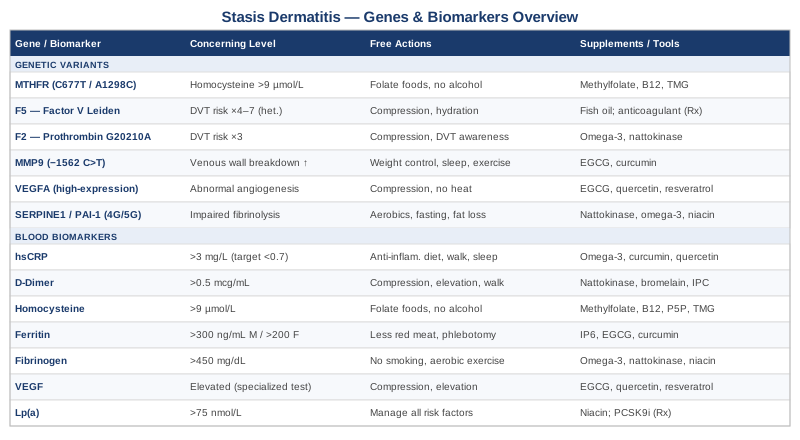
7 Biomarkers to Track When You Have Stasis Dermatitis
The vascular and inflammatory biology of stasis dermatitis leaves a measurable fingerprint in the blood. These seven biomarkers map to the most important mechanisms: venous wall inflammation, clot activity, endothelial damage, iron accumulation, and angiogenic dysregulation. Tracking them gives you a working picture of what is actually driving your condition — and a clearer target for any intervention.
1. High-Sensitivity C-Reactive Protein (hsCRP): The Inflammation Index
Why it matters: hsCRP is produced by the liver in response to tissue inflammation, and it rises reliably in chronic venous insufficiency. Research has consistently found elevated hsCRP levels in patients with advanced venous disease, where it correlates with the severity of skin changes including lipodermatosclerosis — the hardening and discoloration that precedes and accompanies stasis dermatitis. Systemic inflammation accelerates venous wall deterioration and impairs normal healing responses, making this marker one of the most actionable in the panel.
How to measure it
A standard hsCRP blood test costs between $20 and $60 through most labs. Request high-sensitivity CRP specifically, not the standard CRP, as the standard test misses low-grade chronic inflammation. Peter Attia recommends targeting below 0.7 mg/L for optimal vascular health; values above 3.0 mg/L indicate significant inflammatory burden. Multiple studies confirm elevated hsCRP in progressive venous skin disease.
If the score is bad, the plan without supplements
Begin with dietary changes: eliminate ultra-processed foods, seed oils, and refined carbohydrates. Prioritize oily fish — sardines, mackerel, salmon — two to three times per week. Lose visceral fat if present; even a 5% reduction in body weight produces measurable hsCRP reduction. Consistent calf-muscle activity (daily walking, ankle pumps, stair climbing) improves venous return and reduces local inflammatory signaling. Optimize sleep to 7–9 hours, as poor sleep alone drives hsCRP up by 30–40% in controlled studies.
If the score is bad, the plan with supplements or equipment
Omega-3 fatty acids (EPA+DHA): 2–4 g/day with food, ongoing. Most effective single supplement for hsCRP reduction. Side effects: mild GI at high doses, slightly increased bleeding time — caution with anticoagulants. Curcumin with piperine: 500–1000 mg twice daily with meals, ongoing. Piperine boosts curcumin absorption by roughly 20-fold. Side effects: GI discomfort in some; avoid high doses alongside blood thinners. Magnesium glycinate: 300–400 mg before bed, ongoing. Reduces inflammatory cytokine activity. Side effects: loose stools above 600 mg/day. Quercetin: 500 mg twice daily, cycle 8 weeks on / 4 weeks off. Inhibits NF-κB inflammatory pathway. Side effects: mild GI in some individuals.
2. D-Dimer: Tracking Clot Activity in Venous Disease
Why it matters: D-dimer is a degradation product of fibrin clots. When it is chronically elevated, it signals ongoing small-scale clotting and fibrinolysis in the venous system. In stasis dermatitis patients, persistently elevated D-dimer suggests that microthrombi are forming in the lower-leg venous microvasculature, worsening tissue hypoxia and driving skin changes. It also flags elevated deep vein thrombosis risk, which is significantly higher in people with advanced chronic venous insufficiency.
How to measure it
D-dimer is widely available through standard labs; cost is typically $40–$100. Normal values are generally below 0.5 mcg/mL (500 ng/mL) FEU. Note that D-dimer rises with age, recent illness, and surgery — interpret with that context in mind. Research shows chronically elevated D-dimer in patients with chronic venous insufficiency and venous leg ulcers even without acute DVT.
If the score is bad, the plan without supplements
Graded compression stockings (20–30 mmHg or 30–40 mmHg depending on severity) are the most evidence-based mechanical intervention for reducing venous stasis and microclot formation. Leg elevation above heart level for 20–30 minutes three times daily dramatically reduces venous pressure. Avoid prolonged static sitting or standing without movement breaks. Daily walking targeting 30 minutes minimum activates the calf muscle pump, which is more effective than most medications for reducing venous pooling. Adequate hydration reduces blood viscosity throughout the day.
If the score is bad, the plan with supplements or equipment
Nattokinase: 2000 FU/day taken away from food, cycle 3 months on / 1 month off. A fibrinolytic enzyme derived from fermented soy. Side effects: blood-thinning effect — do not combine with anticoagulants without physician oversight; GI discomfort in some. Bromelain: 500 mg between meals twice daily, cycle 6 weeks on / 3 weeks off. Proteolytic enzyme with mild fibrinolytic activity. Side effects: GI discomfort, potential allergy in pineapple-sensitive individuals. Intermittent pneumatic compression (IPC) device: Home-use graduated compression pump devices provide mechanical pressure cycles to move venous blood upward — a practical option for severe cases or those with desk-bound work.
3. Homocysteine: The Hidden Vascular Damage Marker
Why it matters: Elevated homocysteine is directly toxic to the endothelium — the inner lining of blood vessels. It promotes oxidative stress in venous walls, impairs nitric oxide signaling, and accelerates venous valve degeneration. In stasis dermatitis, endothelial damage compounds already-impaired venous return by reducing venous wall elasticity and promoting inflammatory cell recruitment. Peter Attia consistently places homocysteine among the most underappreciated and undertreated vascular risk markers in standard medicine.
How to measure it
A homocysteine blood test costs $30–$80 through most labs and should be drawn fasting. Optimal range: below 9 mcmol/L (Attia's target); values above 12 mcmol/L indicate meaningful risk; above 15 mcmol/L qualifies as hyperhomocysteinemia. Studies link elevated homocysteine to chronic venous disease progression and increased DVT risk, making this an important item to test alongside standard panels.
If the score is bad, the plan without supplements
Dietary changes are meaningful. Increase folate-rich foods — dark leafy greens, legumes, asparagus — to 2–3 cups daily. Reduce animal protein modestly; methionine from meat is the direct precursor to homocysteine. Eliminate alcohol, which depletes the B-vitamin cofactors that clear homocysteine. Avoid synthetic folic acid supplements or fortified foods if MTHFR variants are present (see the genetics section), as they compete with the active folate form at receptor sites.
If the score is bad, the plan with supplements or equipment
Methylfolate (5-MTHF): 800–1600 mcg/day, ongoing. The bioactive form of folate — essential if any MTHFR variant is present. Side effects: at very high doses, may trigger anxiety or unmask B12 deficiency. Methylcobalamin B12: 1000–2000 mcg/day, ongoing. Works synergistically with methylfolate; deficiency alone can raise homocysteine significantly. Side effects: essentially none at standard doses. Pyridoxal-5-phosphate (P5P, active B6): 25–50 mg/day, ongoing. Side effects: peripheral neuropathy only at doses above 200 mg/day — irrelevant at this range. Betaine (TMG): 500–1000 mg twice daily, ongoing. Directly remethylates homocysteine through the BHMT pathway independent of MTHFR. Side effects: mildly fishy odor at very high doses; mild GI in some.
4. Ferritin: What Iron Accumulation Reveals About Your Skin
Why it matters: The brownish discoloration characteristic of stasis dermatitis is literally iron accumulating in the skin after red blood cells break down under elevated venous pressure — a process called hemosiderin deposition. Serum ferritin reflects total-body iron stores and also serves as an acute-phase inflammatory protein that rises with chronic inflammation. Elevated ferritin signals active iron-driven oxidative damage in lower-leg tissues and sustains a pro-inflammatory local environment that impairs healing.
How to measure it
Ferritin is a standard lab test costing $20–$50. Test it alongside iron, TIBC, and transferrin saturation for a complete picture. Optimal ferritin for most adults: 30–150 ng/mL. Values above 300 ng/mL in men or 200 ng/mL in women raise concern independent of inflammation. Research confirms that iron overload worsens inflammatory skin conditions and impairs local tissue healing mechanisms, including in venous skin disease.
If the score is bad, the plan without supplements
Reduce dietary iron: limit red meat to twice weekly maximum, avoid cast iron cookware for acidic foods, and eliminate iron-containing multivitamins. If ferritin is significantly elevated (above 300–400 ng/mL) without obvious acute-phase cause, consider therapeutic phlebotomy — donating blood every 8–12 weeks is the fastest non-pharmacological method of reducing iron stores. Rule out hereditary hemochromatosis with HFE gene testing if ferritin is persistently and significantly elevated.
If the score is bad, the plan with supplements or equipment
IP6 (inositol hexaphosphate): 1–2 g/day on an empty stomach, cycle 3 months on / 1 month off. Natural iron chelator. Side effects: reduces absorption of all minerals taken concurrently — always take fasted and at least two hours away from meals and other supplements. EGCG (green tea extract): 400–600 mg/day with food, ongoing at conservative doses. Mild iron chelation effect plus direct anti-inflammatory activity. Side effects: GI discomfort on empty stomach; hepatotoxicity risk at doses above 1200 mg/day — keep to 400–600 mg maximum. Curcumin: 500–1000 mg twice daily with piperine. Chelates non-heme iron mildly and reduces inflammatory cytokines triggered by iron overload.
5. Fibrinogen: The Coagulation Marker Most Panels Skip
Why it matters: Fibrinogen is the raw material that blood clots are built from, and it doubles as an acute-phase inflammatory protein that rises with vascular injury and infection. In stasis dermatitis, chronically elevated fibrinogen reflects both increased clotting tendency in sluggish venous blood and systemic inflammatory burden. It predicts adverse venous disease outcomes and is independently associated with more severe lipodermatosclerosis and ulceration risk.
How to measure it
Fibrinogen is measured in standard coagulation panels; cost is $30–$70. Normal range is 200–400 mg/dL. Values above 450–500 mg/dL in the context of venous disease are clinically relevant. Multiple studies have found elevated fibrinogen in patients with venous leg ulcers and advanced chronic venous insufficiency stages, where it correlates with both inflammatory and thrombotic risk.
If the score is bad, the plan without supplements
Smoking cessation, if applicable, is the single most effective lifestyle intervention — smoking elevates fibrinogen by 20–30% and is a direct driver of venous disease progression. Aerobic exercise at 30–60 minutes of moderate intensity five days per week reliably lowers fibrinogen over 6–8 weeks. An anti-inflammatory dietary pattern (Mediterranean-style) reduces fibrinogen through cytokine modulation. Weight loss, particularly visceral fat reduction, shows a dose-response effect on fibrinogen levels.
If the score is bad, the plan with supplements or equipment
Omega-3 fatty acids (EPA+DHA): 3–4 g/day, ongoing. Reduces fibrinogen through prostaglandin pathway modulation. Side effects: as above. Nattokinase: 2000 FU/day, cycle 3 months on / 1 month off. Degrades fibrin directly and reduces circulating fibrinogen. Side effects: blood-thinning — do not combine with anticoagulants without physician oversight. Niacin (nicotinic acid form only — not nicotinamide): Start at 100 mg/day with food, titrate to 500 mg/day over 4–6 weeks. Cycle 6 months on, then reassess with labs. Side effects: skin flushing (typically fades after 2–4 weeks of consistent use), potential liver stress at higher doses — monitor liver function tests every 6 months above 500 mg.
6. Vascular Endothelial Growth Factor (VEGF): When Angiogenesis Becomes a Problem
Why it matters: VEGF drives the formation of new blood vessels — a process that becomes dysregulated in chronic venous insufficiency. Elevated circulating VEGF contributes to leaky, dysfunctional microvessels in affected skin, perpetuating fluid leakage, tissue hypoxia, and the chronic wound environment of stasis dermatitis. Unlike the other markers in this list, VEGF is not yet standard in routine labs, but it adds mechanistic clarity in cases where stasis dermatitis is severe or progressing despite standard care.
How to measure it
VEGF testing requires a specialized lab; cost ranges from $100 to $300. Plasma VEGF is more reproducible than serum VEGF, which is artificially elevated by platelet activation during blood draw. Studies confirm significantly elevated VEGF in chronic venous insufficiency patients, with levels correlating with disease severity and wound persistence. This is an advanced marker — consider it when the standard panel does not explain the clinical picture.
If the score is bad, the plan without supplements
Compression therapy is the most mechanically direct way to reduce VEGF-driven microvascular leakage — it lowers venous hypertension, the primary stimulus for VEGF production in affected limbs. Regular moderate physical activity improves microvascular remodeling over time. Routine limb elevation throughout the day (20–30 minutes above heart level, three times daily) reduces both hydrostatic pressure and the hypoxia signal that continuously upregulates VEGF expression.
If the score is bad, the plan with supplements or equipment
EGCG: 400 mg twice daily with food, ongoing. Inhibits VEGF receptor signaling through the PI3K/Akt pathway. Side effects: hepatotoxicity risk at very high doses — stay below 800 mg/day total. Quercetin: 500 mg twice daily, cycle 8 weeks on / 4 weeks off. Anti-angiogenic activity through multiple converging pathways. Side effects: mild GI. Resveratrol: 500 mg/day with a fat-containing meal, ongoing. Inhibits VEGF-driven angiogenesis and reduces parallel inflammatory activity. Side effects: rare; mild blood-thinning effect.
7. Lipoprotein(a) [Lp(a)]: The Underdiagnosed Vascular Risk Factor
Why it matters: Lp(a) is a structurally modified form of LDL cholesterol with unique prothrombotic properties — it inhibits plasminogen activation, directly impairing clot clearance in venous beds. This mechanism matters specifically for stasis dermatitis: when Lp(a) is elevated, the venous system's ability to clear microthrombi is further compromised, deepening the chronic hypoxia and inflammatory signaling that drives skin changes. Thomas Dayspring and Peter Attia both consistently flag Lp(a) as one of the most underdiagnosed vascular risk factors in routine practice.
How to measure it
Lp(a) should be measured in nmol/L rather than mg/dL for the most accurate comparison across labs. Cost: $50–$150. Optimal: below 30 nmol/L; elevated risk: 75–124 nmol/L; high risk: above 125 nmol/L. Critically, Lp(a) is almost entirely genetically determined — lifestyle changes have minimal impact on it, which means knowing your level early guides medical planning rather than triggering futile behavioral changes. Research links elevated Lp(a) to venous thromboembolism risk through its plasminogen-inhibiting mechanism.
If the score is bad, the plan without supplements
If Lp(a) is significantly elevated, the practical response is to become more aggressive about all other modifiable vascular risk factors: maintain optimal blood pressure, eliminate smoking, manage hsCRP aggressively, and use compression stockings without exception. Elevated Lp(a) is primarily a signal to raise vigilance across the full biomarker panel — not a reason to despair, but a reason to act. Low-dose aspirin (under physician direction) is sometimes considered due to antiplatelet effects in high Lp(a) settings; discuss with your doctor.
If the score is bad, the plan with supplements or equipment
Niacin (nicotinic acid form only — not nicotinamide or flush-free niacin): The only widely available supplement with meaningful Lp(a)-lowering evidence, reducing levels by 20–30% in some trials. Start at 100 mg/day with food and increase by 100 mg every two weeks to a target of 1000–2000 mg/day in split doses. Cycle with lab monitoring every 6 months. Side effects: significant flushing (pretreat with 325 mg aspirin 30 minutes before, or use extended-release formulation), GI discomfort, potential hepatotoxicity at high doses — mandatory liver function monitoring above 500 mg/day. PCSK9 inhibitors (prescription only): Reduce Lp(a) modestly (15–20%) alongside substantial LDL reduction — relevant to discuss with a preventive cardiologist or lipidologist if Lp(a) is very high (above 150 nmol/L).
The Genetic Side of Stasis Dermatitis: 6 Variants Worth Knowing
Understanding the biomarkers tells you where you are right now. Genetics tells you why your venous and inflammatory biology is calibrated the way it is — and which compensatory strategies are most relevant for your specific makeup. These six variants appear consistently in research on venous insufficiency, thrombophilia, and inflammatory skin conditions.
1. MTHFR (C677T and A1298C): The Methylation Bottleneck
What it affects: MTHFR encodes the enzyme that converts dietary folate into its active form (5-MTHF) — the molecule required to remethylate homocysteine back to methionine. The C677T variant (especially homozygous TT) reduces enzyme activity by up to 70%, leading to elevated homocysteine and impaired methylation across multiple biological systems. For stasis dermatitis, homocysteine-driven endothelial damage accelerates venous wall degeneration directly. Studies consistently link homozygous C677T to elevated venous thrombosis risk and venous disease severity.
If the gene is bad, the plan without supplements
Prioritize folate-rich whole foods at every meal: dark leafy greens, legumes, asparagus, broccoli — aim for 2–3 cups daily. Eliminate alcohol completely (it depletes folate and B12). Avoid synthetic folic acid in supplements and fortified foods (it competes with 5-MTHF and may paradoxically worsen function in TT carriers). Monitor homocysteine annually or biannually as a functional read-out.
If the score is bad, the plan with supplements or equipment
Methylfolate (5-MTHF): 800–1600 mcg/day ongoing; TT homozygous individuals may need higher doses — confirm with homocysteine testing. Methylcobalamin B12: 1000–2000 mcg/day, ongoing. Riboflavin (B2): 400 mg/day specifically for C677T TT genotype — reduces homocysteine by up to 40% in this subgroup in clinical trials. P5P (active B6): 25–50 mg/day, ongoing. TMG (betaine): 500–1000 mg twice daily, ongoing. Retest homocysteine every 3 months when initiating; once stable, shift to every 6 months. Side effects: methylfolate at high doses may occasionally trigger anxiety in sensitive individuals — reduce dose if this occurs.
2. F5 (Factor V Leiden, G1691A): The Most Common Inherited Thrombophilia
What it affects: Factor V Leiden is the most common inherited thrombophilia in people of European descent. The G1691A mutation creates a Factor V protein resistant to inactivation by activated protein C, resulting in a hypercoagulable state: heterozygous carriers have 4–7× the baseline risk of venous thromboembolism, and homozygous individuals face up to 80× elevated risk. In stasis dermatitis, this variant dramatically worsens the microthrombus burden in lower-leg venous beds, accelerating tissue hypoxia and skin breakdown. Evidence links Factor V Leiden to more severe venous insufficiency outcomes and faster disease progression.
If the gene is bad, the plan without supplements
Compression stockings are non-negotiable for FVL carriers with existing venous disease — minimum 20–30 mmHg, ideally 30–40 mmHg for more advanced stages. Set movement reminders every 45–60 minutes if desk-bound. Maintain excellent hydration throughout the day to reduce blood viscosity. Proactively discuss your genetic status with any surgeon or anesthesiologist before procedures — perioperative thromboprophylaxis protocols may need adjustment for FVL carriers.
If the score is bad, the plan with supplements or equipment
Fish oil (EPA+DHA): 2–3 g/day, ongoing. Antiplatelet activity modulates clotting tendency without eliminating normal coagulation. Side effects: increased bleeding time — caution with prescription anticoagulants. Important note: for FVL homozygous carriers or anyone with a prior DVT, medical anticoagulation under physician supervision is likely appropriate — no supplement replaces this risk reduction. Discuss rivaroxaban or similar options with a hematologist.
3. F2 (Prothrombin G20210A): Elevated Clotting Substrate
What it affects: The G20210A variant of the prothrombin gene leads to elevated circulating prothrombin, increasing thrombin generation and the rate of clot formation. Carriers have approximately 3× elevated venous thrombosis risk compared to the general population. This variant interacts synergistically with FVL — individuals carrying both face dramatically elevated combined risk. The mechanism is directly relevant to stasis dermatitis progression through enhanced venous microthrombus formation in already-compromised lower-leg vasculature.
If the gene is bad, the plan without supplements
Follow the same compression and mobility strategy as FVL. Given the elevated DVT risk, particularly important: inform any perioperative team, proactively discuss thromboprophylaxis for immobilization events (long flights, hospitalizations), and recognize DVT warning signs (unilateral leg swelling, warmth, tenderness) as requiring immediate evaluation rather than watchful waiting.
If the score is bad, the plan with supplements or equipment
Same framework as Factor V Leiden: omega-3s (2–3 g/day, ongoing), nattokinase (2000 FU/day, cycle 3 months on / 1 month off) for mild-risk carriers only — always discontinue before any surgical procedure. If this variant is combined with FVL, prior DVT, or other risk factors, medical anticoagulation becomes the conversation to have with a hematologist, as supplement approaches are insufficient at higher risk tiers.
4. MMP9 (-1562 C>T): Venous Wall Structural Vulnerability
What it affects: MMP9 encodes matrix metalloproteinase-9, an enzyme that degrades collagen and elastin — the structural proteins of venous walls and valves. The -1562 C>T promoter variant increases MMP9 gene expression, resulting in accelerated breakdown of the extracellular matrix in venous tissue. This translates directly to earlier venous valve degeneration, loss of venous wall integrity, and faster progression of chronic venous insufficiency. Research supports this variant as a contributor to primary varicose veins and advanced CVI staging, particularly in younger onset cases.
If the gene is bad, the plan without supplements
Aggressive weight management reduces chronic mechanical venous pressure that amplifies MMP9-driven structural damage. Avoid prolonged standing on hard surfaces. Prioritize sleep quality — growth hormone released during deep sleep stages drives extracellular matrix repair and synthesis. Regular moderate exercise (walking, swimming) improves venous return without the high-pressure mechanical stress of heavy weightlifting or prolonged standing.
If the score is bad, the plan with supplements or equipment
EGCG (green tea extract): 400 mg twice daily with food, ongoing. Direct MMP-9 inhibitor through TIMP-1 upregulation. Side effects: GI discomfort on empty stomach; hepatotoxicity risk at very high doses — cap at 800 mg/day total. Curcumin with piperine: 1000 mg twice daily, ongoing. Inhibits MMP-9 expression at the transcriptional level through NF-κB suppression. Side effects: GI, potential interaction with blood thinners at high doses. Doxycycline at subantimicrobial dose (prescription only): 20–40 mg/day has been studied as a non-antibiotic MMP inhibitor in vascular and periodontal contexts. This requires a physician prescription, is not a first-line long-term solution, and should be specifically discussed in the context of MMP9-driven venous disease.
5. VEGFA (Vascular Endothelial Growth Factor A Gene Variants): Dysregulated Angiogenesis
What it affects: Variants in the VEGFA gene that increase constitutive VEGF expression drive excessive and structurally abnormal angiogenesis in venous tissue. Instead of creating healthy capillaries, chronically elevated VEGF in CVI generates leaky, poorly organized microvessels that worsen edema, perpetuate tissue hypoxia, and sustain the chronic wound environment. Gene studies confirm that high-expression VEGFA variants associate with more severe venous skin changes and impaired wound healing in chronic venous disease.
If the gene is bad, the plan without supplements
Compression therapy and regular limb elevation directly reduce the venous hypertension and hypoxia signaling that continuously drives VEGFA upregulation. Avoid heat exposure to the affected limb — heat vasodilates already-stressed veins and worsens microvascular leakage driven by VEGF. Regular physical activity normalizes angiogenic signaling in venous beds over time with consistent practice.
If the score is bad, the plan with supplements or equipment
The anti-angiogenic supplement protocol mirrors the VEGF biomarker section: EGCG (400 mg twice daily with food, ongoing), quercetin (500 mg twice daily, cycle 8 weeks on / 4 weeks off), and resveratrol (500 mg/day with a fat-containing meal, ongoing). These compounds converge on VEGFR2 and downstream PI3K/Akt/mTOR signaling that drives the abnormal angiogenesis associated with high-expression VEGFA variants.
6. SERPINE1 (PAI-1 4G/5G): Impaired Clot Clearance
What it affects: PAI-1 (plasminogen activator inhibitor-1) is the body's primary brake on fibrinolysis — the process that dissolves clots after they form. The 4G allele in the SERPINE1 gene promoter creates higher PAI-1 expression, meaning clots are cleared significantly more slowly. In the venous system, impaired fibrinolysis allows microthrombi to accumulate in lower-leg venous beds, deepening tissue hypoxia and perpetuating the chronic inflammatory state of stasis dermatitis. Studies link the 4G/4G genotype to elevated venous thrombosis risk and worse venous disease outcomes.
If the gene is bad, the plan without supplements
Aerobic exercise is the most powerful non-pharmacological intervention for PAI-1 reduction — even a single 30-minute session of moderate-intensity exercise acutely lowers circulating PAI-1 levels, and consistent training lowers baseline expression. Intermittent fasting on a 16:8 schedule reduces PAI-1 expression through insulin pathway modulation. Visceral fat loss is especially impactful because adipose tissue is a major endogenous source of PAI-1 production.
If the score is bad, the plan with supplements or equipment
Nattokinase: 2000–4000 FU/day, cycle 3 months on / 1 month off. Directly compensates for the impaired fibrinolysis caused by high PAI-1. Side effects: blood-thinning effect — caution with anticoagulants; always discontinue 7–10 days before surgery. Omega-3 fatty acids: 2–4 g/day, ongoing. Reduce PAI-1 expression through eicosanoid pathway modulation. Side effects: as above. Niacin (nicotinic acid): 500 mg/day with food, cycle 6 months then reassess. Modestly reduces PAI-1 activity alongside fibrinogen. Side effects: flushing protocol as described in the fibrinogen section.
What Peter Attia's Vascular Aging Framework Reveals for People With Stasis Dermatitis
The biomarker and genetics strategies above target specific mechanisms. Peter Attia's book Outlive: The Science and Art of Longevity and his podcast The Drive offer a complementary framework: a comprehensive synthesis of how cardiovascular and metabolic processes age over decades, and how to intervene before disease becomes visible. That framework maps directly onto the vascular biology underlying stasis dermatitis in ways most standard dermatology consultations never reach. Here are ten takeaways from his work that are most relevant.
1. Lp(a) Should Be Tested Once in Every Adult, Full Stop
Attia argues that Lp(a) is almost entirely genetically determined, does not respond meaningfully to most lifestyle interventions, and yet stands as one of the most potent independent predictors of vascular events — yet it is not included in standard lipid panels. For stasis dermatitis patients, knowing your Lp(a) level early determines whether your management protocol needs to be more aggressive from the outset. The cost is modest. The information is permanent and irreplaceable. He recommends testing it once in every adult and acting on the result.
2. hsCRP Below 0.7 mg/L Is the Actual Target — Not 3.0
Most labs flag hsCRP as "normal" at anything below 3.0 mg/L. Attia argues that from a vascular prevention standpoint, the real target is below 0.7 mg/L. This matters practically: a value of 2.0 mg/L may be reported as normal yet still reflect chronic vascular inflammation that accelerates venous wall deterioration and skin changes in stasis dermatitis over time.
3. Homocysteine Is Completely Treatable — and Almost Never Treated
Hyperhomocysteinemia responds reliably to methylated B vitamins at low cost and with minimal side effects — yet Attia notes it is routinely missed in standard panels or reported without any follow-up intervention. For venous disease patients, this represents a directly reversible endothelial damage driver that most people have never been told to address.
4. VO2max Is the Single Strongest Predictor of Long-Term Mortality
Attia's research synthesis consistently returns VO2max — maximal oxygen processing capacity — as the most powerful predictor of all-cause mortality, surpassing even smoking status in some analyses. For stasis dermatitis, this translates into a direct clinical target: improving cardiovascular fitness through consistent aerobic exercise improves peripheral circulation, strengthens the calf muscle pump, and reduces systemic inflammatory biomarkers simultaneously.
5. Zone 2 Training Rebuilds Mitochondrial Function in Peripheral Tissues
Zone 2 cardio — conversational-pace exercise at 60–70% maximum heart rate, sustained for 30–60 minutes — improves mitochondrial density and efficiency in peripheral musculature including the lower-leg calf complex. For stasis dermatitis, better calf muscle function directly drives venous return, reduces pooling, lowers venous hydrostatic pressure, and reduces the VEGF-driven microvascular leakage into affected skin.
6. Insulin Resistance Elevates Inflammatory Biomarkers Upstream of Diet
Attia emphasizes that insulin resistance — even at subclinical levels well before diabetes — drives elevated hsCRP, fibrinogen, and PAI-1 independently of dietary fat intake. For stasis dermatitis patients who have not responded adequately to dietary changes alone, checking fasting insulin, HOMA-IR, and metabolic markers may reveal that metabolic dysfunction is an upstream driver of the inflammatory burden that is worsening their venous skin disease.
7. Sleep Is a First-Class Cardiovascular and Anti-Inflammatory Intervention
Attia cites extensive evidence that poor sleep quality — below 7 hours or fragmented sleep architecture — raises hsCRP by 30–40%, elevates fibrinogen, and impairs vascular repair processes overnight. Sleep apnea in particular, which is disproportionately common in individuals with central adiposity, is a major underdiagnosed driver of chronic inflammatory biomarker elevation that standard dermatology approaches never address.
8. ApoB Is More Meaningful Than LDL for Vascular Risk Assessment
Though primarily a cardiovascular metric, ApoB — the particle count of all atherogenic lipoproteins — reflects vascular injury risk more accurately than LDL alone. Attia recommends keeping ApoB below 60 mg/dL for individuals with established vascular compromise. High ApoB amplifies the endothelial damage that predisposes venous walls to insufficiency and impairs the repair capacity of already-affected tissue.
9. Visceral Fat Produces Inflammatory Molecules That Elevate Every Biomarker on This List
Visceral adipose tissue is metabolically active — it produces IL-6, TNF-alpha, PAI-1, and other inflammatory mediators that directly elevate hsCRP, fibrinogen, and VEGF. Attia frames visceral fat reduction not as a cosmetic goal but as direct treatment of inflammatory and vascular disease. Even modest visceral fat loss (measured by waist circumference or abdominal MRI) produces measurable reductions across the inflammatory biomarker panel within weeks.
10. Acting on Biomarkers Before Ulceration Is the Entire Game
Attia's central clinical argument is that medicine consistently intervenes too late — by the time disease is symptomatic and visible, the underlying biology has been abnormal for years. For stasis dermatitis, this framing is immediately relevant: acting on elevated biomarkers before skin changes advance to lipodermatosclerosis or open venous ulcers is exponentially more effective than managing wounds that have already formed. Early biomarker correction is not prevention in a vague sense — it is the difference between reversible and irreversible tissue damage.
Evidence-Based Complementary Approaches for Chronic Venous Skin Conditions
The biomarker and genetic strategies above work at the biological level. These four complementary approaches improve venous return, reduce local inflammation, and support skin integrity through mechanisms that do not require medications or supplements — and several of them can be integrated immediately with minimal cost or barrier.
Yoga: Activating the Calf Muscle Pump Through Structured Movement
Yoga is relevant for stasis dermatitis through its direct mechanical action on venous return. Multiple postures activate the calf muscle pump — the primary driver of venous blood from the lower legs back to the heart. Inversions (legs-up-the-wall, supported shoulder stand), standing sequences, and ankle-mobilizing flows reduce venous pooling and lower hydrostatic pressure in lower-leg capillaries. Yoga's systemic effects on cortisol and inflammatory cytokines may add modest background anti-inflammatory benefit on top of the mechanical component.
A 2015 randomized controlled study in Phlebology found that structured lower-limb exercise therapy — including movement patterns strongly paralleled by yoga sequences — improved clinical severity scores and reduced edema in patients with chronic venous insufficiency. The legs-up-the-wall posture (Viparita Karani), held for 10–20 minutes, is particularly well-supported as a low-barrier method of using gravity for passive venous drainage. Research in this area consistently favors structured lower-limb movement therapy in venous disease management.
A practical protocol: practice 20–30 minutes of lower-limb-focused yoga 4–5 days per week. Begin with ankle circles (10 rotations each direction), progress to standing calf raises (20–30 repetitions), then spend 15 minutes in supported inversion. Avoid holding standing poses static for more than two minutes without movement if active edema is present. Always wear compression stockings during upright practice, removing them only when fully horizontal.
Manual Lymphatic Drainage Massage: Moving Fluid Out of Stagnant Tissue
Manual lymphatic drainage (MLD) is a specialized massage technique using light, rhythmic strokes designed to stimulate lymphatic vessel contraction and redirect interstitial fluid out of congested tissue. In stasis dermatitis, where venous hypertension drives fluid and inflammatory mediators into the dermis, MLD offers a non-pharmacological method of reducing edema, improving local tissue perfusion, and clearing accumulated inflammatory debris from affected skin. It works best as an adjunct to compression, not a replacement.
A systematic review published in Phlebology confirmed that combined decongestive therapy — including MLD as a component — reduces limb volume and improves quality of life in venous and lymphoedema conditions. Clinical evidence supports two to three MLD sessions per week with a trained therapist during active disease phases, transitioning to one maintenance session per week as edema stabilizes. It is most effective when combined with compression garments worn between sessions.
Self-MLD can be learned and applied daily at home following initial instruction from a trained therapist. The basic technique involves light (not deep-pressure) strokes beginning at the upper-thigh lymph node clusters, working slowly downward toward the ankle in the direction of lymph flow, then reversing to encourage drainage. Sessions run 15–20 minutes. Important caveats: avoid MLD over actively infected or ulcerated skin; stop immediately if warmth or redness worsens; and self-MLD without initial professional guidance often uses incorrect pressure — prioritize one or two training sessions first.
Low-Level Laser Therapy (Photobiomodulation): Light-Based Tissue Repair
Low-level laser therapy (LLLT), also called photobiomodulation, uses specific wavelengths of red and near-infrared light to stimulate cellular ATP synthesis, reduce oxidative stress, and accelerate tissue repair at the mitochondrial level. In stasis dermatitis, where chronic hypoxia and inflammation impair normal skin regeneration, LLLT has been most extensively studied for venous leg ulcers — the advanced complication of uncontrolled stasis dermatitis — but its anti-inflammatory effects are also relevant in earlier-stage disease where skin changes have not yet progressed to open wounds.
A systematic review examining LLLT for venous leg ulcers found evidence supporting improved healing rates compared to standard care alone, with wavelengths of 630–660 nm (red light) and 820–904 nm (near-infrared) applied at fluences of 1–4 J/cm² two to three times per week demonstrating the strongest results. Multiple clinical trials have tested this protocol with healing improvements documented over 4–12 weeks of consistent application. The strongest evidence targets ulcerated tissue; earlier-stage inflammatory skin benefit is mechanistically plausible but has more limited dedicated trial data.
Home LLLT panels in the 630–850 nm range are commercially available ($100–$500 for consumer-grade devices). A realistic protocol for non-ulcerated stasis dermatitis: apply three times per week for 10–15 minutes per session at the manufacturer-recommended distance, to affected lower-leg skin that has been cleaned beforehand. Expect to assess results over 6–12 weeks of consistent use. Avoid application over actively infected areas, open wounds without practitioner guidance, or any suspicious skin lesions. Start conservatively and increase frequency only if no adverse reaction occurs.
Microbiome-Directed Therapies: The Gut-Skin Axis in Chronic Dermatitis
Emerging research has established bidirectional communication between the gut microbiome and systemic skin inflammation — a pathway called the gut-skin axis. In chronic inflammatory skin conditions, intestinal dysbiosis increases gut permeability, allowing bacterial endotoxins (lipopolysaccharides) into systemic circulation and elevating inflammatory cytokines that amplify local skin responses. For stasis dermatitis, where systemic inflammatory burden is already elevated, gut dysbiosis may be an underappreciated upstream amplifier that is rarely addressed in standard care.
A 2023 review in Frontiers in Microbiology and multiple randomized trials in eczema and dermatitis populations found that targeted probiotic interventions — particularly Lactobacillus rhamnosus GG and Bifidobacterium longum strains — reduced systemic inflammatory markers and improved skin barrier function. Human data specific to stasis dermatitis remains limited, but the gut-skin mechanism is well-established in adjacent inflammatory dermatological conditions, making this a low-risk, mechanistically rational adjunct.
A practical protocol: introduce a multi-strain probiotic containing both Lactobacillus and Bifidobacterium species at a minimum of 20 billion CFU/day alongside 10–20 g/day of prebiotic fiber from vegetables, legumes, or psyllium. Eliminate common gut-disrupting inputs: alcohol, unnecessary antibiotics, and regular NSAID use. Incorporate fermented foods (kefir, sauerkraut, kimchi) several times weekly to add microbial diversity. Expect 8–12 weeks before assessing systemic inflammatory effect. Note that evidence in stasis dermatitis specifically is emerging rather than definitive — this approach is low-risk and does not require pausing other interventions.
Conclusion
Stasis dermatitis is not a condition you have to simply manage around indefinitely. Its progression is driven by specific, measurable, and at least partially correctable biological processes — venous inflammation, coagulation imbalances, endothelial damage, and iron accumulation — that show up in blood work before skin changes become irreversible. Knowing your hsCRP, D-dimer, homocysteine, ferritin, fibrinogen, VEGF, and Lp(a) levels gives you a precise personal map rather than a generic management plan.
The genetics layer adds the second dimension: understanding whether MTHFR, Factor V Leiden, PAI-1, or MMP9 variants are shaping your underlying biology allows you to compensate intelligently — with targeted B vitamins, fibrinolytic support, MMP inhibitors, or compression protocols calibrated to your actual risk level rather than population averages.
The concrete next step is straightforward: schedule a panel that includes hsCRP, homocysteine, D-dimer, ferritin, fibrinogen, and Lp(a) — ideally reviewed by a physician who can interpret them in the context of your venous history. If genetic testing is accessible, adding MTHFR and a basic thrombophilia panel (Factor V Leiden, Factor II) is worth doing once. From there, interventions can be matched to mechanisms rather than guessed at. That shift — from symptom management to mechanistic targeting — is where meaningful, lasting improvement becomes a realistic outcome rather than a hope.
Cardiovascular: Blood Vessel Conditions Vascular Conditions
Skin: Inflammatory Skin Conditions
Autoimmune: Inflammatory Conditions