This article contains AI generated content.
Steroid Myopathy Genes And Biomarkers: 5 Genes And 7 Biomarkers To Track
Introduction
If you have been on corticosteroids for more than a few weeks and your legs feel heavier climbing stairs, or standing up from a chair takes real effort, you have probably already heard the standard advice: "take vitamin D, do some light exercise, and mention it at your next appointment." That advice is not wrong. It is just incomplete, and it does not explain why two people on the same prednisone dose can have completely different outcomes for their muscles.
Steroid myopathy is one of those conditions where generic guidance falls short because the mechanism is specific. It is not simple deconditioning, and it is not the same as an inflammatory muscle disease, even though the symptoms can look similar from the outside. The muscle weakness comes from a fairly well-mapped biological pathway involving hormone receptors, protein-degradation genes, and shifts in blood markers that most primary care visits never check.
This article goes deeper than "take your vitamin D and rest." It looks at the actual biomarkers that clinicians and researchers use to detect and monitor steroid-related muscle loss, what the underlying genetics suggest about who is more vulnerable, and what a well-known longevity-focused book gets right about muscle as a health marker. It closes with complementary approaches that have at least some human evidence behind them.
None of this replaces a conversation with the prescriber managing your corticosteroid treatment. But better information changes the quality of that conversation. Knowing which blood tests actually matter, which genes are involved in the biology, and which lifestyle levers have real supporting data gives you a much stronger starting point than "just take it easy and see how you feel."
Summary
Steroid myopathy develops through a fairly specific chain of events: glucocorticoids bind muscle cell receptors, switch on genes that break down muscle protein, and simultaneously suppress the hormones and growth factors that would normally rebuild it. The good news is that several points along this chain can be measured and, to some degree, influenced. Below you will find the seven biomarkers worth tracking if you are on long-term steroids, from the paradoxically "normal" creatine kinase level that actually helps confirm the diagnosis, to muscle mass and strength testing that Peter Attia and other longevity-focused physicians consider more informative than almost any blood panel. You will also find what current genetic research says about genes like NR3C1, FBXO32, and REDD1, a breakdown of the ten most useful ideas from Attia's book Outlive as they apply to steroid-related muscle loss, and a look at complementary approaches like tai chi and photobiomodulation that have some human evidence, though not always steroid-myopathy-specific. Read on for the practical detail behind each one, including how each test or intervention is measured, what it costs, and what a realistic plan looks like with and without supplements or equipment.
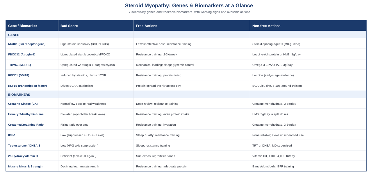
7 Biomarkers That Track Steroid Myopathy Risk And Progression
Steroid myopathy is usually a clinical diagnosis, made by weighing the pattern of proximal weakness (hips, shoulders, thighs) against the duration and type of corticosteroid used. But a handful of biomarkers help confirm the diagnosis, rule out look-alike conditions, and track whether things are getting better or worse. None of these is diagnostic on its own. Together, they build a picture that is far more useful than "you feel weaker."
Creatine Kinase (CK)
Why it matters
CK is usually the first blood marker people think of for muscle problems, and in steroid myopathy it does something counterintuitive: it typically stays normal, or even low, despite real weakness. This is one of the more clinically useful facts about the condition, because inflammatory myopathies like polymyositis or dermatomyositis usually show CK levels several times above normal. A patient with significant proximal weakness and a normal CK is a strong pointer toward steroid myopathy rather than an inflammatory process, especially in someone on chronic glucocorticoids.
How to measure it
CK is a standard blood test, typically 10 to 30 dollars out of pocket, and often bundled into broader metabolic or muscle panels. No fasting is required. Reference ranges vary slightly by lab and sex.
If the score is bad, the plan without supplements
An elevated CK in this context usually means something other than pure steroid myopathy is going on, so the practical step is not "fixing" CK directly but working with the prescriber to rule out an overlapping inflammatory myopathy, statin interaction, or recent intense exercise. If CK is elevated, discuss dose review, alternate-day dosing where clinically appropriate, and resistance training two to three times weekly, which is the single most evidence-backed way to counteract the muscle-wasting signal from glucocorticoids.
If the score is bad, the plan with supplements or equipment
There is no supplement that meaningfully corrects CK as a treatment target. Creatine monohydrate, 3 to 5 grams daily, supports muscle phosphocreatine stores and training capacity in general, but it should be framed as supporting muscle function, not as a way to change a CK reading. It can be taken daily on an ongoing basis without cycling; mild water retention or bloating are the main reported side effects, and people with reduced kidney function should discuss it with their physician first.
Urinary 3-Methylhistidine (3-MH)
Why it matters
3-methylhistidine is released when the contractile proteins actin and myosin are broken down, and it is not reused by the body, which makes it a fairly direct marker of how fast myofibrillar protein is being degraded. In catabolic states, including chronic glucocorticoid exposure, urinary 3-MH excretion rises, reflecting the same protein-breakdown pathways (FBXO32 and TRIM63, discussed later) that drive steroid myopathy at the gene level.
How to measure it
This requires a 24-hour urine collection sent to a specialty or academic laboratory, typically 150 to 300 dollars. It is not a routine primary-care order; it usually needs to come through an endocrinologist, rheumatologist, or a research-affiliated lab, and results need to be interpreted alongside dietary meat intake, since dietary creatine and meat consumption can also affect the reading.
If the score is bad, the plan without supplements
The main non-supplement levers are the same ones that blunt the underlying catabolic gene expression: consistent resistance training, spreading protein intake evenly across the day rather than in one large meal, and working with the prescriber on the lowest effective steroid dose. Correcting steroid-induced hyperglycemia also matters, since poor glycemic control appears to compound muscle protein breakdown.
If the score is bad, the plan with supplements or equipment
HMB (beta-hydroxy-beta-methylbutyrate), typically 3 grams daily split into two or three doses, has shown a modest ability to blunt muscle protein breakdown in other catabolic and disuse states, and is reasonable to discuss as an adjunct. It is well tolerated at standard doses; there is no established need to cycle it, and long-term daily use has not shown significant side effects in the populations it has been studied in (mainly older adults and bed-rest studies rather than steroid myopathy specifically).
Urinary Creatine-to-Creatinine Ratio
Why it matters
This is one of the oldest markers used for steroid myopathy, predating modern imaging. Glucocorticoids impair muscle's ability to take up and phosphorylate creatine, so more of it is excreted unused in urine relative to creatinine. A rising ratio over time can flag worsening myopathy before weakness becomes obvious.
How to measure it
Also a 24-hour urine collection, generally 50 to 150 dollars, somewhat more accessible than 3-MH testing but still typically ordered through a specialist rather than a routine annual physical.
If the score is bad, the plan without supplements
Resistance training and steroid dose optimization remain the foundation. Adequate hydration matters for the accuracy of any urine-based test, and repeat testing under similar conditions (same time of day, similar diet) improves the reliability of comparisons over time.
If the score is bad, the plan with supplements or equipment
Because this ratio reflects impaired creatine handling, some clinicians discuss creatine monohydrate supplementation (3 to 5 grams daily, no loading phase needed) as a way to support intramuscular phosphocreatine despite the handling defect. This should be a conversation with the prescribing physician rather than a self-directed decision, particularly for anyone with reduced kidney function. Side effects are mild and mostly limited to initial water retention; it can be taken indefinitely without cycling.
Insulin-Like Growth Factor 1 (IGF-1)
Why it matters
Glucocorticoids suppress the growth hormone / IGF-1 axis, and IGF-1 is one of the main anabolic signals that normally supports muscle protein synthesis and repair. Lower circulating IGF-1 during steroid treatment removes part of the counterbalance to the muscle-breakdown genes discussed below, which is part of why steroid myopathy tends to worsen the longer treatment continues.
How to measure it
A standard blood test, 80 to 150 dollars, no fasting required, though timing consistency matters since IGF-1 has some day-to-day variability.
If the score is bad, the plan without supplements
Sleep quality has an outsized effect here, since growth hormone is released in pulses during deep sleep, which in turn drives IGF-1 production. Resistance training also produces a local, muscle-specific IGF-1 response independent of the systemic blood level. Adequate total calorie and protein intake matters too, since underfeeding further suppresses this axis.
If the score is bad, the plan with supplements or equipment
There is no supplement that reliably and safely raises systemic IGF-1. Unregulated growth hormone or IGF-1 injections carry real risks, including concerns around cell proliferation, and should not be pursued outside of a legitimate medical indication and supervision. Colostrum supplements have weak, inconsistent evidence for a modest IGF-1 bump and are not something to rely on. The realistic lever here is equipment-based: a structured resistance training program, two to four sessions weekly with progressive overload, is the most defensible way to work with this axis.
Testosterone And DHEA-S
Why it matters
Chronic glucocorticoid use suppresses the hypothalamic-pituitary-gonadal axis, lowering testosterone and DHEA-S in both men and women. Since these hormones support muscle protein synthesis independently of the steroid-myopathy pathway, their suppression compounds muscle loss on top of the direct glucocorticoid effect.
How to measure it
A morning blood draw (testosterone follows a diurnal rhythm, highest in the morning), 50 to 150 dollars depending on whether total, free, and DHEA-S are all included.
If the score is bad, the plan without supplements
Sleep, resistance training, healthy body composition, and reduced alcohol intake all support this axis. Managing stress matters too, since elevated cortisol (whether endogenous or from medication) directly competes with and suppresses these anabolic hormones.
If the score is bad, the plan with supplements or equipment
If levels are clinically low and symptomatic, testosterone replacement therapy is a prescription-only decision that requires ongoing monitoring of hematocrit, PSA (in men), and lipids roughly every three to six months; it is not a self-directed intervention. Over-the-counter DHEA, typically 25 to 50 milligrams daily, is sometimes used with modest evidence, but it changes hormone levels and should be discussed with a physician first, particularly for anyone with a hormone-sensitive condition. It is not something to combine casually with corticosteroid treatment without medical input.
25-Hydroxyvitamin D
Why it matters
Vitamin D deficiency is common in people on long-term steroids, partly from the underlying illness limiting sun exposure and partly from the medications themselves, and low vitamin D is independently associated with proximal muscle weakness. Correcting a deficiency will not reverse steroid myopathy on its own, but it removes a second, additive cause of the same symptom pattern.
How to measure it
A standard blood test, 40 to 80 dollars, widely available and often ordered alongside bone density monitoring, since steroids also increase osteoporosis risk.
If the score is bad, the plan without supplements
Ten to twenty minutes of sun exposure several times a week, depending on skin tone and latitude, along with dietary sources like fatty fish and fortified foods, can help but usually will not fully correct an established deficiency in someone on chronic steroids.
If the score is bad, the plan with supplements or equipment
Vitamin D3, commonly 1,000 to 4,000 IU daily depending on baseline level and physician guidance, with a recheck around three months to confirm the dose is adequate. Side effects are rare at these doses, but megadosing without monitoring risks hypercalcemia, so this should not be an indefinite guess-and-check without follow-up labs. Given that steroids also raise fracture risk, this is often paired with calcium intake and weight-bearing exercise rather than treated in isolation.
Muscle Mass And Strength (DEXA Or Grip Dynamometry)
Why it matters
Every blood marker above is a proxy. This one is the actual outcome. Appendicular lean mass and functional strength are what steroid myopathy ultimately affects, and tracking them directly gives a clearer read on whether the condition is progressing or improving than any single lab value. This is also the category of measurement that longevity-focused physicians like Peter Attia lean on most heavily, on the reasoning that muscle mass and strength predict long-term function and mortality risk better than most conventional panels.
How to measure it
A DEXA scan measuring appendicular lean mass costs roughly 100 to 250 dollars self-pay and is the gold-standard option. A cheaper alternative is a hand-grip dynamometer, around 30 to 60 dollars as a one-time purchase, or a simple sit-to-stand timed test that a physical therapist (or the person themselves) can track at home for free.
If the score is bad, the plan without supplements
Progressive resistance training two to four times weekly targeting the major muscle groups, paired with 1.2 to 1.6 grams of protein per kilogram of body weight spread across meals, is the core intervention. Working with the prescribing physician on dose minimization, alternate-day dosing where appropriate, and correcting any electrolyte imbalance rounds this out.
If the score is bad, the plan with supplements or equipment
Resistance bands or adjustable dumbbells are a practical home setup for people who are deconditioned and need to start light. For those too weak to lift meaningful loads, blood flow restriction (BFR) training, using bands costing roughly 50 to 150 dollars, allows strength gains from very light loads by partially restricting venous return during exercise, and has supporting evidence in other disuse-atrophy populations. Sessions are typically limited to two to three per week with rest days between; mild bruising or discomfort are the main side effects, and it is contraindicated in people with clotting disorders or severe cardiovascular disease, so it should be introduced under professional guidance rather than self-prescribed.
What The Genetic Research Suggests About Steroid Myopathy Susceptibility
The biomarkers above are the more immediately useful, actionable layer. But there is also a genetic layer underneath them that helps explain why some people develop noticeable steroid myopathy on a moderate dose while others tolerate years of treatment with minimal muscle loss. This area is younger and less clinically established than the biomarker panel above, closer to what researchers like Ali Torkamani, who has written extensively about interpreting genomic risk in the context of limited evidence, would describe as "directionally informative rather than diagnostic." Genes here mostly come from strong animal-model evidence with human data still catching up, so treat this section as context rather than a testing checklist.
NR3C1 (The Glucocorticoid Receptor Gene)
NR3C1 encodes the glucocorticoid receptor itself, the protein that steroids bind to before triggering downstream muscle-wasting genes. Certain polymorphisms in this gene, such as BclI and N363S, are associated with altered tissue sensitivity to corticosteroids: some variants appear to increase sensitivity, meaning a person may experience stronger catabolic effects at the same dose others tolerate fine. Human evidence for these variants is stronger in relation to metabolic side effects like weight gain and insulin resistance than for muscle wasting specifically, so this connection should be read as suggestive rather than confirmed.
If the gene is unfavorable, the plan without supplements
If genomic testing suggests higher glucocorticoid sensitivity, or if someone has a personal history of unusually strong steroid side effects at standard doses, the practical response is a conversation with the prescriber about the lowest effective dose, alternate-day dosing where the underlying condition allows it, and steroid-sparing agents. Increasing resistance training frequency provides a compensatory anabolic stimulus regardless of receptor sensitivity.
If the gene is unfavorable, the plan with supplements or equipment
No supplement changes glucocorticoid receptor sensitivity. The realistic option here is medical: discussing steroid-sparing immunosuppressants or biologics with the prescriber to reduce total glucocorticoid exposure, combined with a structured home resistance training setup as the equipment-based lever.
FBXO32 (Atrogin-1)
This gene encodes an E3 ubiquitin ligase that gets switched on by glucocorticoids acting through FOXO transcription factors, and it tags muscle proteins for breakdown. It is one of the most consistently reproduced findings in the animal literature on steroid-induced muscle atrophy, identified in landmark work by Bodine and colleagues on the ubiquitin ligases required for skeletal muscle atrophy. Human muscle biopsy studies during critical illness and steroid exposure show clear upregulation of this pathway as well, described in detail in a widely cited review on glucocorticoid-induced skeletal muscle atrophy.
If the gene is unfavorable, the plan without supplements
Mechanical loading through resistance exercise is the best-documented way to suppress FOXO and atrogin-1 activation, essentially competing directly with the pathway this gene drives. Two to three sessions weekly, even at modest intensity, is more evidence-backed here than any passive intervention.
If the gene is unfavorable, the plan with supplements or equipment
Leucine-rich protein or HMB supplementation (3 grams daily) has shown some ability to blunt atrogene expression in other catabolic states. This is early-to-moderate evidence rather than definitive, well tolerated, and does not require cycling.
TRIM63 (MuRF1)
MuRF1 is a second E3 ligase, also FOXO- and glucocorticoid-responsive, that works alongside atrogin-1 but targets myofibrillar structural proteins like myosin heavy chain more specifically. It tends to show up in the same studies as atrogin-1 and is considered part of the same core atrophy signature.
If the gene is unfavorable, the plan without supplements
The same mechanical-loading principle applies. Sleep quality matters here too, since recovery windows are when this pathway is downregulated, and controlling steroid-induced hyperglycemia helps, since poor glycemic control appears to compound the gene-expression signal.
If the gene is unfavorable, the plan with supplements or equipment
Omega-3 fatty acids, roughly 2 to 3 grams of combined EPA/DHA daily, have shown modest anti-catabolic signaling effects in some human muscle-protein-turnover research, though not specifically in steroid myopathy. Mild gastrointestinal upset is the main side effect, and anyone on blood thinners should discuss dosing with their physician first because of the mild blood-thinning effect at higher doses.
REDD1 / DDIT4
REDD1 is directly induced by glucocorticoids and works by inhibiting mTOR signaling, which blunts muscle protein synthesis at the source rather than just accelerating breakdown. Animal knockout studies show this gene is specifically important in steroid-induced atrophy in a way that distinguishes it from ordinary disuse atrophy, which makes it one of the more mechanistically interesting genes here. Human evidence remains early-stage, so treat this one as a promising research lead rather than an established clinical marker.
If the gene is unfavorable, the plan without supplements
Resistance training remains one of the few known ways to push back against mTOR suppression, alongside careful protein timing around workouts to maximize the anabolic window that does exist.
If the gene is unfavorable, the plan with supplements or equipment
No supplement directly blocks REDD1. Leucine has some early evidence for partially activating mTOR signaling independent of REDD1-driven inhibition, but this should be framed clearly as preliminary rather than a solved problem.
KLF15
KLF15 is a transcription factor induced by glucocorticoids that redirects muscle metabolism toward breaking down branched-chain amino acids, depleting the very building blocks muscle needs for repair. The rodent evidence for this pathway is strong; human data is still developing.
If the gene is unfavorable, the plan without supplements
Spreading adequate protein intake evenly across the day, rather than concentrating it in one meal, helps offset the increased amino acid catabolism this gene drives.
If the gene is unfavorable, the plan with supplements or equipment
Branched-chain amino acid or leucine supplementation, often 5 to 10 grams around training sessions, is sometimes used to counter KLF15-driven amino acid depletion. Evidence here is early-stage rather than conclusive; side effects are minimal at normal doses, though anyone with kidney disease should avoid adding a large extra protein load without medical guidance.
Ten Ideas From Peter Attia's Outlive That Apply To Steroid-Related Muscle Loss
Peter Attia's book Outlive: The Science and Art of Longevity is not written about steroid myopathy specifically, but its central argument, that muscle mass and strength are among the most important predictors of long-term function, maps directly onto what someone managing chronic steroid use needs to hear. Attia's broader framework challenges the more reactive style of conventional medicine ("Medicine 2.0," in his terms) in favor of proactive tracking and intervention, which is exactly the mindset this article is built around.
Muscle Is An Organ, Not An Accessory
Attia frames skeletal muscle as a metabolically active organ that influences glucose regulation, hormone balance, and mortality risk, not just an aesthetic feature. For someone on chronic steroids, this reframes muscle loss from a cosmetic concern into a systemic one worth actively monitoring.
Marginal Decade Thinking
He argues that the real goal of longevity work is preserving function in the last decade of life, not just adding years. Someone experiencing steroid myopathy in their 40s or 50s should think about today's muscle loss in terms of the physical independence it threatens decades from now, which is a strong motivator for consistent resistance training rather than waiting for weakness to become severe.
Exercise Is The Most Powerful Longevity Drug We Have
Attia repeatedly makes the case that no pharmaceutical intervention comes close to matching structured exercise for reducing all-cause mortality risk. This directly supports the resistance-training recommendations that appear throughout the biomarker and gene sections above; it is not a supplementary nice-to-have, it is the primary lever.
Strength Training As A Non-Negotiable
He treats strength training as equally important to cardiovascular exercise, specifically because grip strength, leg strength, and the ability to carry load correlate strongly with independence in later life. This lines up precisely with the muscle mass and strength biomarker described earlier as the most direct outcome measure for steroid myopathy.
Stability And Movement Quality Matter Before Load
Attia emphasizes building a stable movement foundation before adding heavy load, particularly for people returning to exercise after a period of weakness or illness. For someone with active steroid myopathy, this supports starting with lighter resistance, bands, or bodyweight work before progressing, rather than jumping straight into heavy lifting.
VO2 Max As An Underrated Marker
He considers cardiorespiratory fitness one of the strongest predictors of longevity, often stronger than traditional risk factors. While VO2 max is not muscle-specific, steroid myopathy often coincides with reduced exercise tolerance, so tracking this alongside strength gives a fuller picture of functional decline or recovery.
Protein Intake Needs Are Higher Than Conventional Guidelines Suggest
Attia argues that standard protein recommendations underestimate what is needed to preserve muscle, particularly in anyone dealing with a catabolic stressor. This directly supports the 1.2 to 1.6 grams per kilogram target mentioned throughout the biomarker plans above.
The "Four Horsemen" Framing Of Chronic Disease
He organizes chronic disease risk around cardiovascular disease, cancer, neurodegenerative disease, and metabolic dysfunction. Steroid myopathy does not fit neatly into this list, but the metabolic dysfunction category overlaps meaningfully, since chronic glucocorticoid use worsens insulin resistance, which in turn compounds muscle protein breakdown.
Medicine 3.0: Proactive Rather Than Reactive Care
Attia's core critique of conventional medicine is that it waits for disease to become symptomatic before acting. Applied here, this means tracking the biomarkers in this article before weakness becomes severe, rather than waiting for a clinical diagnosis of myopathy to prompt action.
Personalization Over Population Averages
He repeatedly stresses that population-level guidelines are a starting point, not a personal prescription, and that individual data (labs, genetics, functional testing) should refine the plan. This is the same argument underlying this entire article: generic steroid-myopathy advice is a reasonable floor, not a ceiling.
Complementary Approaches Worth Considering
None of the approaches below reverse steroid myopathy on their own, and the human evidence specific to this exact condition is limited. Most of the supporting research comes from populations who are on corticosteroids for another reason (asthma, COPD, rheumatoid arthritis) rather than people with a formal steroid myopathy diagnosis, so these should be read as reasonable adjuncts rather than core treatment.
Yoga
Yoga combines gentle resistance against body weight, controlled breathing, and balance work, all of which are relevant to someone with proximal muscle weakness who also needs to avoid falls and maintain functional mobility. It is also low-impact enough to be appropriate for people who cannot yet tolerate conventional resistance training.
Several trials in asthma and COPD patients, many of whom are on inhaled or oral corticosteroids, have shown improvements in muscle strength and functional capacity with structured yoga programs, generally two to three sessions weekly over eight to twelve weeks. Evidence specific to a steroid myopathy diagnosis is limited, and results in the broader literature are mixed in magnitude, so this should be read as supportive rather than definitive; searchable summaries of this research area are available through PubMed.
A realistic starting point is a beginner or therapeutic yoga class, twice weekly, focused on standing poses and gentle strength work rather than deep stretching, since overstretching a weakened muscle carries little benefit and some injury risk. It pairs well with, rather than replaces, the resistance training described in the biomarker section above.
Tai Chi
Tai chi is a slow, controlled-movement practice that builds lower-body strength, balance, and proprioception, which matters directly for steroid myopathy given that proximal leg weakness is one of its hallmark features and a major fall-risk contributor.
Randomized trials in older adults and in rheumatoid arthritis populations, many on corticosteroids, have shown improvements in lower-extremity strength and balance with tai chi practiced two to three times weekly over several months. As with yoga, direct steroid-myopathy-specific trials are scarce; the evidence base sits mainly in adjacent steroid-treated populations, searchable through PubMed.
A practical approach is a beginner tai chi class or a well-produced instructional video, done two to three times weekly for fifteen to twenty minutes, ideally alongside a physical therapist's input if balance is already compromised.
Photobiomodulation (Low-Level Laser Therapy)
Photobiomodulation uses low-level laser or LED light applied to muscle tissue, with proposed effects on mitochondrial function and oxidative stress that could plausibly support muscle recovery and reduce fatigue, both relevant to someone whose muscles are already under catabolic stress from steroids.
Human trials, largely in athletic and general muscle-fatigue populations rather than steroid myopathy specifically, have shown reduced markers of muscle damage and improved recovery when applied before or after exercise; this line of research is summarized in reviews searchable through PubMed. Evidence directly in steroid-treated muscle is essentially absent, so this should be considered exploratory in this specific context.
Devices range from clinic-based treatments to consumer-grade at-home units; realistic use would be short sessions two to three times weekly around resistance training days, with no established need to cycle, and minimal reported side effects beyond mild, temporary warmth at the treatment site.
EMG Biofeedback
Electromyography (EMG) biofeedback gives real-time visual or audio feedback on muscle activation, which can help someone with weakened or deconditioned muscles learn to recruit them more effectively during rehabilitation exercises, a common issue after any period of significant muscle weakness.
This technique has documented use in rehabilitation after disuse atrophy and critical-illness-related weakness, a population that substantially overlaps with steroid myopathy since high-dose steroids are a major contributor to ICU-acquired weakness; supporting literature on EMG biofeedback in muscle rehabilitation is searchable through PubMed.
In practice, this usually requires working with a physical therapist who has EMG biofeedback equipment, typically for a defined block of sessions (six to twelve, once or twice weekly) rather than indefinite use, with the goal of transferring improved muscle activation patterns into an independent home exercise routine.
Conclusion
Steroid myopathy is not a mystery condition, even though it often gets treated like one in routine care. There is a fairly clear biological chain from glucocorticoid exposure to receptor activation, to specific muscle-breakdown genes, to measurable changes in blood and urine markers, and finally to the muscle mass and strength that actually determine how someone functions day to day. Tracking the right points along that chain, rather than waiting for weakness to become obvious, is the practical edge this article has tried to offer.
If you take one step away from this, make it a concrete one: ask your prescriber which of these biomarkers, starting with CK, vitamin D, and a baseline strength or muscle mass measurement, makes sense to check given your specific steroid regimen and duration. Pair that with a resistance training routine you can actually sustain, even a modest one. That combination of better data and consistent mechanical loading is the most evidence-backed lever available, and it is one you can start building on well before your next appointment.
Musculoskeletal Endocrine & Metabolic
Musculoskeletal: Muscle Conditions
Endocrine & Metabolic: Adrenal Conditions