This article contains AI generated content.
· UpdatedStevens-Johnson Syndrome - 5 Genes and 6 Biomarkers to Track
Introduction
Stevens-Johnson Syndrome is not a diagnosis anyone expects. One week you are taking a drug your doctor prescribed with confidence; the next week you are in a burn unit watching your skin detach in sheets. For people who survive it, the aftermath — scarred eyes, damaged airways, permanent sensitivity to light, months of wound care — is often worse than any prior health experience. What makes it even harder is that the medical system, after performing the genuinely difficult work of keeping you alive, rarely explains why this happened to you specifically. "Document the allergy and avoid the drug" is clinically correct but emotionally and practically hollow.
The standard framing treats SJS as an unpredictable reaction. The science tells a different story. Specific genetic variants — primarily in the human leukocyte antigen (HLA) system — predict SJS susceptibility for particular drug-ethnicity combinations with a precision that approaches binary. If you carry HLA-B*15:02 and take carbamazepine, your risk of SJS is roughly 1 in 10. Without the allele, it is approximately 1 in 10,000. That is not unpredictability — that is a 100-fold difference that has been known since 2004. The genetic signal for allopurinol, abacavir, and several anticonvulsants is equally strong.
Beyond genetics, the acute episode leaves measurable biological traces in your blood. Biomarkers like granulysin, serum albumin, LDH, and liver enzymes are not just numbers — they reflect the extent of immune activation, tissue destruction, and organ strain in ways that directly affect clinical decisions and long-term outcomes. Tracking them during and after a reaction tells you things that no symptom checklist can.
This article walks through both dimensions. The primary section covers six biomarkers worth tracking — what they measure, how to obtain them, what an abnormal result means, and what evidence exists for improving them. A following section covers five genetic variants that define susceptibility, with practical guidance on what to do if you carry them. There are also complementary approaches for the long-term sequelae of SJS, and a synthesis of the pharmacogenomics thinking that is quietly changing how this condition is prevented before it starts. The goal is not to replace your medical team. It is to give you the information needed to work with that team more effectively.
Summary
Six biomarkers — granulysin, high-sensitivity CRP, LDH, liver enzymes, serum albumin, and complete blood count — reveal how severe a Stevens-Johnson reaction was, how much organ damage occurred, and whether recovery is proceeding normally. Five genetic variants, principally HLA-B*15:02, HLA-B*57:01, HLA-A*31:01, HLA-B*58:01, and CYP2C9, determine who is likely to develop SJS on specific drugs and why — and most are testable through standard pharmacogenomics panels available today. This article explains each biomarker and gene in full, including how to test for them, what a bad result means, and what interventions — with and without supplements — have evidence for improving them. It also covers complementary recovery approaches backed by clinical trials and summarizes the pharmacogenomics framework from Ben Lynch's Dirty Genes, which offers a practical model for reducing background immune reactivity even when you cannot change your HLA alleles. If you have had SJS or carry known risk genes, this is the level of detail the standard medical visit rarely provides.
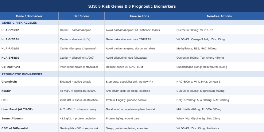
6 Biomarkers to Track in Stevens-Johnson Syndrome
SJS biomarkers serve three distinct functions depending on when they are measured. During an acute episode, they assess severity and predict mortality. In the weeks immediately after, they track organ recovery and help calibrate the transition out of intensive care. Over the long term, they offer a window into immune normalization — and can catch residual dysfunction before it compounds. Understanding which markers matter most, what they actually reflect biologically, and how to act on an abnormal result transforms these numbers from abstract reference ranges into a practical monitoring system.
Biomarker 1: Granulysin
Why It Matters
Granulysin is the most SJS-specific biomarker currently described in the scientific literature. It is a small cytotoxic protein released by CD8+ T lymphocytes and natural killer cells when they target cells for destruction. In SJS and its more severe variant, toxic epidermal necrolysis (TEN), granulysin is the primary weapon that CD8+ T cells deploy against keratinocytes — the structural cells of the skin. Elevated serum granulysin precedes visible skin detachment and distinguishes SJS/TEN from other severe drug reactions such as DRESS syndrome (drug reaction with eosinophilia and systemic symptoms) and staphylococcal scalded skin syndrome, which require entirely different clinical management.
Research published by Wen-Hung Chung and colleagues confirmed that serum granulysin is dramatically elevated in SJS/TEN and that levels correspond with disease severity (Chung et al., Nature Medicine, 2008). The same group demonstrated that granulysin collected from blister fluid could reproduce keratinocyte death when applied directly to skin cells, confirming a causal rather than merely correlational role.
How to Measure It
Granulysin is measured in serum or blister fluid by enzyme-linked immunosorbent assay (ELISA). It is currently a hospital and research laboratory test — not routinely available at commercial lab chains. In major academic medical centers and hospital dermatology units, testing is generally accessible during acute admissions. Cost ranges from approximately $80 to $200 depending on the facility. Rapid point-of-care granulysin detection kits have been developed for clinical triage and are increasingly available in Asia, where SJS incidence is higher due to the prevalence of HLA-B*15:02.
If Granulysin Is Elevated: The Plan Without Supplements
Elevated granulysin during an acute episode is a direct indicator of active cytotoxic immune assault on keratinocytes. The immediate non-pharmacological priorities are: discontinue the triggering drug immediately if still being administered, ensure wound care in a specialist burns or dermatology unit, implement adequate nutritional support to preserve immune competence without further activating the immune cascade, and eliminate any additional immunological triggers — infections, unnecessary new drug exposures, elective vaccinations during the acute window.
Over the medium term, a confirmed history of high granulysin during an acute episode warrants full pharmacogenomics evaluation (HLA typing, CYP2C9 genotyping) to identify future drug risks before they materialize. Immunology or allergy consultation should be standard in the recovery phase.
If Granulysin Is Elevated: The Plan With Supplements and Equipment
No supplement directly suppresses granulysin the way pharmaceutical immunosuppressants do, and during the acute phase, supplements must never replace medical care. During recovery and for long-term immune calibration, several agents have evidence for modulating cytotoxic T-cell activity and reducing baseline inflammatory tone.
N-acetylcysteine (NAC): 600 mg twice daily. NAC replenishes glutathione and reduces oxidative-stress-driven immune activation. It has a direct cytoprotective effect on epithelial cells and is used in some SJS protocols during the acute phase. Reduce to once daily after three months if inflammatory markers normalize. Side effects: nausea at higher doses; rare bronchospasm in asthmatics.
Vitamin D3 with K2: 5,000 IU D3 plus 100 mcg MK-7 K2 daily. Vitamin D is a potent immunomodulator that shifts immune balance toward regulatory T-cell activity and away from cytotoxic responses. Deficiency is extremely common among people with hypersensitivity conditions. Monitor 25-OH vitamin D quarterly; target 60–80 ng/mL. Side effects: well tolerated at these doses when K2 is co-administered; monitor calcium if maintaining over 5,000 IU long-term.
Omega-3 fatty acids: 2–4 g of EPA plus DHA daily from a high-quality fish oil. Omega-3s shift immune signaling toward resolvins and protectins — molecules that actively resolve inflammatory cascades rather than merely suppressing them. Take with meals to reduce GI side effects. Use with caution if on anticoagulants.
Biomarker 2: High-Sensitivity C-Reactive Protein (hsCRP)
Why It Matters
C-reactive protein is a liver-synthesized acute-phase reactant that rises in response to any significant inflammatory or tissue-damage signal. In SJS, hsCRP elevates sharply during the acute episode and provides a longitudinal tracking measure for systemic inflammatory burden. While less specific to SJS than granulysin, hsCRP is universally available, inexpensive, and sensitive enough to detect ongoing inflammation during recovery that might otherwise be missed clinically. Persistently elevated levels months after discharge suggest incomplete immune normalization and warrant investigation.
CRP levels also have a direct pharmacological dimension. Elevated CRP suppresses the activity of several CYP450 enzymes, including CYP2C9 — the enzyme that metabolizes phenytoin, sulfonamides, and several NSAIDs associated with SJS. Inflammation-driven reduction in CYP2C9 activity can cause drug accumulation even at standard doses, representing a mechanism by which intercurrent infection might tip a borderline drug exposure toward a full reaction in a susceptible individual.
How to Measure It
High-sensitivity CRP is a standard test available at virtually any clinical laboratory. Cost: $15–$40 standalone. Optimal range: below 1.0 mg/L. A level of 1–3 mg/L indicates moderate systemic inflammation; above 3 mg/L indicates significant inflammation requiring investigation. During acute SJS, CRP typically exceeds 50 mg/L and should be monitored every 48–72 hours to track trajectory. During recovery monitoring, recheck every 6–8 weeks until levels are consistently below 1.0 mg/L.
If hsCRP Is Elevated: The Plan Without Supplements
Address root causes first. The most impactful non-supplement interventions for chronically elevated hsCRP are: an anti-inflammatory dietary pattern (Mediterranean-style, eliminating refined carbohydrates, seed oils, and ultra-processed foods), consistent sleep of 7.5–9 hours (poor sleep is among the strongest independent elevators of inflammatory cytokines), 150–200 minutes weekly of aerobic exercise, and smoking cessation where applicable. These interventions collectively reduce hsCRP by 30–60% in most populations over 8–12 weeks, with effects that rival pharmaceutical interventions in primary prevention populations.
If hsCRP Is Elevated: The Plan With Supplements and Equipment
High-bioavailability curcumin: 500–1,000 mg daily of a formulation with demonstrated bioavailability (BCM-95 or Longvida). Meta-analyses consistently show reductions of 2–6 mg/L in baseline CRP. A 3-month-on, 1-month-off cycle is a common precautionary approach. Side effects: GI discomfort at higher doses; potential interaction with blood thinners.
Magnesium glycinate: 400–500 mg at night. Magnesium deficiency — extremely prevalent in general populations — independently elevates CRP. Magnesium is also involved in over 300 enzymatic reactions relevant to immune function and drug metabolism. Side effects: loose stools at higher doses; otherwise well tolerated.
Infrared sauna (if available): 3–4 sessions weekly, 20 minutes at 140–160°F. Repeated mild heat stress reduces inflammatory cytokines including IL-6 and TNF-alpha and improves CRP in populations with cardiovascular and rheumatic conditions. Begin cautiously post-SJS — particularly if mucosal healing is incomplete or ocular complications are active.
Biomarker 3: Lactate Dehydrogenase (LDH)
Why It Matters
Lactate dehydrogenase is an enzyme present in nearly all cells of the body. When cells are destroyed — by any mechanism — they release LDH into circulation. In SJS, where keratinocytes are being killed en masse and internal organs may also be involved, LDH rises steeply and tracks the cumulative extent of tissue destruction. It contributes prognostic weight in modified SCORTEN severity scoring for SJS/TEN, independent of the body surface area involved.
Elevated LDH also signals mitochondrial dysfunction in surviving cells — mitochondria are both a major LDH source and primary targets of oxidative damage during severe inflammatory episodes. Monitoring LDH recovery over weeks post-discharge gives a rough measure of cellular repair kinetics that clinical examination alone cannot provide.
How to Measure It
LDH is a standard chemistry marker available at any laboratory for $10–$30. Normal range: approximately 140–280 U/L, though this varies by assay. During acute SJS/TEN, LDH commonly exceeds 500 U/L and tracks disease burden. Measuring LDH every 2–4 weeks post-discharge provides an affordable confirmation that tissue damage has plateaued and cellular repair is occurring.
If LDH Is Elevated: The Plan Without Supplements
The foundation is ensuring the immune cascade has been adequately controlled — no secondary triggers, no residual drug exposures — and providing sufficient substrate for cellular repair. Prioritize protein intake (1.4–1.8 g per kilogram body weight daily) to support keratinocyte and epithelial regeneration. Confirm iron-replete status to support tissue oxygenation. Control blood glucose and avoid prolonged fasting, both of which increase metabolic burden on already-stressed cells.
If LDH Is Elevated: The Plan With Supplements and Equipment
Ubiquinol (CoQ10): 200–400 mg daily. Ubiquinol is the active form of CoQ10, with superior bioavailability. It is essential for mitochondrial electron transport and cellular energy production; replenishing it reduces LDH release by supporting cellular membrane integrity under oxidative stress. Benefits accumulate over 4–8 weeks. Side effects: generally well tolerated; take earlier in the day to avoid sleep disruption.
Alpha-lipoic acid (ALA): 300–600 mg daily. ALA is a mitochondrial antioxidant that regenerates vitamins C and E and directly reduces intracellular LDH release by protecting cell membranes from lipid peroxidation. Cycle: 3 months on, 1 month off. Side effects: mild GI effects; theoretical concern in thiamine-deficiency states.
N-acetylcysteine (NAC): 600 mg twice daily (as discussed under granulysin). NAC's glutathione-raising effect also reduces LDH by protecting mitochondrial membrane integrity.
Biomarker 4: Liver Function Panel (ALT, AST, Bilirubin)
Why It Matters
Hepatic involvement occurs in a significant minority of SJS cases and is not always clinically obvious. Elevated ALT, AST, and bilirubin indicate hepatocyte damage from the triggering drug itself, from immune-mediated hepatitis driven by the same CD8+ T-cell activation that attacks the skin, or from secondary infection. Liver injury changes drug metabolism — impaired CYP450 activity means that drugs at standard doses may reach toxic plasma concentrations in a compromised liver. Understanding hepatic status after SJS is therefore not just about tracking organ damage; it directly informs future drug safety decisions.
Persistent enzyme elevation beyond 6–8 weeks post-acute phase warrants gastroenterology review. Drug-induced autoimmune hepatitis can occasionally be triggered by the same immunological event that caused SJS and may require treatment independent of the skin condition.
How to Measure It
A comprehensive metabolic panel (CMP) includes ALT, AST, bilirubin, and alkaline phosphatase for $20–$50 at standard labs. Conservative optimal targets: ALT below 25 U/L in women, below 35 U/L in men (thresholds used by lipidologists including Thomas Dayspring); AST similar; total bilirubin 0.3–1.2 mg/dL. During acute SJS, recheck every 3–5 days while hospitalized. After discharge: at 4 weeks, 8 weeks, and 6 months.
If Liver Enzymes Are Elevated: The Plan Without Supplements
Remove all hepatotoxic exposures first: any suspected triggering drugs, alcohol (complete abstinence during recovery), over-the-counter acetaminophen (which reaches toxic concentrations in an already-stressed liver at standard doses), and unnecessary herbal supplements that add metabolic load. A low-fat, low-fructose dietary pattern reduces hepatic metabolic demand — the same evidence base used for managing non-alcoholic fatty liver disease applies here. Weight-bearing exercise and aerobic activity improve hepatic insulin sensitivity and reduce liver fat accumulation independent of steatosis levels.
If Liver Enzymes Are Elevated: The Plan With Supplements and Equipment
Milk thistle (silymarin): 420 mg daily from a standardized extract (70–80% silymarin). Multiple randomized trials demonstrate reductions in ALT and AST in drug-induced liver injury and chronic liver conditions. Mechanism: direct antioxidant protection of hepatocytes and inhibition of inflammatory Kupffer cell signaling. Safe for long-term use; 3–6 month treatment cycles are typical. Side effects: mild laxative effect; rare allergic reactions in those sensitive to Asteraceae family plants.
TUDCA (tauroursodeoxycholic acid): 500–750 mg daily. TUDCA is a bile acid derivative with strong hepatoprotective properties — it stabilizes the mitochondrial membrane under toxic stress and reduces hepatocyte apoptosis. It is used in transplant medicine and increasingly in chronic liver conditions with a favorable safety profile. Cycle: 2–3 months on, 1 month off. Side effects: minor GI effects.
Phosphatidylcholine: 400–500 mg daily. Choline deficiency independently causes elevated liver enzymes and hepatic fat accumulation, and is commonly depleted during inflammatory illness. It is among the most overlooked nutrients in liver recovery. Side effects: fishy odor at high doses; not recommended for those with elevated cardiovascular TMAO concerns without medical supervision.
Biomarker 5: Serum Albumin
Why It Matters
Albumin is the most abundant protein in the bloodstream, synthesized exclusively in the liver, and serves as the primary transport vehicle for drugs, fatty acids, hormones, and electrolytes. Low albumin is a reliable indicator of either hepatic synthetic failure or significant protein loss — both of which occur in SJS. Serum albumin is one of the original seven parameters in the SCORTEN severity scoring system for SJS/TEN, where a level below 3.0 g/dL is independently associated with substantially higher mortality. Beyond prognosis, depleted albumin has a direct pharmacological consequence: when albumin falls, drug-protein binding is reduced, meaning free drug concentrations rise unpredictably. In SJS patients where existing medications or new exposures carry high risk, this pharmacokinetic shift is particularly dangerous.
Both Peter Attia and Thomas Dayspring emphasize albumin not simply as a nutrition marker but as a global indicator of physiological reserve — lower albumin, even within the conventional "normal" range, consistently tracks with worse outcomes across a range of acute and chronic conditions.
How to Measure It
Albumin is included in a comprehensive metabolic panel at no additional cost ($20–$50 for CMP). Optimal range: 4.0–5.0 g/dL. A level below 3.5 g/dL indicates significant depletion and warrants both dietary and clinical intervention. Below 2.5 g/dL, intravenous albumin infusion may be indicated. During acute SJS hospitalization, albumin should be checked on admission and every 48–72 hours thereafter.
If Albumin Is Low: The Plan Without Supplements
The non-supplement foundation is dietary protein adequacy. During SJS recovery, protein requirements increase substantially due to protein-rich wound exudate, tissue loss, and the metabolic demands of epithelial regeneration. Targets: 1.6–2.0 g of complete protein per kilogram body weight daily, from high-quality sources (eggs, poultry, fish, dairy, well-combined plant proteins). Reducing ongoing protein losses is equally important: optimize wound care to minimize exudate, control systemic inflammation, and treat any secondary infection promptly.
If Albumin Is Low: The Plan With Supplements and Equipment
Whey protein isolate: 25–40 g daily. Whey provides the highest leucine density of any dietary protein and directly stimulates hepatic albumin synthesis through mTOR pathways. Divide into two doses for best absorption. If GI intolerance occurs, switch to a pea-and-rice protein blend. Side effects: GI discomfort in lactose-sensitive individuals with concentrate formulations; isolate is better tolerated.
Glycine: 3–5 g before bed. Glycine is conditionally essential during acute tissue damage, depleted faster than most amino acids in severe injury, and directly incorporated into albumin's structural framework. It also improves sleep quality, which compounds recovery benefits. Side effects: very well tolerated.
Zinc bisglycinate: 25–30 mg daily with food. Zinc is required for albumin synthesis and for multiple steps in immune regulation; it is commonly depleted in inflammatory states. Cycle: 4 months on, 1 month off to prevent copper displacement. Side effects: nausea if taken on an empty stomach.
Biomarker 6: Complete Blood Count with Differential
Why It Matters
A complete blood count with differential provides a comprehensive snapshot of the hematopoietic immune response. In SJS, three specific values carry the most clinically actionable information. Lymphocyte count reflects the CD8+ T-cell and NK-cell populations responsible for keratinocyte destruction — monitoring their trajectory tracks immune normalization. Eosinophil count rises in drug hypersensitivity reactions and, when persistently elevated, raises concern for overlap with DRESS syndrome or ongoing drug reaction. Neutrophil count, which falls during severe immune dysregulation, is the most immediately dangerous abnormality — neutropenia below 500 cells/μL predicts susceptibility to bacterial sepsis, which is the leading cause of SJS mortality.
Lymphopenia in the subacute recovery phase indicates immune depletion. Persistent eosinophilia beyond the acute episode warrants systematic review of all current medications and, in some cases, further immunological workup.
How to Measure It
CBC with differential is included in most standard blood panels and costs $10–$30 standalone. Reference ranges relevant to SJS monitoring: lymphocytes 1,000–4,800 cells/μL (lower bound during recovery); eosinophils 0–500 cells/μL; neutrophils above 1,500 cells/μL. During acute SJS hospitalization, check daily. Post-discharge: at 2 weeks, 6 weeks, and 3 months.
If CBC Values Are Abnormal: The Plan Without Supplements
Neutropenia below 500 cells/μL requires immediate infectious disease consultation and protective isolation protocols — this is not a scenario managed with dietary changes alone. For lymphopenia and eosinophilia during recovery, the most impactful non-supplement measures are: confirmed elimination of all offending agents, protein and micronutrient nutritional rehabilitation, and sleep optimization (7.5–9 hours — lymphocyte proliferation occurs predominantly during sleep). Moderate aerobic exercise, beginning conservatively at weeks 4–6 of recovery, has demonstrated effects on natural killer cell recovery and T-cell homeostasis normalization.
If CBC Values Are Abnormal: The Plan With Supplements and Equipment
Vitamin D3 with K2: As described above. Vitamin D directly promotes regulatory T-cell differentiation and attenuates cytotoxic T-cell overactivity. Deficiency is independently associated with lymphopenia in autoimmune and hypersensitivity conditions.
Zinc bisglycinate: 25 mg daily. Zinc is required for T-cell maturation in the thymus and for NK cell cytotoxic function. It is the most consistent nutritional predictor of lymphopenia in depleted patients. Cycle: 4 months on, 1 month off.
Multi-strain probiotics: 10+ billion CFU daily with Lactobacillus and Bifidobacterium strains. Gut-immune axis disruption following severe inflammatory episodes and antibiotic treatment is nearly universal in SJS patients. Correcting dysbiosis supports T-regulatory cell populations and reduces systemic immune activation. Begin 48–72 hours after completing antibiotics. Side effects: transient bloating; well tolerated in most individuals.
The Genetic Architecture of Stevens-Johnson Syndrome: 5 Alleles That Predict Susceptibility
The genetics of SJS represent one of the most striking stories in pharmacogenomics. Unlike most drug adverse reactions, where genetic risk is modest and multifactorial, several SJS gene-drug associations approach deterministic prediction. The underlying mechanism involves the HLA system — a collection of genes encoding cell-surface proteins that present peptide fragments to cytotoxic T cells. When a specific HLA variant presents drug-derived peptides in a conformation that triggers massive CD8+ T-cell activation, SJS follows with a rapidity and severity that conventional dose adjustments cannot prevent.
What makes these findings practically important is that they have already been translated into policy. Taiwan mandated HLA-B*15:02 screening before carbamazepine prescribing in 2010; Thailand, Hong Kong, and Singapore followed. The FDA updated carbamazepine labeling to recommend HLA-B*15:02 testing in patients of Asian ancestry. The science is not theoretical — it has been implemented at population scale and has measurably reduced SJS incidence in screened populations.
Gene 1: HLA-B*15:02
What the Gene Does
HLA-B*15:02 is a variant of the human leukocyte antigen B gene encoding a cell-surface protein that presents peptide fragments to CD8+ cytotoxic T cells. The *15:02 variant appears to present carbamazepine-derived peptides in a structural conformation that triggers massive T-cell activation — the immunological cascade directly responsible for SJS.
The landmark study by Wen-Hung Chung and colleagues, published in Nature in 2004, identified HLA-B*15:02 in 100% of Han Chinese patients who developed SJS on carbamazepine, compared to approximately 3% of carbamazepine-tolerant controls (Chung et al., Nature, 2004). This remains one of the strongest drug-gene associations ever reported in pharmacogenomics. Prevalence: 8–10% in Han Chinese populations, 6–8% in Thai and Malaysian populations, approximately 2–3% in South Asian populations, and under 1% in most European populations.
If You Carry HLA-B*15:02: The Plan Without Supplements
Testing and permanent documentation are the non-negotiable foundation. HLA-B*15:02 genotyping is available through comprehensive pharmacogenomics panels (Invitae, GeneSight, Genomind, and others) at $100–$400 depending on the panel scope and insurance coverage. Once identified, the allele status must be permanently recorded in your medical record and communicated proactively to every prescribing clinician at every clinical encounter. A medical alert bracelet documenting the allele is strongly recommended.
Avoid carbamazepine, oxcarbazepine, and phenytoin. Work with your neurologist to identify safe alternatives — valproate and levetiracetam do not carry HLA-B*15:02-mediated SJS risk. Never start a new aromatic anticonvulsant without explicit confirmation from your pharmacist and neurologist that it is safe for HLA-B*15:02 carriers.
If You Carry HLA-B*15:02: The Plan With Supplements and Equipment
No supplement neutralizes an HLA-B*15:02 allele. The genetic risk is structural — the variant HLA protein presents drug peptides in a way that triggers the immune system, and no currently available compound modifies that protein-level interaction. What supplements can do is reduce the baseline inflammatory tone that may lower the activation threshold for immune reactions.
Quercetin: 500–1,000 mg daily. Quercetin functions as a mast-cell stabilizer and broad anti-inflammatory. It does not block HLA-mediated T-cell activation, but may reduce overall hypersensitivity reactivity. Cycle: safe for long-term use; 3 months on, 1 month off is commonly recommended. Side effects: generally well tolerated; potential mild estrogen-like effects at very high doses.
Vitamin D3 with K2: As described under biomarkers. Adequate vitamin D shifts immune balance toward tolerance and reduces CD8+ T-cell cytotoxicity. This does not eliminate HLA-B*15:02 risk, but supports a more calibrated immune baseline. Monitor serum levels quarterly.
Gene 2: HLA-B*57:01
What the Gene Does
HLA-B*57:01 is the critical genetic risk factor for abacavir-induced SJS and hypersensitivity syndrome — primarily relevant to HIV-positive individuals receiving antiretroviral therapy. The association is robust enough that HLA-B*57:01 testing before abacavir prescription is now mandated in many countries and recommended by WHO guidelines. The mechanism involves abacavir molecules inserting into the HLA-B*57:01 peptide-binding groove, altering the set of self-peptides presented to CD8+ T cells and triggering autoreactive cytotoxic responses — a model known as the altered self-peptide hypothesis.
Prevalence: approximately 5–8% in European populations, 3–4% in African Americans, and under 1% in most Asian populations. Testing is available through the same pharmacogenomics panels used for other HLA alleles and is medically necessary for any HIV patient being considered for abacavir.
If You Carry HLA-B*57:01: The Plan Without Supplements
Never take abacavir. Alternative antiretroviral backbone drugs not associated with HLA-B*57:01-mediated hypersensitivity include tenofovir disoproxil fumarate (TDF) and tenofovir alafenamide (TAF)-based combinations — effective, widely available, and not linked to this genetic risk. Document the allele status permanently in your HIV care record and all other medical records. For HIV-positive carriers on ARV therapy, CBC with differential and hsCRP every 6 months constitute a reasonable immune monitoring baseline.
If You Carry HLA-B*57:01: The Plan With Supplements and Equipment
The same framework applies as for HLA-B*15:02 carriers: no supplement blocks the genetic mechanism, but vitamin D optimization, omega-3 supplementation (2–4 g EPA+DHA daily), quercetin (500 mg daily), and zinc (25 mg cycled) provide a reasonable anti-inflammatory foundation. The HIV-positive context adds specific considerations: antiretroviral therapy itself is immunomodulatory, and supplement interactions with ARVs should be reviewed with an HIV specialist pharmacist before initiating any new regimen.
Gene 3: HLA-A*31:01
What the Gene Does
HLA-A*31:01 is the primary genetic risk factor for carbamazepine-induced adverse reactions — including SJS — in European and Japanese populations, functioning as the counterpart to HLA-B*15:02 in non-Asian patients. While HLA-B*15:02 accounts for nearly all carbamazepine-SJS in Southeast Asia, HLA-A*31:01 is the allele European-ancestry prescribers need to screen. Prevalence: 2–5% in Northern European populations, up to 9% in Japanese.
A landmark European pharmacogenomics study published in the New England Journal of Medicine identified HLA-A*31:01 as a significant risk factor across multiple types of carbamazepine-related adverse reactions, including SJS, DRESS, and maculopapular exanthem (McCormack et al., NEJM, 2011). The relative risk for SJS is somewhat lower than for HLA-B*15:02 in Asian populations, but clinically significant.
If You Carry HLA-A*31:01: The Plan Without Supplements
HLA-A typing is included in comprehensive pharmacogenomics panels. European and Japanese patients prescribed carbamazepine should explicitly discuss HLA-A*31:01 testing with their neurologist. While the association is not as absolute as HLA-B*15:02 in Asian populations, the risk elevation is sufficient to warrant alternative anticonvulsant consideration whenever feasible. Document the allele permanently. Valproate and levetiracetam remain appropriate alternatives and are not associated with HLA-A*31:01-mediated risk.
If You Carry HLA-A*31:01: The Plan With Supplements and Equipment
Optimize methylation status in addition to the standard anti-inflammatory protocol described above. Folate (as methylfolate, 400–800 mcg daily) and B12 (as methylcobalamin, 500–1,000 mcg daily) support immune gene expression through methylation pathways, which directly influence T-cell differentiation. These are low-risk interventions with broad benefits. NAC (600 mg twice daily) provides cytoprotective cover if any new drug exposure is unavoidable. Side effects of methylfolate: rare paradoxical anxiety in individuals with specific MTHFR variants — reduce dose if this occurs.
Gene 4: HLA-B*58:01
What the Gene Does
HLA-B*58:01 is the primary genetic risk factor for allopurinol-induced SJS and TEN, which is the most common cause of drug-induced SJS in Taiwan and several East Asian countries. Allopurinol is among the most widely prescribed drugs globally for gout and hyperuricemia, making this allele particularly relevant. SJS risk in HLA-B*58:01 carriers taking allopurinol approaches 1 in 200 — an extraordinarily high risk for a routine prescription medication in an outpatient setting. Prevalence: 6–9% in Han Chinese populations; lower in other Asian groups; very low in Europeans.
Taiwan has implemented population-level HLA-B*58:01 screening before allopurinol prescribing, resulting in a dramatic, documented reduction in drug-induced SJS incidence. This is among the clearest demonstrations in all of medicine that pharmacogenomics screening at scale can prevent a life-threatening condition.
If You Carry HLA-B*58:01: The Plan Without Supplements
Avoid allopurinol entirely. Alternative urate-lowering therapies without HLA-B*58:01 risk include febuxostat (a non-purine xanthine oxidase inhibitor) and probenecid (a uricosuric agent). Both are effective for gout management and are the appropriate first-line choices in confirmed carriers. If you are of Southeast or East Asian ancestry and are being prescribed allopurinol, HLA-B*58:01 testing is medically essential before starting — request it explicitly if it has not been offered.
If You Carry HLA-B*58:01: The Plan With Supplements and Equipment
Managing uric acid without allopurinol requires a combination of dietary modification and targeted supplementation. Reduce purine-rich foods: organ meats, shellfish, red meat in excess, beer and spirits. Maintain hydration of at least 2.5–3 L of water daily to support uric acid excretion. Tart cherry extract: 480 mg standardized anthocyanins daily has modest clinical evidence for reducing serum urate and gout flare frequency. Quercetin: 500–1,000 mg daily inhibits xanthine oxidase in vitro and has some human evidence for modest urate reduction. These are complementary to pharmaceutical alternatives (febuxostat), not replacements. Quercetin cycling: 3 months on, 1 month off. Side effects: well tolerated.
Gene 5: CYP2C9 (Variants *2 and *3)
What the Gene Does
Unlike HLA alleles, which operate through immune recognition, CYP2C9 affects SJS risk through drug metabolism. CYP2C9 is a cytochrome P450 enzyme responsible for metabolizing several drugs implicated in SJS, including phenytoin, some sulfonamides, piroxicam, and diclofenac. The *2 and *3 variants significantly reduce CYP2C9 enzymatic activity, classifying carriers as intermediate metabolizers (*2) or poor metabolizers (*3 homozygotes).
In poor metabolizers, standard doses of CYP2C9-substrate drugs accumulate to 2–5 times normal plasma concentrations, substantially increasing the drug-tissue interaction that drives SJS. Prevalence: CYP2C9*2 has approximately 15% carrier frequency in Europeans; CYP2C9*3 is approximately 5–7% in Europeans, with variable prevalence in other populations.
If You Carry CYP2C9*2 or *3: The Plan Without Supplements
CYP2C9 genotyping is included in most pharmacogenomics panels and is also identifiable through 23andMe or Ancestry raw data analysis using pharmacogenomics interpretation tools (Promethease, GenomeLink). With a confirmed intermediate or poor metabolizer result, inform all prescribing clinicians. For drugs that are CYP2C9 substrates, request dose reductions (typically 25–50% for *2 heterozygotes; 50% for *3 heterozygotes) and therapeutic drug monitoring where available. This is especially critical for phenytoin, which has a narrow therapeutic window and reaches toxic levels in poor metabolizers within days of standard dosing.
If You Carry CYP2C9*2 or *3: The Plan With Supplements and Equipment
Sulforaphane (50–100 mg daily from broccoli sprout extract): sulforaphane induces phase 1 and phase 2 detoxification enzyme pathways broadly and has a plausible rationale for supporting CYP enzyme activity in intermediate metabolizers. Cycle: 8–12 weeks on, then assess with liver enzymes before continuing. Side effects: GI bloating; those with hypothyroidism should monitor TSH, as cruciferous compounds have mild thyroid effects at high doses.
Resveratrol (250–500 mg daily of trans-resveratrol): modestly induces CYP enzyme pathways in some study populations. The effect is inconsistent across individuals and should not substitute for dose adjustment, but it represents a reasonable adjunct. Cycle: 3 months on, 1 month off. Side effects: generally well tolerated; potential interaction with blood thinners; avoid high doses during pregnancy.
The most important supplement strategy for CYP2C9 carriers is actually a list strategy: maintain a clearly documented record of all CYP2C9-substrate drugs and share it proactively with every prescribing clinician and pharmacist.
What Dirty Genes by Ben Lynch Reveals About Drug Hypersensitivity
Ben Lynch's 2018 book Dirty Genes challenges the clinical assumption that genetic variants are fixed determinants of disease. His framework — drawn from biochemistry, epigenetics, and clinical observation — proposes that gene variants can behave more or less problematically depending on how the body's overall biochemical environment is maintained. While Lynch does not write about SJS directly, his model maps directly onto understanding why HLA risk allele carriers and CYP2C9 reduced-function carriers face such disproportionate risk — and what can be modified in the surrounding biochemical context even when the gene itself cannot be changed. The following are the ten most clinically relevant insights from Lynch's framework as applied to SJS susceptibility.
1. Born Dirty vs. Acting Dirty
Lynch distinguishes between a "born dirty" gene — a fixed genetic variant like HLA-B*15:02, which cannot be changed — and a gene that is "acting dirty" because of poor nutritional or lifestyle support. For SJS-relevant genes like CYP2C9 and methylation enzymes, the magnitude of functional impairment depends heavily on cofactor availability: B vitamins, zinc, magnesium, antioxidants. A CYP2C9*2 carrier with optimized nutrient status may metabolize drugs closer to normal than a wildtype individual on a nutrient-depleted diet.
2. Methylation Is the Master Regulator of Immune Gene Expression
Lynch identifies methylation as a core process controlling whether immune genes are expressed appropriately or become hyperactivated. Hypomethylation of certain cytotoxic immune genes can promote the CD8+ T-cell overactivity that defines SJS. MTHFR variants C677T and A1298C, which reduce folate-driven methylation by 30–70%, are worth assessing in anyone with a history of drug hypersensitivity. Methylfolate and methylcobalamin supplementation provides a direct intervention on this modifiable background factor.
3. GST Variants Determine Reactive Metabolite Burden
Glutathione S-transferase (GST) genes encode enzymes that conjugate reactive drug metabolites for excretion. GSTT1 null and GSTM1 null variants — both at 30–50% carrier frequency in general populations — reduce the body's ability to clear these reactive metabolites before they bind HLA proteins and potentially trigger immune recognition. Supporting glutathione through NAC, whey protein, and cruciferous vegetables reduces the reactive metabolite pool — a plausible mechanism for lowering the activation threshold even in confirmed HLA risk carriers.
4. DAO Variants Create a Histamine Amplification Loop
Diamine oxidase (DAO) degrades dietary histamine. DAO gene variants reduce enzyme activity and allow histamine to accumulate, amplifying mast cell activity and lowering the threshold for immune hypersensitivity reactions broadly. Lynch recommends DAO enzyme supplements taken with histamine-rich meals, alongside vitamin B6 and copper as DAO cofactors. In the SJS context, a chronically elevated histamine baseline may lower the immune activation threshold for HLA-mediated reactions — a potentially modifiable factor distinct from the HLA allele itself.
5. MAOA Variants Sustain Chronic Low-Grade Inflammation
Monoamine oxidase A (MAOA) regulates neurotransmitter degradation but also intersects with inflammatory signaling pathways. Lynch identifies slow MAOA variants as a pattern associated with chronic low-grade inflammation that can amplify hypersensitivity responses. For SJS survivors with persistent post-recovery inflammatory symptoms, assessing MAOA status through SNP analysis of existing genetic data (23andMe, Ancestry raw data) may reveal an actionable target. Riboflavin (B2), exercise, and circadian rhythm optimization are Lynch's primary interventions for slow MAOA.
6. Sleep Is Biochemically Non-Negotiable
Lynch devotes significant attention to sleep as a master regulator of gene expression — particularly for immune and detoxification genes. During sleep, the glymphatic system clears drug metabolites from the central nervous system, cytokine profiles shift toward resolution (increased IL-10, reduced TNF-alpha), and T-regulatory cells proliferate. For SJS survivors with sleep disruption from pain, scarring, or ocular complications, addressing sleep quality is not optional — it is a biochemical requirement for immune normalization with more impact than most supplements.
7. Chronic Infections Prime the HLA System
Lynch emphasizes that the HLA system evolved specifically to respond to infections — and remains in a hyperactivated state when chronic infections are present. Unresolved EBV, CMV, or Mycoplasma infections keep HLA-presenting machinery primed and may lower the threshold for drug-triggered immune reactions. This is clinically relevant to SJS: concurrent infection is documented at the time of reaction in some cases. Managing chronic viral burden is therefore not tangential to SJS risk reduction — it is part of the biological context that determines whether a drug exposure triggers a catastrophic response.
8. The Stacking Principle: One Intervention at a Time
Lynch is emphatic that introducing multiple supplements simultaneously creates unpredictable interactions in individuals with gene variants affecting nutrient metabolism. His protocol: introduce one intervention, assess response over 2–4 weeks, then add the next. This is especially important in the SJS population, where immune hyperreactivity to new compounds is an established concern. The supplemental protocols throughout this article are written with this sequencing principle in mind — begin with the lowest-risk, highest-impact interventions (vitamin D, magnesium, zinc) before adding more complex agents.
9. Comprehensive Panels Reveal What Single-Gene Testing Misses
Lynch argues that isolated gene testing produces an incomplete picture. A pharmacogenomics panel limited to HLA typing identifies immune recognition risk but misses the CYP2C9 metabolic context and the GST detoxification capacity that together determine whether a specific drug exposure is dangerous for a specific individual. Comprehensive testing (Invitae, GeneSight, or equivalent) costs $300–$600 and provides substantially more actionable information than targeted HLA testing alone.
10. Epigenetic Optimization Is the Practical Goal
Lynch's core message is practical: you cannot change your HLA alleles, but you can change how your immune system responds to triggers by optimizing the epigenetic environment in which your immune genes operate. Consistent methylation support (methylfolate, B12), adequate antioxidant defense (glutathione, NAC, vitamin C), reduced inflammatory load (anti-inflammatory diet, sleep, structured exercise), and systematic avoidance of the specific drug classes your HLA type makes dangerous — together, these interventions do not eliminate genetic risk, but they may raise the activation threshold enough that a borderline exposure does not become a catastrophic one.
Complementary Approaches With Clinical Evidence for Stevens-Johnson Syndrome
SJS is an acute condition, but its aftermath stretches across months and years. Ocular scarring, skin complications, mucosal damage, and residual immune dysregulation are common long-term sequelae that conventional follow-up rarely addresses comprehensively. The approaches below have meaningful human clinical evidence for either the immune mechanisms involved in SJS or its specific long-term complications. They are not proposed as treatments for the acute phase — that requires medical care — but as evidence-based additions to a recovery and prevention protocol.
The Autoimmune Protocol (AIP) — Sarah Ballantyne's Framework
The Autoimmune Protocol, developed by Sarah Ballantyne, PhD, and detailed in The Paleo Approach, is a structured elimination and reintroduction dietary framework designed to reduce immune hyperreactivity. SJS is not a classical autoimmune disease — it is a drug-triggered immune reaction — but it shares the fundamental pathology of aberrant cytotoxic T-cell activation against host tissue. The AIP targets the dietary contributors to immune dysregulation: intestinal permeability, molecular mimicry, and pro-inflammatory nutrients that prime innate immune pathways and lower the threshold for adaptive immune reactions. For SJS survivors with evidence of ongoing immune dysregulation, this framework provides a systematic dietary tool.
A randomized controlled trial by Konijeti et al. (2017) in Inflammatory Bowel Diseases found significant reductions in inflammatory markers and disease activity scores in IBD patients on AIP, with 73% achieving clinical remission after 6 weeks (Konijeti et al., 2017). The immune pathways targeted — gut barrier integrity and innate-to-adaptive immune transition — are directly relevant to reducing background immune activation in SJS-susceptible individuals. The protocol removes grains, legumes, nightshades, dairy, eggs, nuts, seeds, alcohol, and processed foods for 4–8 weeks, then reintroduces foods systematically.
For SJS survivors, the AIP is most appropriate in the subacute recovery phase — 8 or more weeks post-discharge — when the immune system is normalizing but systemic inflammation may persist. Monitor hsCRP and CBC at 6-week intervals to track immune normalization during the protocol. Work with a registered dietitian familiar with AIP to maintain nutritional adequacy — particularly protein, calcium, and fat-soluble vitamins — during the elimination phase. The reintroduction phase matters as much as the elimination: it provides personalized information about individual immune triggers that no panel test can fully replicate.
Low-Level Laser Therapy (LLLT / Photobiomodulation) for Skin and Mucosal Repair
Low-level laser therapy (LLLT), also called photobiomodulation (PBM), uses specific wavelengths of red and near-infrared light (typically 630–850 nm) to stimulate cellular energy production through cytochrome c oxidase, reduce inflammation, and accelerate tissue repair. SJS survivors frequently face chronic wound-healing challenges — scarred skin, oral mucosal damage, esophageal complications — where conventional wound care reaches a plateau. LLLT addresses the cellular bioenergetics of repair directly: supporting keratinocyte proliferation, collagen remodeling, and mucosal regeneration in damaged tissue.
Multiple randomized trials have demonstrated that LLLT significantly accelerates oral mucositis healing — the mucosal damage pattern most analogous to SJS oral complications. A specific randomized controlled trial by Ottaviani and colleagues found that LLLT reduced duration and severity of chemotherapy-induced oral mucositis by approximately 50% compared to controls. For skin wound healing, multiple RCTs confirm accelerated wound closure and reduced scar formation. Clinical protocols typically apply 830 nm at 30–60 J/cm² over affected tissue, 3–5 sessions per week for 4–6 weeks.
For SJS survivors with oral mucosal scarring or stalled skin repair, LLLT represents a safe, non-pharmacological acceleration strategy. Devices are available in physiotherapy, dermatology, and dentistry settings. Consumer-grade panels (PlatinumLED BioMax, Joovv) allow home application for skin areas: 10–20 minutes at 2–4 inches distance, 3–4 times weekly. Always avoid applying light to areas with active infection. Wear protective goggles during any device use near the face. Consult your dermatologist before applying LLLT to areas with incomplete epithelialization or active inflammation.
Ocular Rehabilitation: The David De Angelis Vision Approach
Ocular complications are among the most devastating long-term sequelae of SJS. The conjunctival inflammation and scarring that occur during the acute episode can cause symblepharon (adhesion of the eyelid to the eyeball), corneal opacification, and a form of chronic dry eye syndrome that is substantially more severe and treatment-resistant than routine dry eye. David De Angelis's vision rehabilitation approach, described in The Secret of Perfect Vision, focuses on ciliary muscle retraining, relaxation of accommodative spasm, and restoration of dynamic visual function. While his methods were primarily developed for myopia, the principles of visual system rehabilitation — reducing habitual muscular tension, restoring accommodative range, improving peripheral awareness — are applicable to the functional vision impairment that accompanies SJS-related corneal and conjunctival damage. The foundational science includes primate studies by Frank Young demonstrating that visual stress patterns could induce myopia in rhesus monkeys, confirming that the visual system responds dynamically to functional input.
For SJS-specific ocular complications, specialized ophthalmological care is the primary intervention: amniotic membrane transplantation during the acute phase, scleral lens fitting for chronic dry eye and corneal irregularity, and aggressive lubrication therapy. The De Angelis relaxation exercises — accommodative convergence training, palming, peripheral awareness drills — can serve as gentle adjunctive rehabilitation approximately 3–4 months post-acute, once the ocular surface has stabilized. Twenty to thirty minutes daily. This should be coordinated with your ophthalmologist: mechanical pressure or prolonged near-focus exercises are contraindicated in eyes with active corneal compromise or incomplete scarring. High-dose omega-3 fatty acids (3–4 g EPA+DHA daily) have strong, independent clinical evidence for reducing dry eye severity and complement any topical or rehabilitative approach.
Mindfulness-Based Stress Reduction (MBSR)
MBSR — the structured 8-week program developed by Jon Kabat-Zinn at the University of Massachusetts Medical School — trains attentional regulation and stress response through formal meditation and body scan practices. Its relevance to SJS is bidirectional. Psychologically, SJS survivors face significant trauma, body image disruption, and hypervigilance around medications — patterns with features of post-traumatic stress that require specific attention beyond wound care. Physiologically, chronic psychological stress elevates cortisol, suppresses regulatory T-cells, and raises inflammatory cytokines — exactly the pattern that increases immune hyperreactivity and impairs recovery.
A landmark randomized controlled trial by Kabat-Zinn et al. demonstrated that MBSR practice accelerated rates of skin clearing in psoriasis patients undergoing phototherapy — a condition sharing key T-cell pathology with SJS — compared to phototherapy alone (Kabat-Zinn et al., Psychosomatic Medicine, 1998). Subsequent meta-analyses have confirmed MBSR reductions in cortisol, IL-6, and CRP across populations with immune and inflammatory conditions. The immune-modulating mechanism is well-characterized: mindfulness practice reduces amygdala reactivity, lowering the hypothalamic-pituitary-adrenal axis output that suppresses T-regulatory cell proliferation.
The MBSR program is available through trained instructors, online platforms (Palouse Mindfulness, UCSF Health), and hospital-based stress reduction clinics. For SJS survivors, begin no earlier than 4–6 weeks post-discharge, when physical recovery has stabilized. Formal program: 8 weeks of 2–2.5 hour weekly classes with 30–45 minutes of daily home practice. The trauma dimension of SJS may require integration with PTSD-specific therapy (EMDR or somatic experiencing) alongside mindfulness practice — mindfulness alone may be insufficient for processing the visceral trauma of a severe skin reaction.
Microbiome-Directed Therapies
The gut microbiome is now recognized as a central regulator of systemic immune function. The composition and diversity of intestinal microbiota directly influences T-cell differentiation — particularly the balance between pro-inflammatory effector T-cells and anti-inflammatory regulatory T-cells. In SJS, where the pathological mechanism is cytotoxic T-cell overactivation against host tissue, dysbiosis represents a potentially modifiable background factor. SJS treatment typically involves broad-spectrum antibiotics, proton pump inhibitors, and prolonged hospitalization — all of which severely disrupt the microbiome and can create a post-treatment state of heightened immune dysregulation that extends recovery time.
Randomized controlled trials confirm that high-potency probiotic supplementation after antibiotic exposure significantly reduces dysbiosis and supports regulatory immune function. Specific strains with evidence for T-regulatory cell promotion include Lactobacillus rhamnosus GG, Bifidobacterium longum, and Lactobacillus plantarum. Prebiotic fiber supplementation — inulin, fructooligosaccharides (FOS), partially hydrolyzed guar gum — has additional evidence for Bifidobacterium recovery and Treg promotion. Fecal microbiota transplantation (FMT), now established for recurrent C. difficile infection, is being studied for immune dysregulation conditions more broadly, though evidence specific to SJS sequelae does not yet exist.
Begin probiotic repletion as soon as oral intake is tolerated post-SJS — ideally within 48–72 hours of completing antibiotic courses. Use high-potency, multi-strain formulations (30–50 billion CFU daily) including Lactobacillus acidophilus, L. rhamnosus, Bifidobacterium longum, and B. bifidum for 3 months, then reduce to 10–15 billion CFU for maintenance. Pair with prebiotic fiber (10–15 g daily) and dietary diversification targeting 30 or more distinct plant foods weekly. For SJS patients with persistent immune dysregulation 6 or more months post-recovery, a gastroenterology consultation specifically addressing microbiome rehabilitation and intestinal permeability is warranted.
Conclusion
Stevens-Johnson Syndrome follows biological patterns that are now well-mapped, not a random misfortune without explanation. Knowing your HLA status — especially HLA-B*15:02 if you are of Southeast or East Asian ancestry, or HLA-B*58:01 if allopurinol has been or may be prescribed to you — is one of the highest-leverage, lowest-cost actions available in modern preventive medicine. For those who have already experienced SJS, tracking the six biomarkers described here provides a structured framework for understanding how the recovery is progressing and identifying residual immune or organ-level risks before they compound into chronic complications.
The next practical step is specific: ask your physician about a comprehensive pharmacogenomics panel that includes HLA typing and CYP2C9 genotyping; review your most recent CRP and albumin levels if recent labs are available; and share the HLA risk information in this article with your prescribing clinicians so drug selection decisions are made with full genetic context. None of this replaces medical care — it improves it by giving you and your team more of the information that already exists.
Digestive: Liver & Gallbladder Conditions
Skin: Inflammatory Skin Conditions
Autoimmune: Autoimmune Skin Conditions