This article contains AI generated content.
· UpdatedStickler Syndrome Genes Biomarkers – 6 Genes And 7 Biomarkers To Track
Introduction
Living with Stickler syndrome means navigating a set of overlapping uncertainties — vision that fluctuates with the structure of your eye, joints that complain unpredictably, hearing that may be quietly shifting, and a persistent awareness that something foundational in your connective tissue is not working the way it should. Because the condition presents differently even within the same family, advice built around a generic "connective tissue disorder" framework rarely maps cleanly onto your actual experience.
Most clinical conversations focus, reasonably, on diagnosis and damage control: retinal detachment repair, hearing aids, joint management. These matter enormously. But there is a quieter, underexplored layer of monitoring and targeted intervention that rarely comes up in appointment rooms — tracking the biological signals your body is already sending, and responding to them before they cross into crisis territory.
The missing ingredient for most people is precision. Not just knowing that a collagen gene is mutated, but understanding which downstream processes are running quietly in the wrong direction, and which specific habits or interventions can meaningfully slow them. That is what this article is about. Not a cure, not a protocol with miracle claims — but better information, applied consistently, leading to better decisions.
Two angles run through this article. The first maps the seven most clinically useful biomarkers to track — measurable signals that reveal how Stickler syndrome is expressing itself in your body right now. The second unpacks the six key genes driving the condition, with specific compensating strategies tied to each mutation profile. Together, they give you a framework that is more actionable than either approach alone.
7 Biomarkers to Monitor When You Have Stickler Syndrome
Stickler syndrome originates in the genome, but it expresses itself through the body in measurable, trackable ways. The biomarkers below are not diagnostic tools for Stickler — genetic testing remains the standard for that. What they do is illuminate the condition's downstream activity: joint inflammation, cartilage breakdown, eye pressure, hearing trajectory, collagen metabolism. Most can be measured affordably. Several are routinely recommended by longevity-focused clinicians like Peter Attia and Thomas Dayspring for general health monitoring — here they carry specific relevance to Stickler pathology.
1. Intraocular Pressure (IOP)
Why it matters: Stickler syndrome is one of the connective tissue conditions most consistently associated with secondary glaucoma. Abnormal vitreous architecture and early-onset high myopia both raise the risk of elevated intraocular pressure. Glaucomatous optic nerve damage can progress silently for years before vision loss becomes apparent — and it is irreversible once it occurs.
What it reveals: IOP above 21 mmHg is considered suspicious; readings consistently above 24 mmHg warrant active intervention in most guidelines. In people with Stickler syndrome, normal-tension glaucoma — where optic nerve damage occurs despite IOP within the technically normal range — is also documented, meaning pressure targets may need to be more conservative than the standard threshold.
How to measure it: Standard tonometry performed by an ophthalmologist or optometrist. Goldmann applanation tonometry is the reference method. Cost: typically included in annual eye exams ($80–$200), often free through specialist follow-up. Frequency: minimum annually; every 6 months if myopia is severe (above –8 diopters) or family history of glaucoma is present.
If the score is bad — the plan without supplements: Consistent aerobic exercise lowers IOP by 2–4 mmHg in studies of moderate-intensity training; sustained over months, this is a meaningful contribution. Reduce caffeine — multiple studies show transient IOP elevation following caffeine intake. Sleeping with the head slightly elevated (15–20°) reduces IOP compared to flat positioning. Apply the 20-20-20 rule rigorously (every 20 minutes of near work, look 20 feet away for 20 seconds). If IOP is elevated, urgent ophthalmology referral should not wait.
If the score is bad — the plan with supplements or equipment: Ginkgo biloba (120 mg/day standardized to 24% flavone glycosides) has shown modest IOP-lowering and neuroprotective properties in small trials. Magnesium glycinate (300–400 mg/day) improves ocular blood flow. Neither replaces prescription drops; discuss with your ophthalmologist before adding either. Home tonometry devices (iCare HOME2, approximately $650) allow high-frequency personal monitoring — a worthwhile investment for anyone with documented pressure variability or Stickler-related glaucoma risk.
2. Vitamin D (25-Hydroxyvitamin D)
Why it matters: Vitamin D is not only a bone health marker — it plays a direct role in collagen hydroxylation, matrix metalloproteinase regulation, and immune modulation. For people with Stickler syndrome, where collagen is already structurally compromised at the gene level, vitamin D deficiency adds a compounding problem: the quality of whatever collagen the body can still produce is further impaired. Low vitamin D is also independently associated with increased joint pain, muscle weakness, and accelerated sensorineural hearing loss.
What it reveals: Levels below 20 ng/mL indicate deficiency. Most functional medicine clinicians, including Peter Attia, target 40–60 ng/mL as an optimal range. Levels above 100 ng/mL can be harmful — supplementation should always be guided by actual blood results, not assumptions.
How to measure it: Standard serum 25-OH vitamin D blood test, available through any primary care physician or direct-to-consumer labs. Cost: $30–$60 without insurance; commonly covered. Frequency: twice yearly — end of summer and end of winter to capture seasonal variation.
If the score is bad — the plan without supplements: Ten to twenty minutes of midday sun exposure on the arms and legs raises vitamin D levels meaningfully. Dietary sources make a modest contribution: wild-caught salmon (600–1,000 IU per serving), sardines, eggs, and UV-exposed mushrooms. For Stickler patients with significant photosensitivity or retinal concerns, morning diffuse light is preferable to midday direct sun.
If the score is bad — the plan with supplements or equipment: Vitamin D3 (not D2) at 2,000–5,000 IU/day paired with vitamin K2 as MK-7 (90–200 mcg/day) to guide calcium appropriately. Take with the fattiest meal of the day for best absorption. If levels remain persistently low despite supplementation, assess magnesium status — magnesium is an essential cofactor for vitamin D activation and is frequently depleted. Retest after 12 weeks. Side effects: rare at doses below 10,000 IU/day; if supplementing above 5,000 IU/day long-term, monitor serum calcium annually.
3. High-Sensitivity C-Reactive Protein (hsCRP)
Why it matters: Chronic low-grade inflammation is a consistent companion to connective tissue disorders. In Stickler syndrome, joint degeneration is partly inflammatory in nature — degraded collagen fragments act as local irritants, triggering ongoing inflammatory cascades in cartilage and synovial tissue. hsCRP is the most accessible blood marker for systemic inflammation and tracks the pace of that degeneration more sensitively than imaging alone.
What it reveals: Optimal hsCRP is below 1 mg/L. Values between 1 and 3 mg/L indicate moderate inflammatory burden; above 3 mg/L signals high systemic inflammation. Both Dayspring and Attia flag anything above 2 mg/L as warranting investigation. Acute illness can temporarily spike CRP — always retest after recovery if a result seems unexpectedly elevated.
How to measure it: The high-sensitivity CRP test is required for meaningful low-grade monitoring — standard CRP misses the relevant range. Cost: $10–$30 at most labs. Frequency: twice yearly, or 8–12 weeks after any significant lifestyle intervention to assess impact.
If the score is bad — the plan without supplements: The most potent non-pharmacological CRP reducer is a Mediterranean-style dietary pattern: extra-virgin olive oil, fatty fish 3 times weekly, abundant colorful vegetables, and reduced refined carbohydrates. Sleep quality is equally important — poor sleep elevates CRP more than most dietary interventions can lower it. Target 7–9 hours with consistent timing. Structured exercise (Zone 2 aerobic + resistance training) independently reduces inflammatory markers within 6–8 weeks.
If the score is bad — the plan with supplements or equipment: Omega-3 fatty acids (EPA+DHA combined, 2–4 g/day from high-quality fish oil or algal oil) reduce hsCRP reliably in clinical trials. Curcumin in a bioavailable formulation — BCM-95 or Meriva, 500–1,000 mg/day — shows consistent anti-inflammatory effects in joint conditions. Cycling note: omega-3 can be taken continuously; curcumin can also be maintained daily. Side effects: fish oil at high doses may modestly thin blood — relevant if anticoagulants are prescribed.
4. Cartilage Oligomeric Matrix Protein (COMP) and C-Telopeptide (CTX)
Why it matters: COMP is a glycoprotein found in cartilage, tendons, and ligaments — precisely the tissues most affected by Stickler syndrome's collagen mutations. Elevated serum COMP indicates active cartilage breakdown occurring right now, often before symptoms worsen and years before radiographic changes appear. CTX measures type I collagen degradation from bone and joint tissue, adding a second dimension to connective tissue turnover monitoring.
What it reveals: Elevated COMP has been documented in early osteoarthritis when imaging still appears clean. It is used by rheumatologists and sports medicine specialists to track joint degradation velocity. CTX above 0.573 ng/mL in adults suggests elevated bone and connective tissue turnover. In combination, these two markers give a real-time picture of how aggressively the extracellular matrix is being dismantled.
How to measure it: COMP requires a specialty lab order — not all standard labs stock this assay; hospital labs and academic medical centers typically can run it. Cost: $50–$150. CTX is widely available at $30–$80. Blood draw should be fasting and in the morning for CTX (it shows significant diurnal variation). Frequency: annually, or every 6 months if joint symptoms are progressing.
If the score is bad — the plan without supplements: Shift from high-impact activities (running, jumping) to low-impact alternatives: swimming, cycling, elliptical. Contrary to intuition, well-designed resistance training with controlled loads stimulates healthy collagen synthesis and reduces joint degradation markers — the key is avoiding end-range loading of hypermobile joints. Zone 2 aerobic exercise (moderate intensity, conversational pace) improves joint tissue perfusion and nutrient delivery. Avoid prolonged immobility — even mild, regular movement is measurably protective.
If the score is bad — the plan with supplements or equipment: Hydrolyzed collagen peptides (10–15 g/day, taken 30–60 minutes before exercise with vitamin C to support collagen synthesis) show reduction in joint pain and COMP-related markers in athletic populations. Glucosamine sulfate (1,500 mg/day) has the most robust long-term evidence among joint supplements. Boswellia serrata extract (200–400 mg/day in an AKBA-enriched formulation) reduces inflammatory cartilage markers in randomized trials. Cycling: collagen peptides can be taken continuously; consider cycling boswellia 8 weeks on, 4 weeks off. Low-level laser therapy applied to affected joints is covered in the complementary section below.
5. Audiometric Thresholds
Why it matters: Sensorineural hearing loss affects 30–80% of people with Stickler syndrome depending on the genetic subtype, and it is typically progressive. By the time hearing loss is noticeable in daily conversation, significant threshold shift may already have occurred at higher frequencies. Serial audiometry is one of the clearest ways to track progression, determine subtype-specific risk, and time interventions before irreversible function is lost.
What it reveals: Pure-tone audiometry maps hearing sensitivity across 250 Hz to 8,000 Hz. A shift of 10 dB or more in high-frequency thresholds (4,000–8,000 Hz) on serial testing indicates progression. High-frequency loss is the characteristic early pattern and typically precedes any impairment in the speech frequencies that daily communication relies on.
How to measure it: Performed by an audiologist. Cost: $100–$300 without insurance; commonly covered through specialist referral. Frequency: annually from childhood; every 6 months if progressive loss is documented on prior exams.
If the score is bad — the plan without supplements: Protect existing hearing aggressively. Hearing protection rated NRR 25 or above for concerts, sporting events, and occupational noise is essential — noise-induced damage compounds genetic hearing loss without warning. Avoid ototoxic medications when alternatives exist, including high-dose NSAIDs and certain aminoglycoside antibiotics. Consider cochlear implant evaluation early if sensorineural loss progresses to moderate-severe levels — outcomes are substantially better when intervention timing is right.
If the score is bad — the plan with supplements or equipment: N-acetylcysteine (NAC) at 600 mg twice daily has been studied as a cochlear protectant against noise-induced damage — evidence is primarily preventive rather than restorative. Magnesium (400–500 mg/day elemental) has clinical support for reducing noise-induced hearing loss progression. Alpha-lipoic acid (600 mg/day) is used in some European ENT protocols for sensorineural hearing loss with some supporting evidence. These are low-risk additions; discuss with an audiologist or ENT. Modern behind-the-ear hearing aids with directional microphone systems provide significant quality-of-life improvement and should be fitted without delay when indicated.
6. IGF-1 (Insulin-like Growth Factor 1)
Why it matters: IGF-1 is the primary anabolic signal for connective tissue repair. It drives collagen synthesis in chondrocytes, fibroblasts, and osteoblasts — the exact cell types responsible for producing the structurally compromised collagen in Stickler syndrome. When IGF-1 levels are suboptimal, the body synthesizes collagen more slowly and with reduced organizational quality. Low IGF-1 is a quiet accelerant of tissue deterioration that rarely makes it onto the standard monitoring list.
What it reveals: Optimal IGF-1 for adults varies by age and sex, but most longevity-focused clinicians including Attia target the 150–250 ng/mL range in middle-aged adults. Levels below 100 ng/mL indicate significant deficiency and warrant investigation. Levels above 350 ng/mL may carry elevated cancer risk — the goal is optimization within range, not maximization.
How to measure it: Standard blood test, ordered by any physician. Cost: $40–$80. Frequency: annually; every 6 months while actively modifying relevant lifestyle variables.
If the score is bad — the plan without supplements: Resistance training is the most powerful natural IGF-1 stimulator — specifically compound movements (squats, rows, presses) at moderate-to-high intensity over 3–4 sessions per week. For people with Stickler syndrome, this must be adapted for joint protection: controlled range of motion, avoiding end-range loading of hypermobile joints. Deep sleep is when growth hormone (the upstream signal for IGF-1) is predominantly released — sleep optimization is inseparable from IGF-1 optimization. Adequate dietary protein, 1.6–2.2 g/kg body weight, provides the necessary substrate.
If the score is bad — the plan with supplements or equipment: Zinc (15–25 mg/day elemental) supports IGF-1 synthesis and is frequently deficient in people with absorption issues. Ashwagandha (600 mg/day KSM-66 extract) has shown IGF-1 increases in resistance-training contexts in randomized trials. Colostrum (2–4 g/day) contains natural IGF-1 precursors and has some supporting evidence for connective tissue applications. Cycling: ashwagandha 8 weeks on, 4 weeks off as a precaution; zinc can be maintained daily at lower doses. Side effects: occasional GI upset with ashwagandha; zinc above 40 mg/day long-term can deplete copper.
7. Homocysteine
Why it matters: Elevated homocysteine directly impairs collagen crosslinking — the biochemical process that gives collagen fibers their structural tensile strength. In Stickler syndrome, where the collagen triple helix is already architecturally compromised at the gene level, elevated homocysteine degrades whatever functional crosslinking remains. It is modifiable, inexpensive to test, and consistently overlooked in connective tissue management.
What it reveals: Optimal homocysteine is below 9 µmol/L. Levels above 15 µmol/L indicate hyperhomocysteinemia, most commonly driven by B12, B6, or folate insufficiency — or by MTHFR gene variants that impair methylation. Even the 9–15 µmol/L range warrants action in the context of a connective tissue condition where collagen quality is already compromised at baseline.
How to measure it: Standard blood test, fasting preferred. Cost: $20–$50. Frequency: annually; retest 8–12 weeks after initiating B-vitamin supplementation.
If the score is bad — the plan without supplements: Increase dietary B vitamins: leafy greens for folate, eggs and meat for B12, poultry and fish for B6. Reduce alcohol, which depletes B vitamins at the cellular level. Regular physical activity lowers homocysteine independently of diet. If absorption is a concern — particularly B12, which requires intrinsic factor — investigate gut health and consider sublingual or injectable B12 as an alternative route.
If the score is bad — the plan with supplements or equipment: The combination of methylfolate (400–800 mcg/day as 5-MTHF, not folic acid), methylcobalamin B12 (500–1,000 mcg/day), and P5P-form of B6 (25–50 mg/day) reliably lowers homocysteine in most cases. Methylated forms are important — particularly if MTHFR variants are present, which standard folic acid cannot properly process. Betaine/TMG (1,000–2,000 mg/day) provides an alternative methylation pathway and further reduces homocysteine. Side effects: generally very well tolerated; avoid high-dose pyridoxine HCl (not P5P) as prolonged high doses can cause peripheral neuropathy.
Understanding your biomarkers tells you what is happening in your body right now. The following section tells you why it is wired a particular way — and how different genetic subtypes call for different compensating strategies.
The 6 Key Genes Behind Stickler Syndrome
Stickler syndrome is caused by variants in genes encoding the collagen scaffolding that forms the vitreous humor, articular cartilage, and inner ear structures. Unlike polygenic traits where dozens of small-effect variants accumulate, Stickler syndrome typically involves a single dominant mutation — or in rarer cases, two recessive variants — in one of six known genes. Knowing your specific gene matters because the clinical profile, the complication hierarchy, and therefore the monitoring priorities differ meaningfully between subtypes.
This section draws on the framework outlined in the GeneReviews entry on Stickler Syndrome maintained by the NCBI Bookshelf, one of the most comprehensive clinical genetics references available.
COL2A1 — Type II Collagen Alpha-1 (Stickler Type 1)
What this gene does: COL2A1 encodes the alpha-1 chain of type II collagen — the primary structural collagen of hyaline cartilage and the vitreous humor of the eye. It is the most commonly affected gene in Stickler syndrome, accounting for roughly 75% of all cases. Pathogenic variants cause a structurally abnormal vitreous with a characteristic membranous pattern visible on examination, alongside high myopia that is present from birth or early infancy.
What the variant may affect: Severe myopia (typically above –8 diopters), progressive vitreoretinal degeneration, significantly elevated retinal detachment risk, premature osteoarthritis, and in some families, sensorineural hearing loss and mitral valve prolapse.
If the gene is bad — the plan without supplements: Annual dilated retinal examination by an ophthalmologist experienced with Stickler syndrome is not optional — it is the single most impactful action a COL2A1 carrier can take. Retinal detachment can occur without warning and is vision-threatening within hours. Prophylactic laser demarcation of lattice degeneration or retinal tears is offered at specialist centers; discuss candidacy with a vitreoretinal surgeon. Avoid contact sports and activities with significant concussive force — basketball, martial arts, diving, and boxing all create vitreous traction that increases tear risk on an already fragile retina. For joints: prioritize low-impact exercise, aquatic exercise for joint decompression, and weight management to reduce cartilage load per step.
If the gene is bad — the plan with supplements or equipment: Vitamin C (500–1,000 mg/day) is foundational for collagen hydroxylation — the post-translational modification step that even structurally abnormal collagen chains require to fold correctly. Hydrolyzed type II collagen peptides (10–15 g/day) or undenatured type II collagen (UC-II, 10 mg/day) both have mechanistic rationale and some clinical support for cartilage preservation. High-index glasses or specialty contact lenses from an optician experienced with high myopia (above –8D) improve safety and visual quality. Orthokeratology lenses worn overnight can slow myopia progression and improve uncorrected daytime vision in appropriately screened candidates.
COL11A1 — Type XI Collagen Alpha-1 (Stickler Type 2 and Marshall Syndrome)
What this gene does: COL11A1 encodes a component of heterotrimeric type XI collagen, which co-assembles with type II collagen fibrils in cartilage and vitreous. Type XI collagen regulates fibril diameter — without it, fibrils are disorganized and structurally weaker than normal. COL11A1 mutations cause Stickler Type 2 (with a beaded vitreous pattern rather than membranous) and, at the more severe end of the allelic spectrum, Marshall syndrome — characterized by more prominent midfacial hypoplasia and early-onset lens opacities.
What the variant may affect: High myopia and vitreoretinal changes (as in Type 1), but with notably higher rates of sensorineural hearing loss, more frequent cleft palate, and — in Marshall syndrome overlap — significant early posterior subcapsular cataracts.
If the gene is bad — the plan without supplements: Annual audiological evaluation is a critical priority specifically for COL11A1 carriers — hearing loss in this subtype can be more severe and earlier in onset than the Stickler average. Implement hearing protection from childhood. Ophthalmological screening should include slit-lamp examination targeting early posterior subcapsular lens changes, which can be subtle in the early stages. Speech-language therapy referral in childhood if hearing loss is identified.
If the gene is bad — the plan with supplements or equipment: Same foundational collagen support protocol as COL2A1 (vitamin C, collagen peptides). For hearing protection: NAC (600 mg twice daily) and magnesium (400 mg/day) as discussed in the audiometric section above. Early hearing aid fitting should be considered earlier than standard age-matched norms — untreated hearing loss carries independent cognitive consequences that compound over time. Modern bilateral hearing aids with directional microphone systems substantially improve signal-to-noise performance in real-world listening environments.
COL11A2 — Type XI Collagen Alpha-2 (Non-Ocular Stickler, Type 3)
What this gene does: COL11A2 encodes the alpha-2 chain of type XI collagen. The critical distinction is that this gene is not expressed in the vitreous humor — making Type 3 Stickler syndrome (previously called OSMED syndrome or non-ocular Stickler) fundamentally different in its clinical hierarchy. The retinal and vitreous abnormalities that define Types 1 and 2 are absent.
What the variant may affect: Hearing loss, often more severe and earlier in onset than Types 1 and 2; joint and skeletal involvement; midfacial hypoplasia; and cleft palate. The absence of vitreoretinal disease changes everything about monitoring priorities.
If the gene is bad — the plan without supplements: With vitreoretinal risk effectively absent, standard-frequency eye exams are appropriate — and the clinical focus should shift decisively toward audiological surveillance. COL11A2 patients can experience significant sensorineural hearing loss by the second decade of life. Annual pure-tone audiometry, regular ENT involvement, and speech pathology support are the highest-leverage interventions. Redirect medical attention and personal monitoring bandwidth accordingly.
If the gene is bad — the plan with supplements or equipment: Hearing protection and NAC protocol identical to the COL11A1 approach. For skeletal and joint manifestations: collagen support (vitamin C + peptides), glucosamine, and adapted resistance training per the protocols in the biomarker section. Cochlear implant candidacy evaluation should be initiated without delay when hearing loss approaches severe-profound levels — COL11A2 mutations do not typically affect cochlear nerve integrity in ways that would contraindicate implants, and outcomes are significantly better with timely surgery.
COL9A1, COL9A2, COL9A3 — Type IX Collagen (Stickler Types 4, 5, and 6)
What these genes do: The three alpha chains of type IX collagen are encoded by COL9A1, COL9A2, and COL9A3. Unlike the autosomal dominant genes above, mutations in these three genes cause autosomal recessive Stickler syndrome — two defective copies are required for the condition to manifest. Type IX collagen acts as a bridging molecule that links type II collagen fibrils to proteoglycans and other matrix components. It is expressed in cartilage, vitreous, and the inner ear.
What the variants may affect: Clinical phenotype is generally milder than Types 1 and 2 for vitreoretinal findings, though changes are still present. Joint disease may be comparatively more prominent relative to ocular manifestations. Hearing loss is variable across families. These subtypes are rarer and clinical data is more limited than for the dominant subtypes — which means individual variation is wide and genetic counseling is particularly valuable.
If the gene is bad — the plan without supplements: Because both alleles are non-functional in autosomal recessive disease, no wildtype type IX collagen is being produced — making compensating strategies for matrix quality particularly relevant. Joint protection is paramount: aquatic exercise, controlled resistance training, and regular physical therapy guidance on joint-sparing movement patterns. Genetic counseling is important both for understanding recurrence risk and for extended family testing. Vitreoretinal screening should still be maintained; annual dilated exams are appropriate even if the risk is lower than in Types 1 and 2.
If the gene is bad — the plan with supplements or equipment: Chondroitin sulfate (1,200 mg/day) and glucosamine sulfate (1,500 mg/day) provide substrate for the proteoglycan matrix that type IX collagen normally helps stabilize. Undenatured type II collagen (UC-II, 10 mg/day) is particularly relevant here — it works via an immune-tolerance mechanism distinct from structural supplementation and has randomized trial data for joint pain reduction. Vitamin C is foundational. A consultation with a physical therapist experienced in connective tissue hypermobility conditions is worth the investment for establishing a safe and progressive loading program.
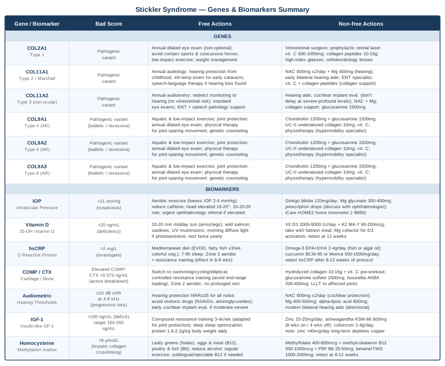
The table below brings together all six genes and seven biomarkers in a single reference, organized by what a problematic result looks like and what response it calls for — both at no cost and with targeted tools or supplements.
What the Huberman Lab's Vision Science Episodes Mean for Stickler Syndrome
Dr. Andrew Huberman, professor of neurobiology at Stanford, has devoted multiple Huberman Lab podcast episodes to the science of vision — myopia mechanisms, retinal health, photobiology, and ocular neuroprotection. Because high myopia and vitreoretinal degeneration are defining features of Stickler syndrome in its most common forms, his research synthesis is unusually actionable for this community. The following distills 10 of the most evidence-dense insights from his vision-focused episodes, applied specifically to Stickler syndrome.
1. Time Outdoors Is the Most Consistent Myopia Intervention in the Literature
Children who spend two or more hours per day outdoors show substantially lower rates of myopia progression — a finding replicated across multiple large epidemiological cohorts in Asia, Europe, and North America. The mechanism appears to involve broad-spectrum, high-intensity outdoor light stimulating retinal dopamine release, which inhibits axial elongation. In Stickler syndrome, the genetically driven component of myopia cannot be prevented this way, but slowing the additional acquired layer through outdoor light exposure is biologically plausible and practically free.
2. Indoor Light Is Far Dimmer Than the Eye Expects
Most indoor environments run at 200–500 lux. Natural outdoor light ranges from 10,000 to 100,000 lux. This mismatch chronically suppresses retinal dopamine and creates the biological conditions for ongoing axial elongation. Huberman recommends deliberate bright light exposure, particularly in the morning. For Stickler patients with photosensitivity, diffuse morning light is preferable to midday direct sun — it delivers the luminance benefit without the UV exposure risk at peak hours.
3. The Real Problem Is the Absence of Distance Vision, Not Near Work Itself
A paradigm-shifting reframing: near work does not cause myopia — the absence of periodic stimulation at optical infinity does. The eye requires regular visual input at distance to regulate axial length. For patients with Stickler syndrome who already have long axial lengths, building in deliberate distance-viewing breaks throughout the day targets the actual mechanism rather than just reducing screen time.
4. Red Light at 670 nm Has Emerging Evidence for Retinal Protection
A randomized trial by Jeffery and colleagues at University College London demonstrated that brief daily exposure to 670 nm red light improved retinal mitochondrial function and reduced cone photoreceptor loss in aging eyes. The effect is likely mediated through mitochondrial cytochrome c oxidase stimulation. For Stickler syndrome, where retinal tissue is under chronic structural stress, two to three minutes of 670 nm light exposure in the morning represents a low-risk, mechanistically grounded supportive intervention. Consumer devices are available at $150–$400.
5. DHA Is Structural, Not Optional, in the Retina
DHA is the dominant omega-3 fatty acid in photoreceptor outer segment membranes — it is a structural component, not merely an anti-inflammatory cofactor. Huberman cites consistent evidence that dietary DHA supports retinal integrity over time. For Stickler syndrome patients, where retinal tissue is already under structural stress, maintaining retinal DHA through fatty fish consumption (3× weekly) or algal oil supplementation (1–2 g DHA/day) is a low-friction protective strategy.
6. Lutein and Zeaxanthin Are the Retina's Own Filter
These two xanthophyll carotenoids form the macular pigment optical density — the eye's endogenous filter against high-energy blue light reaching the photoreceptors. The body cannot synthesize them; they must come from diet or supplementation. A well-studied protocol based on the AREDS2 framework is 20 mg lutein + 4 mg zeaxanthin daily, taken with a fat-containing meal. Eggs and dark leafy greens are the strongest dietary sources.
7. Sleep Is When the Eye Recovers
The glymphatic system clears metabolic waste from neural tissue — including the retina and optic nerve — during deep sleep. Chronic sleep deprivation elevates intraocular pressure and accelerates retinal ganglion cell loss. No supplement or behavioral intervention compensates for inadequate sleep in terms of visual system maintenance. For Stickler patients monitoring IOP, sleep quality is a direct determinant of their morning pressure baseline.
8. Accommodation Is a Trainable Skill
The ciliary muscle contracts and relaxes to shift focus from near to far distances. Huberman discusses evidence that this flexibility atrophies with sedentary visual habits — predominantly near-distance screen time with little variation. Exercises that deliberately cycle focus between near and far targets maintain ciliary muscle function and accommodation range. For Stickler patients with lens involvement or dislocation, individualize this to ophthalmologist guidance before attempting intensive accommodation training.
9. Blue Light at Night Disrupts Retinal Repair Cycles
Evening blue light from screens and LED lighting suppresses melatonin and disrupts circadian input to retinal cell biology — affecting both retinal repair cycling and the circadian IOP rhythm. Practical protocol: dim screens after sunset, use warm-spectrum lighting in the home during evening hours, and consider amber-tinted glasses for screen use after 9 pm. This is a zero-cost change with meaningful downstream benefit for long-term ocular health maintenance in a population already carrying elevated retinal risk.
10. The Vestibular and Visual Systems Share More Infrastructure Than Most Realize
Huberman has discussed the vestibulo-ocular reflex and how gaze stabilization, balance training, and walking in varied environments support shared neural circuits between the visual and vestibular systems. For Stickler syndrome patients with both hearing loss and ocular abnormalities — both of which can subtly compromise vestibular processing — this creates a potential dual-benefit intervention. Gaze stabilization exercises (tracking a finger held at arm's length while rotating the head) are free, brief, and have no downside risk.
Complementary Approaches With Meaningful Clinical Support
The following modalities have evidence relevant to Stickler syndrome's primary manifestations — joint pain, myopia progression, and connective tissue maintenance. None replace conventional care; all can reasonably complement it when applied with appropriate caution.
Low-Level Laser Therapy and Photobiomodulation
Photobiomodulation uses specific wavelengths of red and near-infrared light (typically 630–850 nm) to stimulate mitochondrial activity in cells — including chondrocytes, fibroblasts, and retinal ganglion cells. For Stickler syndrome, where cartilage integrity and collagen quality are structurally compromised, LLLT offers a non-pharmacological route to upregulating cellular energy production and collagen synthesis in affected joints. At the 670 nm wavelength, it also has documented applications in retinal neuroprotection, as noted in Strategy 3 above.
A randomized controlled trial by Alfredo et al. (2012) in Lasers in Medical Science demonstrated significant pain reduction and functional improvement in knee osteoarthritis patients receiving LLLT versus sham. While not Stickler-specific, knee osteoarthritis involves articular cartilage pathology directly analogous to the joint degeneration in Stickler syndrome.
Practical protocol: Apply an FDA-cleared LLLT device to affected joints for 10–15 minutes daily at 100–300 mW/cm² across the red and near-infrared spectrum (660 nm + 850 nm combination). For retinal protection, 670 nm light aimed at closed eyes for 2–3 minutes in the morning follows the UCL protocol studied by Jeffery et al. Consumer devices range $200–$700; clinical LLLT sessions cost $50–$150 each. No significant side effects at recommended parameters — avoid direct, close-range eye exposure with high-power units unless using a device specifically designed for that application.
The David De Angelis Method for Myopia
Given that high myopia is the most functionally impactful daily feature of Stickler syndrome in most cases, the approach described by David De Angelis in The Secret of Perfect Vision: How You Can Prevent and Reverse Nearsightedness warrants serious attention. De Angelis argues that a functional layer of myopia — driven by chronic ciliary muscle contraction from near-work habits and full optical overcorrection — sits on top of any structural component. Addressing the functional layer through active distance training and reduced optical correction can arrest and in some cases partially reverse the acquired component.
The foundational animal evidence cited by De Angelis derives from the landmark form-deprivation and lens-induced myopia research using primate models. Studies published in Science by Wallman et al. (1987) established that myopia induced through optical manipulation is reversible when the deprivation is removed — demonstrating biological plasticity in the emmetropization mechanism. This evidence base underlies modern myopia control research including orthokeratology and low-dose atropine protocols.
For Stickler syndrome patients, full reversal is unlikely given the irreducible genetic and structural component of axial elongation. However, preventing additional acquired myopia progression is clinically meaningful. Protocol: use the weakest correction that allows safe daily function rather than maximum optical correction, take consistent distance-viewing breaks throughout the day, and practice near-to-far focus cycling for 10–15 minutes daily. Critical caveat: do not alter optical correction strategies without first consulting your retinal surgeon, given the elevated retinal detachment risk in Stickler syndrome. Any significant refractive change can alter vitreous traction dynamics.
Progressive Muscle Relaxation and Mindfulness-Based Stress Reduction
Joint pain in Stickler syndrome is often chronic, variable, and unpredictable — the psychological burden of that unpredictability is real. Neurological amplification of pain through central sensitization is increasingly documented in connective tissue disorders, creating a feedback loop where anxiety about pain can lower the threshold for experiencing it. Mindfulness-based stress reduction (MBSR) and progressive muscle relaxation (PMR) directly target this amplification mechanism.
A meta-analysis by Goyal et al. (2014) published in JAMA Internal Medicine reviewed 47 randomized trials and found that mindfulness meditation programs produced moderate improvement in pain, anxiety, and depression across chronic pain populations — with effects persisting at long-term follow-up.
Practical protocol: An 8-week MBSR program — available in-person through many hospital wellness centers or via the structured free curriculum at Palouse Mindfulness — followed by 10–15 minutes of daily maintenance practice. PMR: systematically tense and hold each major muscle group for 5–7 seconds, then release over 20–30 seconds, moving from feet to head. Practice nightly, particularly on high-pain days. Combine with a 4-count inhale, 6-count exhale breathing rhythm for enhanced parasympathetic effect. Avoid PMR of acutely inflamed or unstable joints — adapt the protocol to your current physical state.
Breathing-Based Therapies for Systemic Inflammation and Pain Tolerance
Slow, controlled breathing at 5–6 cycles per minute directly upregulates vagal tone and reduces systemic inflammatory markers including hsCRP and IL-6. For Stickler syndrome patients managing chronic joint pain and elevated inflammation, this represents a zero-cost, genuinely physiological intervention with measurable effects on the same biomarkers tracked in this article.
A systematic review by Zaccaro et al. (2018) in Frontiers in Human Neuroscience confirmed that slow breathing at 5–6 cycles per minute consistently increases heart rate variability and reduces sympathetic nervous system activation — effects that translate to measurably reduced inflammatory tone and improved pain threshold over regular practice.
Practical protocol: Coherent breathing — 5.5-second inhale, 5.5-second exhale, through the nose — practiced for 10–20 minutes daily. Any low-activity period works. A pacing app (Breathwrk, Insight Timer) helps maintain the rhythm without effort. On high-pain days, box breathing (4-4-4-4 count) provides faster acute relief. One caution specific to Stickler syndrome: avoid hyperventilation-based protocols (rapid Wim Hof cycles) — any breathing technique that substantially elevates intracranial or intraocular pressure carries risk in the context of retinal detachment vulnerability and glaucoma monitoring. Stick with slow, controlled, normoventilatory patterns.
Conclusion
Stickler syndrome cannot be erased by lifestyle choices or supplementation. But how quickly it progresses, how well you function day to day, and how many of its complications are caught early — all of these sit within your meaningful influence.
The seven biomarkers in this article — intraocular pressure, vitamin D, hsCRP, COMP/CTX, audiometric thresholds, IGF-1, and homocysteine — form a practical monitoring framework. Not every one needs to be tested immediately, but building a baseline and tracking changes over time gives you information that no single specialist appointment can replicate.
The six genes tell you where in the collagen architecture the fault lies, which downstream systems are at greatest risk, and which compensating strategies are most relevant to your subtype — from retinal surveillance priorities for COL2A1 carriers to aggressive early hearing protection for COL11A1 and COL11A2.
The next smart step is a concrete one: bring this framework to your next specialist visit, ask for a baseline panel of any biomarkers you have not yet tested, and work with your clinical team — geneticist, vitreoretinal surgeon, or audiologist — to build a monitoring schedule calibrated to your specific mutation and presentation. Better information, applied consistently, is the most durable advantage available.
Musculoskeletal: Joint Conditions
Autoimmune: Inflammatory Conditions Connective Tissue Conditions
Eye: Retinal Conditions Glaucoma & Pressure Conditions
Ear, Nose & Throat: Hearing & Balance Conditions