This article contains AI generated content.
· UpdatedSubchondral Cyst Genes and Biomarkers: 7 Biomarkers and 6 Genes to Track
Introduction
Finding out you have a subchondral cyst — often from an incidental MRI finding or after joint pain finally prompted imaging — tends to come with a frustratingly vague explanation. You're told it's related to joint wear, that you should manage your symptoms, and perhaps that you might eventually need intervention if things worsen. What almost never comes with that conversation is any meaningful investigation into why your joint reached that state, or what measurable signals might tell you whether the underlying process is accelerating or stabilizing.
Subchondral cysts are not random structural anomalies. They form at the junction of failing cartilage and stressed bone, driven by a specific chain of biological events: cartilage matrix breakdown, altered bone remodeling, pathological fluid dynamics, and chronic low-grade inflammation. Each of those processes leaves traceable marks — in your bloodstream, in inflammatory markers, in enzymes, and in the genetic variants that shaped your joint architecture from the beginning.
The problem with generic advice — rest, anti-inflammatories, physiotherapy — is that it treats the symptom layer without addressing the drivers. Two people with identical MRI findings may have very different underlying profiles: one driven primarily by systemic inflammation and poor metabolic health, another by inherited cartilage vulnerability with otherwise normal inflammatory markers. The interventions that help one may do little for the other.
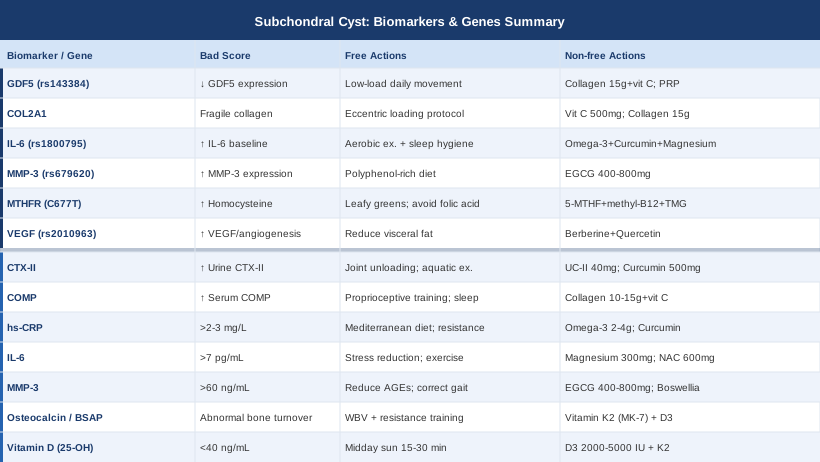
This article offers a more precise approach. The primary section covers seven biomarkers you can actually measure, what each one reveals about the mechanisms behind subchondral cyst formation, and what the evidence supports for improving each one — with and without supplements. The genetics section follows as a useful complement: six key genetic variants that affect joint vulnerability and what you can do if your profile is unfavorable. A book-based evidence synthesis and a set of complementary therapies with genuine clinical backing round out the picture. The goal is not to replace your physician — it is to help you arrive better informed, with more useful questions and more relevant data.
7 Biomarkers That Illuminate the Biology Behind Subchondral Cysts
Subchondral cysts form at the intersection of cartilage degradation, bone remodeling dysregulation, and chronic inflammation. The seven biomarkers below each reflect a different layer of that process. Tracking even three or four of them longitudinally — at baseline and then every three to six months — gives you a functional map of where the problem is most active and whether your interventions are having any effect.
1. CTX-II — Direct Signal of Cartilage Collagen Breakdown
Why it matters: CTX-II (C-terminal crosslinking telopeptide of type II collagen) is among the most specific available markers of articular cartilage collagen degradation. When matrix metalloproteinases and inflammatory enzymes break down type II collagen — the structural backbone of cartilage — CTX-II fragments are released and excreted in urine. Chronically elevated CTX-II reflects ongoing cartilage matrix loss, which is the primary precondition for the fluid intrusion into subchondral bone that creates and expands cysts. In longitudinal OA studies, elevated CTX-II predicts radiographic progression independent of baseline symptoms.
How to measure it
CTX-II is measured in a second-morning urine sample using an ELISA test. It is not widely available at standard labs but is orderable through specialty labs and many functional medicine providers. Cost typically ranges from $80 to $180. Values are normalized to urinary creatinine to correct for dilution. Repeat testing at three to six month intervals is where the real value lies — a trend is more meaningful than any single reading.
If the score is high: the plan without supplements
Elevated CTX-II signals active cartilage destruction, and the first response should be mechanical unloading of the affected joint. For weight-bearing joints, this means eliminating high-compressive loading (impact running, loaded squats with poor alignment) and replacing it with low-impact alternatives: swimming, cycling, or elliptical training at 30 to 60 minutes, four to five times per week. Aquatic exercise in particular reduces joint compressive forces dramatically while maintaining synovial fluid circulation — which is critical for nutrient delivery to avascular cartilage.
A biomechanical assessment with a physiotherapist or gait analyst is worth pursuing if the cyst is in a lower-limb joint. Subtle asymmetries in gait, foot strike, or pelvic positioning concentrate stress at specific points in the joint surface. Correcting these through targeted exercise and movement coaching is a meaningful, zero-cost intervention that can reduce cartilage stress over weeks. Biomarker retesting at three to six months is needed to assess objective effect.
If the score is high: the plan with supplements or equipment
Undenatured type II collagen (UC-II): 40 mg per day, taken on an empty stomach. UC-II works through oral tolerance — modulating the immune-mediated attack on joint collagen — rather than simply providing building blocks. Clinical trials in OA patients show meaningful reductions in knee pain and stiffness at this dose. Cycling is not required; trials have run for up to six months without issues. Side effects: rare and mild, occasional mild GI discomfort.
Curcumin with piperine: 500 to 1000 mg curcuminoids per day, split into two doses with food. Curcumin inhibits NF-κB signaling and reduces MMP expression, directly targeting collagen degradation pathways. Piperine or bioperine increases bioavailability significantly. Use for 8 to 12 weeks, then reassess CTX-II. Side effects: loose stools at higher doses; avoid at medicinal doses during pregnancy; may interact with anticoagulants.
Unloading braces or orthotics: for knee cysts associated with medial compartment loading, an unloading brace shifts force to the lateral compartment, reducing compression at the site of the lesion. Available from orthotists and sports medicine physicians at $200 to $600. Wear during weight-bearing activities; not required at rest.
2. COMP — Cartilage Matrix Integrity Signal
Why it matters: Cartilage Oligomeric Matrix Protein (COMP) is a non-collagenous structural protein that organizes the collagen network and acts as a scaffolding molecule within articular cartilage. When the cartilage matrix is mechanically stressed or degraded, COMP is released into the joint fluid and bloodstream. Serum COMP is elevated in both rheumatoid arthritis and osteoarthritis, and rising levels over time predict structural deterioration. For subchondral cysts specifically, elevated COMP reflects the matrix instability that allows abnormal fluid passage into subchondral bone — a key step in cyst initiation.
How to measure it
COMP is measured in serum via ELISA. It is available through rheumatology-associated labs and some specialty functional medicine providers, but is not a routine offering at most general labs. Cost: $100 to $250. The primary value is tracking change over time within the same individual, as reference ranges vary between labs.
If the score is elevated: the plan without supplements
Elevated COMP in the setting of subchondral cysts points to active cartilage matrix disruption. Proprioceptive and neuromuscular training is one of the highest-leverage non-pharmacological interventions: when the joint is precisely controlled through movement, peak compressive forces are distributed more evenly across the cartilage surface, reducing matrix stress. Frequency: two to three physiotherapy sessions per week for eight to twelve weeks minimum.
Sleep quality is directly relevant here and consistently overlooked. Growth hormone — the primary anabolic signal for cartilage matrix repair — is predominantly secreted during slow-wave sleep. Consistently poor sleep (under seven hours, or fragmented by alcohol or screen use) impairs this nightly repair signal. Addressing sleep hygiene is free and has measurable downstream effects on inflammatory markers and tissue repair rates within weeks.
If the score is elevated: the plan with supplements or equipment
Hydrolyzed collagen peptides with vitamin C: 10 to 15 g of collagen peptides combined with 500 mg of vitamin C, taken approximately 30 to 60 minutes before exercise. This timing is intentional — post-exercise connective tissue uptake of collagen-derived amino acids (glycine, proline, hydroxyproline) is increased when loading follows ingestion. Evidence from trials in athletes with connective tissue injuries suggests this approach meaningfully increases collagen synthesis. Safe for continuous long-term use.
Glucosamine sulfate: 1500 mg per day as a single dose or split. Evidence is mixed in large trials overall, but a meaningful subset of patients — particularly those with moderate-to-severe OA — show clinical benefit at this dose. It may support cartilage proteoglycan stability and slow COMP release over time. Use for at least twelve weeks before assessing response. Side effects: mild GI effects in some; avoid formulations derived from shellfish if allergic.
3. hs-CRP — The Systemic Inflammation Gauge
Why it matters: High-sensitivity C-reactive protein (hs-CRP) is the most widely accessible marker of low-grade systemic inflammation. While it is not joint-specific, it reflects the inflammatory environment that accelerates both cartilage loss and subchondral bone remodeling. Even modest chronic elevations — above 2 to 3 mg/L — are consistently associated with faster OA progression and greater cartilage volume loss in longitudinal studies. It is also a cornerstone biomarker for longevity-oriented clinicians such as Peter Attia and Thomas Dayspring, who treat it as a primary metabolic risk signal, not simply a downstream finding.
How to measure it
hs-CRP is included in many standard blood panels and is one of the cheapest available tests — typically $10 to $40. Target: below 1.0 mg/L for optimal joint and metabolic health. Levels consistently above 3.0 mg/L indicate significant systemic inflammation requiring investigation.
If the score is elevated: the plan without supplements
Dietary pattern is the highest-leverage single intervention for hs-CRP. A Mediterranean-style diet — rich in vegetables, extra-virgin olive oil, fatty fish, legumes, and nuts, and low in refined carbohydrates and ultra-processed foods — has robust randomized evidence for reducing hs-CRP by 20 to 40% within three months. This is not a soft recommendation — dietary inflammation is one of the most potent upstream drivers of elevated CRP, more modifiable than genetics and faster-acting than most supplements.
Resistance training two to three times per week consistently reduces hs-CRP over weeks, partly through improved insulin sensitivity and reductions in visceral adiposity — both of which are independent drivers of systemic inflammation. For affected joints, exercise selection matters: focus on strengthening the muscles around the joint without high compressive loading at the lesion site.
Sleep consistency: even a single night of partial sleep deprivation can acutely elevate hs-CRP by 40 to 60%. Prioritizing seven to nine hours of quality, consistent sleep is one of the most reliably anti-inflammatory behavioral changes available, at zero cost.
If the score is elevated: the plan with supplements or equipment
Omega-3 fatty acids (EPA + DHA): 2 to 4 g per day of combined EPA and DHA from fish oil or algal oil, taken with food. This is among the most evidence-supported anti-inflammatory supplements, with multiple meta-analyses confirming meaningful hs-CRP reduction at these doses. Long-term use at 4 g/day should be reviewed with a physician, as it may affect platelet function. At 2 g/day, continuous long-term use is generally well-tolerated. Side effects: fishy aftertaste, occasional loose stools.
Curcumin with piperine: 500 to 1000 mg/day. Curcumin's inhibition of NF-κB is a primary mechanism for hs-CRP reduction, supported by multiple RCTs in OA and metabolic syndrome populations. Use for 8 to 12 weeks, then reassess. Side effects: loose stools at higher doses; caution with anticoagulant medications.
4. IL-6 — The Joint Inflammation Driver
Why it matters: Interleukin-6 (IL-6) is a pro-inflammatory cytokine that plays a direct mechanistic role in joint destruction — not simply a marker of it. In the context of subchondral cysts, IL-6 drives synovial inflammation, activates osteoclasts (bone-resorbing cells), and upregulates matrix metalloproteinases that break down cartilage collagen. Elevated serum IL-6 is associated with worse OA outcomes, and elevated synovial fluid IL-6 is routinely found in joints with active subchondral changes. The important distinction from hs-CRP is that IL-6 is a cause — it is part of the mechanism degrading the joint, not simply a downstream reflection of it.
How to measure it
Serum IL-6 is available at most labs as a standalone test, cost: $50 to $150. It is less commonly included in routine panels but is orderable by most physicians and through functional medicine labs. Note: IL-6 rises acutely with exercise or active infection — test only when resting and well. Normal serum values are typically below 7 pg/mL.
If the score is elevated: the plan without supplements
Stress reduction has a more direct effect on IL-6 than is widely appreciated. Chronic psychological stress dysregulates cortisol pulsatility, which in turn sustains elevated IL-6. Structured autonomic interventions — mindfulness practice, HRV biofeedback, or even consistent low-intensity walking — can meaningfully reduce IL-6 over weeks. Wearable devices with HRV tracking (Oura Ring, WHOOP, Garmin) provide objective recovery data that can guide training load and stress management decisions.
Reducing visceral adipose tissue is the highest-leverage intervention for chronically elevated IL-6. Visceral fat is a primary secretory source of IL-6 — it produces it continuously and independently of any specific inflammatory trigger. Even a 5 to 10% reduction in body weight achieved through caloric moderation and resistance training leads to measurable decreases in serum IL-6 within weeks to months.
If the score is elevated: the plan with supplements or equipment
Magnesium glycinate or malate: 300 to 400 mg elemental magnesium per day, taken in the evening. Magnesium deficiency — extremely common in Western diets — is associated with elevated IL-6 and NF-κB activation. Repleting magnesium in deficient individuals consistently reduces inflammatory cytokines. Start at 200 mg and titrate up to avoid loose stools. Long-term use at these doses is safe. Side effects: GI discomfort and loose stools at higher doses.
NAC (N-acetylcysteine): 600 to 1200 mg per day, taken in the morning or split into two doses. As a glutathione precursor with direct antioxidant and anti-inflammatory properties, NAC has been shown to reduce IL-6 in inflammatory conditions. Use for 8 to 12 weeks; some practitioners recommend cycling (4 to 8 weeks on, 2 weeks off) as a precautionary practice, though this is convention rather than firmly established evidence. Side effects: nausea at higher doses — take with food.
5. MMP-3 — The Matrix Degradation Amplifier
Why it matters: Matrix Metalloproteinase-3 (MMP-3, stromelysin-1) is an enzyme that degrades multiple extracellular matrix components including proteoglycans, fibronectin, and collagen types II and IV. It also activates other MMPs, making it a key amplifier of joint tissue breakdown. In both OA and RA, elevated MMP-3 in serum and synovial fluid correlates with erosive joint damage. For subchondral cysts, MMP-3 is directly implicated in the cartilage matrix dissolution that creates pathways for pressurized fluid to penetrate into bone — the defining event in cyst formation.
How to measure it
Serum MMP-3 is available through rheumatology-associated labs and specialty functional medicine panels, though not widely offered at general labs. Cost: $100 to $250. It is more commonly monitored in RA than OA, but its relevance extends clearly to both. Normal values are generally below 60 ng/mL, though ranges vary by lab.
If the score is elevated: the plan without supplements
Reducing advanced glycation end products (AGEs) in the diet is a practical and direct approach to lowering MMP-3. AGEs — abundant in ultra-processed foods, charred meats, and foods cooked at very high dry heat — activate RAGE receptors, which upregulate MMP expression. Switching to lower-temperature cooking methods (steaming, poaching, slow cooking) and dramatically reducing ultra-processed food intake reduces AGE load meaningfully within weeks.
Correcting movement asymmetries: localized mechanical stress caused by gait abnormalities, muscle imbalances, or leg length discrepancy upregulates local MMP production in stressed tissue. A physiotherapy assessment focused on biomechanical correction — at no out-of-pocket cost if covered — is a practical intervention that targets the mechanical trigger of MMP overexpression.
If the score is elevated: the plan with supplements or equipment
EGCG (green tea extract): 400 to 800 mg per day of standardized extract containing 45 to 50% EGCG. EGCG inhibits MMP-1, MMP-3, and MMP-13 through multiple pathways. Early clinical evidence in RA patients shows reductions in serum MMP-3 levels. Human joint-specific evidence is still largely preliminary, so modest expectations are appropriate. Take with food; avoid on an empty stomach. Cycle every 8 to 12 weeks with a four-week break — consistent use above 800 mg/day without cycling carries low but non-zero risk of hepatic stress. Side effects: caffeine effects unless decaffeinated; potential hepatotoxicity only at very high long-term doses.
Boswellia serrata extract (AKBA form): 100 to 200 mg of AKBA-standardized extract per day. Boswellia's primary mechanism is 5-lipoxygenase inhibition, which reduces leukotriene synthesis and downstream MMP activity in joint tissue. Multiple clinical trials in OA patients show pain reduction and functional improvement within four to eight weeks. Well tolerated for three to six months of continuous use. Side effects: occasional GI discomfort; avoid during pregnancy.
6. Osteocalcin and Bone-Specific Alkaline Phosphatase — The Bone Remodeling Panel
Why it matters: Subchondral bone remodeling is not merely a consequence of subchondral cysts — it is a driver of their formation and expansion. Osteocalcin reflects osteoblast (bone-building) activity and functions as a metabolic hormone influencing insulin sensitivity. Bone-specific alkaline phosphatase (BSAP) reflects overall bone formation activity. Together, they help identify whether subchondral bone is in a state of pathological turnover — either insufficient formation contributing to cyst expansion, or excessive and disorganized remodeling creating structural weakness. Abnormal bone turnover markers have been documented in OA joints even before cartilage changes are apparent on imaging.
How to measure it
Both tests are available through blood draw. Osteocalcin: $30 to $80. BSAP: $40 to $100. For a more complete picture of bone turnover, some clinicians add serum beta-CTX (a bone resorption marker) at an additional $30 to $80. These can be ordered by primary care physicians, endocrinologists, or functional medicine providers.
If the values are abnormal: the plan without supplements
Weight-bearing and vibration exercise is the most reliable mechanical stimulus for osteoblast activity. For patients with subchondral cysts in weight-bearing joints, the challenge is stimulating bone without overloading cartilage. Whole-body vibration training (WBV) at 25 to 40 Hz, 10 to 20 minutes per session, three to five times per week, stimulates osteoblast activity with minimal cartilage compressive stress. Home platforms are commercially available at $200 to $800 and have evidence for improving bone density metrics in OA populations. Low-load resistance training (resistance bands, bodyweight) targeting muscles around the affected joint is also appropriate.
If the values are abnormal: the plan with supplements or equipment
Vitamin K2 (MK-7 form): 100 to 200 mcg per day with a fat-containing meal. K2 activates osteocalcin through carboxylation — without adequate K2, osteocalcin is produced but cannot bind calcium to bone matrix effectively. The MK-7 form has the longest half-life and best clinical evidence for bone health. Long-term use is safe. Important side note: K2 interacts with vitamin K antagonist anticoagulants (warfarin) — do not use without physician guidance if anticoagulated.
Vitamin D3 and K2 in combination: D3 upregulates osteocalcin gene expression; K2 activates the resulting protein. These two work synergistically. Standard dosing: 2000 to 5000 IU D3 daily with 100 to 200 mcg K2 MK-7. Add magnesium (300 to 400 mg) as a cofactor — D3 metabolism is magnesium-dependent. This combination is affordable, practical, and addresses multiple layers of bone metabolism simultaneously.
7. 25-OH Vitamin D — The Bone and Immune Modulator
Why it matters: Vitamin D deficiency is one of the most prevalent nutrient deficiencies globally and directly affects bone mineral metabolism, immune regulation, and inflammatory signaling — all three of which are relevant to subchondral cyst formation and progression. Low vitamin D levels are associated with greater OA severity, faster cartilage volume loss, and worse pain outcomes in longitudinal cohorts. Vitamin D also regulates RANK/RANKL signaling — the key system controlling osteoclast activation — which directly governs subchondral bone resorption rates.
How to measure it
The 25-OH vitamin D blood test is among the least expensive lab tests available — $30 to $60 — and is orderable by any physician or through direct-to-consumer lab services. Target range: 40 to 60 ng/mL (100 to 150 nmol/L). Most people without supplementation fall significantly below this, particularly in northern latitudes.
If the level is low: the plan without supplements
Midday sun exposure — 15 to 30 minutes of UVB exposure to large skin areas (arms, legs, back) during peak hours — produces meaningful vitamin D synthesis depending on latitude, season, and skin pigmentation. For latitudes above 35°N, UVB intensity between October and March is insufficient for synthesis regardless of exposure duration. Sun exposure remains the most bioavailable source where it is practically achievable.
Dietary sources — fatty fish (salmon, mackerel, sardines), egg yolks, and fortified dairy — provide modest amounts but are generally insufficient to correct a meaningful deficiency. They can be part of a maintenance strategy once levels are optimized.
If the level is low: the plan with supplements or equipment
Vitamin D3 supplementation: 2000 to 5000 IU per day to reach the optimal range. For confirmed deficiency (below 20 ng/mL), higher initial loading doses of 5000 to 10,000 IU daily for eight to twelve weeks may be used under physician supervision, followed by a lower maintenance dose. Retest at three months to assess response. Take with a fat-containing meal for optimal absorption. Pair with K2 (see above) to direct calcium appropriately. Side effects at standard doses are minimal; doses above 10,000 IU daily without monitoring can cause hypercalcemia over time.
Narrowband UVB phototherapy lamp: commercially available home devices (such as those from Sperti or Kernel) provide targeted UVB that synthesizes vitamin D through the skin during low-sun months. Cost: $250 to $500. Use two to four minutes per session on exposed skin, three to five days per week. Highly effective when geographic or lifestyle factors limit sun exposure. Side effects: skin erythema if overexposed — start minimal and increase gradually.
With a clear picture of these seven biomarkers, you can identify which mechanisms are most active in your specific situation. The next layer — genetics — helps explain the structural predispositions that may have set the stage.
6 Genetic Variants Worth Understanding for Subchondral Cyst Risk
Genetics does not determine outcome, but it does set the baseline. Researchers like Ali Torkamani at Scripps Research have advanced polygenic risk modeling for musculoskeletal conditions, while practitioners like Gary Brecka have brought practical attention to how specific variants — particularly in methylation pathways — affect inflammation and tissue repair. The following six variants are among the most relevant to subchondral cyst formation and joint degeneration.
1. GDF5 (rs143384) — The Cartilage Formation Gene
What it does: Growth Differentiation Factor 5 (GDF5) is critical for joint formation, cartilage development, and post-injury repair. The rs143384 T allele reduces GDF5 expression in joint tissue. This is one of the most robustly replicated genetic risk factors for osteoarthritis, identified across multiple genome-wide association studies in both Asian and European populations. Reduced GDF5 means the joint has diminished capacity to form and maintain cartilage — making subchondral failure more likely under mechanical stress that other joints might tolerate.
If the gene is bad: the plan without supplements
Low-load, high-frequency movement becomes essential when GDF5 expression is reduced. Cyclical joint loading at moderate intensity — aquatic exercise, cycling, elliptical training five days per week — provides the mechanical stimulation that drives residual chondrocyte activity without exceeding what a GDF5-compromised joint can absorb. Avoid extended joint immobilization: rest accelerates cartilage atrophy in GDF5-compromised joints. Keep the joint moving, within pain limits, every day.
If the gene is bad: the plan with supplements or equipment
Hydrolyzed collagen peptides (15 g/day) + vitamin C (500 mg) taken before exercise provides substrate for matrix synthesis even where GDF5 signaling is reduced. This compensates on the structural input side when growth factor signaling is limited. Continuous use; safe long-term. No formal cycling required.
PRP (Platelet-Rich Plasma) injections: medically administered, PRP delivers concentrated autologous growth factors — including TGF-β and PDGF — that partially compensate for reduced GDF5 activity. Evidence for OA pain and cartilage support is promising but heterogeneous across studies. Discuss with an orthopedic or sports medicine physician. Cost: $500 to $2,000 per injection series, typically not covered by insurance.
2. COL2A1 — The Type II Collagen Structural Gene
What it does: COL2A1 encodes the alpha-1 chain of type II collagen, the primary structural protein in articular cartilage. Pathogenic mutations cause severe skeletal dysplasias, but common functional variants reduce the mechanical quality of cartilage collagen — affecting cross-linking efficiency and matrix stiffness. This makes cartilage more vulnerable to fatigue damage and enzymatic breakdown under normal loading, setting a lower threshold for cartilage failure and the subchondral changes that follow.
If the gene is bad: the plan without supplements
Eccentric loading protocols applied carefully to periarticular muscles and tendons — rather than directly to the damaged cartilage — improve overall joint mechanics and reduce cartilage compressive peaks without requiring intact collagen quality. Supervised eccentric exercise protocols (such as decline squat protocols for knee involvement), performed three times per week for twelve weeks, have strong evidence for improving pain and function in OA-related joint conditions.
If the gene is bad: the plan with supplements or equipment
Vitamin C: 500 to 1000 mg per day. Vitamin C is the essential cofactor for prolyl hydroxylase, the enzyme responsible for the hydroxyproline cross-links that give collagen tensile strength. When COL2A1 variants reduce collagen structural quality, maximizing cross-linking efficiency is a practical compensatory strategy. Continuous use is safe; doses above 2000 mg/day may cause GI discomfort. Side effects: loose stools at higher doses.
Hydrolyzed collagen peptides: 15 g/day providing proline, glycine, and hydroxyproline — the amino acids that serve as collagen synthesis substrates. Combined with pre-exercise timing and vitamin C co-administration, this supports whatever collagen-producing capacity the joint retains. Safe for continuous long-term use.
3. IL-6 Variant (rs1800795) — The Inflammatory Threshold Gene
What it does: The rs1800795 polymorphism in the IL-6 promoter region is associated with higher baseline IL-6 expression. Carriers of the C allele produce more IL-6 in response to inflammatory stimuli than non-carriers. Since IL-6 drives synovitis, osteoclast activation, and MMP upregulation — all central to subchondral bone pathology — this variant effectively lowers the threshold at which inflammatory joint damage is triggered and sustained.
If the gene is bad: the plan without supplements
Consistent moderate aerobic exercise (150 or more minutes per week at moderate intensity) is one of the few interventions demonstrated to durably reduce baseline IL-6 in high-expression variant carriers. The net anti-inflammatory effect of regular exercise — mediated in part through IL-6's paradoxical anti-inflammatory myokine function in muscle — is well-established. Sleep hygiene is particularly critical for IL-6 variant carriers: sleep disruption amplifies IL-6 release, and high-expression carriers amplify this response further. Consistent schedule, seven to nine hours, minimal alcohol, are non-negotiable baseline practices for this genotype.
If the gene is bad: the plan with supplements or equipment
The omega-3, curcumin, and magnesium combination described in the biomarker section is especially relevant for IL-6 variant carriers. Omega-3 at 2 to 4 g EPA+DHA/day, curcumin at 500 to 1000 mg/day, magnesium at 300 to 400 mg/day — these three address IL-6 signaling through complementary mechanisms. For carriers of this variant, this stack is better viewed as a long-term metabolic maintenance strategy than a short-term correction.
4. MMP-3 Variant (rs679620) — The Matrix Degradation Gene
What it does: The rs679620 variant (related to the functionally studied 5A/6A MMP-3 promoter polymorphism) is associated with higher MMP-3 expression in synovial tissue. Because MMP-3 degrades aggrecan, fibronectin, and multiple collagen types, and activates downstream MMPs including MMP-1, MMP-9, and MMP-13, carriers of the high-expression variant have a structurally amplified capacity for cartilage matrix destruction under inflammatory conditions. This is directly relevant to subchondral cyst progression.
If the gene is bad: the plan without supplements
A consistently polyphenol-rich diet is a practical targeted response. Quercetin (onions, capers, apples), resveratrol (dark berries, red grapes), and EGCG (green tea) naturally modulate MMP-3 gene expression through epigenetic mechanisms. Early clinical evidence in RA patients shows that consistent green tea polyphenol consumption measurably reduces serum MMP-3. This is most effective as a daily dietary habit, not an episodic intervention.
If the gene is bad: the plan with supplements or equipment
EGCG supplement: 400 to 800 mg/day of standardized green tea extract. For MMP-3 variant carriers, this is a targeted intervention addressing the specific enzymatic excess. Cycle 8 to 12 weeks on, four weeks off to limit cumulative hepatic exposure at higher doses.
Low-dose doxycycline (medical prescription only): at sub-antibiotic doses (20 mg twice daily), doxycycline functions as an MMP inhibitor — a mechanism well-established in periodontitis and studied in OA contexts. This is mentioned as a medically supervised option only, not for self-administration. Require physician oversight and liver monitoring. Duration is determined by clinical response and physician assessment.
5. MTHFR (C677T, A1298C) — The Methylation and Homocysteine Gene
What it does: The MTHFR gene encodes methylenetetrahydrofolate reductase — the enzyme required to convert dietary folate into 5-MTHF, the active form that drives the methylation cycle. The C677T and A1298C variants, widely discussed through Gary Brecka's practical methylation framework, reduce enzyme activity and cause homocysteine accumulation. Elevated homocysteine is pro-inflammatory, impairs collagen cross-linking (directly relevant to cartilage integrity), damages vascular endothelium, and increases oxidative stress — all of which contribute to the tissue environment in which subchondral cysts form and expand. MTHFR is often overlooked in orthopedic and rheumatology contexts but is highly relevant to the upstream inflammatory and tissue quality issues.
If the gene is bad: the plan without supplements
Prioritize dietary methylfolate sources: leafy greens (spinach, romaine, kale), beets, lentils, and eggs all provide natural methyl donors and folate that can partially compensate for reduced MTHFR activity. Daily inclusion of these foods is meaningful, particularly when combined with avoiding synthetic folic acid, which MTHFR variant carriers have difficulty converting and may actually accumulate in counterproductive form. Actively avoid fortified foods using folic acid (look for the word folic acid in the ingredient list of cereals, energy drinks, and multivitamins).
If the gene is bad: the plan with supplements or equipment
5-MTHF (methylfolate): 400 to 1000 mcg per day. Start low — 400 mcg — as some individuals experience stimulatory side effects (mood changes, headache) when beginning methylation support. This is the bioavailable active form of folate that entirely bypasses the MTHFR enzyme defect. Pair with methylcobalamin (methyl B12) at 500 to 1000 mcg per day, which is necessary for the homocysteine-to-methionine conversion step. Long-term continuous use is appropriate for confirmed variant carriers. Side effects: in sensitive individuals (particularly those with existing anxiety), excess methyl donors can cause irritability or jitteriness — reduce dose if this occurs.
Betaine (TMG — trimethylglycine): 500 to 1500 mg per day with food. Betaine provides an alternative methylation pathway (the BHMT pathway) that is entirely independent of MTHFR. It effectively lowers homocysteine through a different route, providing meaningful redundancy. Safe for long-term use. Side effects: occasional fishy body odor at high doses due to TMAO metabolism in some individuals.
6. VEGF (rs2010963) — The Subchondral Vascularization Gene
What it does: Vascular Endothelial Growth Factor (VEGF) drives angiogenesis — the formation of new blood vessels. In healthy joints, articular cartilage is avascular by design. But in OA and in subchondral cyst formation, VEGF-driven neovascularization invades the calcified cartilage and subchondral bone in a pathological pattern. This abnormal vascularization is directly implicated in cyst initiation and expansion: new blood vessels bring inflammatory cells and alter bone fluid dynamics. The rs2010963 (+405 G/C) variant is associated with higher VEGF expression and has been linked to more aggressive synovial inflammation and worse joint pathology outcomes.
If the gene is bad: the plan without supplements
Visceral fat reduction is the most practical systemic intervention for excess VEGF expression. Adipose tissue is a prolific source of VEGF, and visceral fat reduction through caloric moderation and consistent exercise directly and measurably reduces circulating VEGF. Monitoring DEXA scan results for visceral fat area (rather than relying on BMI) provides a more accurate target for this intervention.
If the gene is bad: the plan with supplements or equipment
Berberine: 500 mg two to three times per day with meals. Berberine activates AMPK signaling, which has downstream anti-angiogenic effects including VEGF pathway inhibition. It is additionally beneficial for insulin sensitivity and metabolic health — directly relevant co-factors in joint pathology. Cycle every 8 weeks with a 4-week break to maintain AMPK sensitivity and avoid microbiome disruption from extended continuous use. Side effects: GI discomfort (nausea, loose stools) particularly at initiation — start with 250 mg twice daily and titrate. May interact with diabetes medications and certain CYP3A4 substrates; verify with a physician.
Quercetin: 500 to 1000 mg per day with food. Quercetin inhibits VEGF receptor signaling and has anti-angiogenic properties demonstrated in preclinical models. Direct human evidence for joint-specific VEGF inhibition remains limited; this should be considered a supportive measure alongside berberine rather than a standalone primary intervention. Safe for long-term use. Side effects: rare; occasional headache at high doses.
Understanding which of these variants you carry — through a SNP report from services such as 23andMe analyzed via tools like Genetic Genie, or through clinical genetic testing — helps you prioritize which supplements and strategies are most relevant to your specific biology. The next section zooms out to a broader evidence framework.
What Outlive by Peter Attia Reveals About Joint Longevity
Peter Attia's Outlive: The Science and Art of Longevity (2023) is one of the most study-dense health books published in recent years, drawing on hundreds of peer-reviewed studies across metabolism, cardiology, oncology, and musculoskeletal medicine. Though not written about subchondral cysts specifically, its framework for how chronic disease develops — and why the conventional medical system consistently misses the window for intervention — is directly applicable to joint degeneration. Here are ten of the most impactful insights from Outlive as applied to this condition.
1. Inflammation Is the Root Driver, Not a Downstream Finding
Attia frames chronic low-grade inflammation as the upstream cause of most chronic disease, not a consequence of it. For joints, this means that reducing hs-CRP and IL-6 through diet, exercise, and sleep is foundational prevention — not adjunct care layered on top of structural treatment.
2. The Conventional Medical System Waits Too Long
Attia's central argument is that standard medical practice is optimized for treating disease after it is established, not preventing it years or decades earlier. For subchondral cysts, this means the cartilage changes, bone remodeling alterations, and inflammatory drivers have typically been active for years before any imaging is ordered. Biomarker tracking beginning in the 30s and 40s — before symptoms appear — creates a window for earlier intervention that imaging cannot provide.
3. Zone 2 Cardio Is His Most-Discussed Anti-Inflammatory Tool
Attia consistently returns to Zone 2 aerobic training (65% of maximum heart rate, 45 to 60 minutes, four to five times per week) as the single most broadly beneficial non-pharmacological intervention. At this intensity, mitochondrial efficiency improves, insulin sensitivity rises, and inflammatory cytokines decrease — without the oxidative stress of high-intensity training. For joint patients, Zone 2 in the pool or on a stationary bike avoids cartilage impact while delivering these systemic benefits.
4. Muscle Mass Is the Foundation of Joint Protection
Maintaining and building muscle mass is, in Attia's framework, the highest-priority physical adaptation for healthy aging — and it directly protects joints. Periarticular muscle absorbs compressive forces that would otherwise concentrate on cartilage and subchondral bone. Resistance training three to four times per week is non-negotiable, with exercise selection modified (not eliminated) when joints are compromised.
5. Sleep Is a Biological Necessity with Measurable Consequences When Deficient
Attia cites extensive evidence showing that inadequate or fragmented sleep elevates inflammatory markers, impairs growth hormone secretion, reduces cartilage repair signaling, and raises cortisol. For subchondral cysts, chronic sleep impairment sustains the very inflammatory environment driving joint damage. He recommends treating sleep as a structured variable — tracked, assessed, and actively improved — rather than a lifestyle afterthought.
6. Insulin Resistance Drives Systemic Inflammation Even Before Diagnosis
Elevated fasting insulin and early glucose dysregulation — well before HbA1c rises into diabetic range — sustain low-grade systemic inflammation that accelerates joint damage. Attia recommends tracking fasting insulin and HOMA-IR as early, actionable markers. Dietary carbohydrate moderation combined with resistance training is his most consistent strategy for improving insulin sensitivity and, downstream, inflammatory load.
7. Visceral Fat Is the Most Metabolically Destructive Tissue
Visceral adipose tissue continuously secretes IL-6, TNF-α, and VEGF — all directly implicated in the biomarker and genetic discussion above. Attia frames visceral fat reduction as a first-order health priority. DEXA scans (not BMI) provide the most accurate visceral fat assessment. For joint health specifically, each kilogram of visceral fat lost measurably reduces the cytokine burden on the joint.
8. Omega-3 Fatty Acids Are One of His Few Consistent Supplement Recommendations
Attia is notably skeptical of most supplements and supplements categories. Omega-3 fatty acids at 2 to 4 g EPA+DHA per day are among his consistent recommendations specifically because of their robust anti-inflammatory mechanism and evidence base. This aligns directly with the hs-CRP and IL-6 biomarker sections above.
9. Vitamin D Is Ubiquitously Under-Corrected
Attia regularly highlights that most people's vitamin D levels sit in ranges that impair immune function, bone metabolism, and muscle function. His target (40 to 60 ng/mL) matches the clinical evidence and is significantly above what most people achieve without supplementation. He emphasizes testing before supplementing and adjusting doses based on lab results — not using fixed population doses.
10. You Are the Primary Actor in Your Health Trajectory
Outlive's conclusion is that longevity medicine requires patients to be active agents — not passive recipients of reactive care. For subchondral cysts, this means using biomarker testing to create a personalized inflammatory and cartilage health profile, making evidence-based lifestyle changes before symptoms force the issue, and engaging with medical providers with specific, trackable questions rather than general complaints.
Complementary Approaches with Clinical Evidence
The following modalities have meaningful human clinical evidence in the context of joint pain, cartilage health, and OA management — the clinical category most directly relevant to subchondral cysts. These are not replacements for biomarker-guided intervention, but they can meaningfully reduce pain and preserve function while underlying processes are addressed.
Tai Chi
Tai chi is a mind-body practice built around slow, controlled, flowing movements that combine weight shifting, balance training, and low-impact joint loading. It is particularly relevant for subchondral cyst management because it simultaneously improves proprioception, reduces joint loading asymmetry, strengthens periarticular musculature, and reduces psychological stress — all contributors to joint degeneration. Unlike higher-impact exercise, it avoids the compressive stress that can aggravate damaged cartilage.
A landmark randomized controlled trial by Wang et al. published in Annals of Internal Medicine compared tai chi to physical therapy in 204 patients with knee osteoarthritis and found equivalent pain relief and functional improvement, with additional psychological benefits. This is a well-powered, high-quality trial with direct relevance to OA-associated joint pathology.
Practically, tai chi is accessible at community centers and through online programs at all skill levels. A beginner protocol of two to three sessions per week for twelve weeks is a practical starting point. For those with significant joint pain, modified seated or wall-supported tai chi is available and has been shown to produce benefits even in lower-mobility individuals. Benefits accumulate over months of consistent practice.
Low-Level Laser Therapy (Photobiomodulation)
Low-level laser therapy (LLLT), also called photobiomodulation, uses near-infrared light (typically 810 to 1064 nm) to stimulate mitochondrial function via cytochrome c oxidase, reduce local inflammation, and promote tissue repair at the cellular level. For joints, LLLT has been studied specifically in OA contexts and shows consistent evidence for pain reduction and modest improvements in functional outcomes with a highly favorable safety profile.
A systematic review and meta-analysis by the Ottawa Panel evaluated LLLT for knee OA and found moderate-quality evidence supporting its use for pain and disability reduction. More recent device-specific trials in knee and hip OA have reinforced these findings. Near-infrared home devices (100 to 500 mW, 810 to 980 nm range) cleared for home use are available at $200 to $800 from manufacturers including Kineon and Mito Red Light.
For subchondral cysts, LLLT is applied directly over the affected joint, probe or panel held against the skin without pressure. A typical protocol is 10 to 20 minutes per session, three to five times per week for four to eight weeks. Pain reduction is the most consistently reported outcome; structural repair evidence remains preliminary. The safety profile is excellent when used as directed — avoid direct eye exposure.
Mindfulness-Based Stress Reduction (MBSR)
Structured mindfulness meditation — particularly the eight-week MBSR program developed by Jon Kabat-Zinn — has a growing evidence base for chronic pain conditions including OA and inflammatory arthritis. Its relevance to subchondral cysts spans multiple mechanisms: it reduces cortisol-mediated inflammation, lowers IL-6 through autonomic nervous system regulation, improves sleep architecture, and modifies the psychological relationship with pain — reducing catastrophizing, which amplifies perceived pain and disability independent of structural findings.
A randomized trial published in Annals of the Rheumatic Diseases showed that a mindfulness-based intervention significantly reduced pain interference and fatigue in arthritis patients compared to controls. While not conducted specifically in subchondral cyst patients, chronic joint pain mechanisms are functionally comparable across OA-related diagnoses.
Practically, MBSR courses are available online — including the free, evidence-based Palouse Mindfulness program modeled directly on Kabat-Zinn's curriculum. Daily practice of 20 to 40 minutes, within an eight-week structured format, produces the most robust outcomes. Effects on inflammatory markers and pain perception typically become evident after four to six weeks of consistent practice, and — unlike most interventions — the benefits tend to persist long after the formal course ends.
Chinese Herbal Medicine — Targeted Botanical Evidence
The botanical pharmacopeia used in traditional Chinese medicine includes several compounds with meaningful modern clinical evidence for joint inflammation. Boswellia serrata — while Indian in origin, its active compound AKBA is used across Asian traditional medicine systems — is a well-characterized 5-lipoxygenase inhibitor that reduces leukotriene synthesis and MMP activity in joint tissue, directly addressing two of the mechanisms described in the biomarker section.
A double-blind randomized controlled trial by Gupta et al. (2011) published in International Journal of Medical Sciences found that standardized Boswellia extract providing 100 mg AKBA per day significantly reduced knee pain and improved physical function versus placebo over 90 days in OA patients, with onset of benefit by week seven. This is one of the more methodologically sound botanical trials in the joint health space.
Practically, target a standardized Boswellia extract specifying its AKBA content (not just total extract weight) at 100 to 200 mg AKBA per day, taken with a fat-containing meal. Use for three to six months. It combines safely with omega-3 and curcumin, providing complementary anti-inflammatory mechanisms through distinct pathways. Side effects are generally mild (GI discomfort in some); avoid during pregnancy.
Conclusion
Subchondral cysts are a signal that multiple biological processes have gone wrong together — cartilage matrix breakdown, subchondral bone remodeling dysregulation, and chronic inflammation do not occur in isolation. The good news is that most of these processes are measurable and, to meaningful degrees, modifiable.
Starting with three or four of the biomarkers described here — particularly CTX-II, hs-CRP, vitamin D, and either COMP or MMP-3 depending on what your physician can order — creates a functional baseline that most clinical consultations do not provide. Adding genetic context through SNP analysis helps explain why your joints responded the way they did and which strategies are most likely to move the needle in your case.
The next smart step is to bring this framework to a physician, rheumatologist, or functional medicine practitioner willing to order these tests and track trends longitudinally. Progress is measured in the direction of change over three to six months — not in a single data point. The goal is not a perfect result but a clear trajectory toward one, supported by evidence and monitored over time.
Musculoskeletal: Bone Conditions Joint Conditions
Autoimmune: Inflammatory Conditions Connective Tissue Conditions