This article contains AI generated content.
· UpdatedSubchondral Insufficiency Fracture of the Knee: 5 Genes and 7 Biomarkers to Track
Introduction
A subchondral insufficiency fracture of the knee (SIFK) is not a traumatic injury in the conventional sense. It happens when the bone just beneath the cartilage — the subchondral plate — can no longer tolerate normal, everyday mechanical load. It cracks under the weight of regular activity, not a fall or a collision. Most patients are told to rest, use crutches, and wait. Some are given a follow-up MRI. Very few are ever asked why this bone failed in the first place, and that question matters more than it is typically given credit for.
Generic bone health advice — calcium, vitamin D, weight-bearing exercise — is not wrong, but it is often too broad to be useful for someone with SIFK. The subchondral bone has a specific biology: it is highly vascularized, metabolically active, and particularly sensitive to the balance between bone formation and resorption. When that balance tilts toward excessive resorption, or when mineralization is impaired due to nutritional deficiencies or hormonal shifts, the subchondral plate becomes a structural weak point. Understanding why this happened requires looking at the individual's biochemical environment, not just their imaging report.
This article approaches SIFK from two angles that are rarely discussed in clinical settings. The first examines seven specific biomarkers — measurable values in the blood or urine — that can reveal the metabolic conditions driving bone fragility. The second looks at five genetic variants that may predispose someone to poor bone quality, excessive resorption, or impaired repair, and what can actually be done if those variants are present. Neither approach promises a cure, but together they offer something more valuable than passive waiting: a clearer understanding of where the problem originates and what levers are available to address it.
Better information leads to better decisions. If your vitamin D is chronically low, your bone resorption markers are elevated, and you carry a variant in the gene that controls how your body responds to vitamin D, that is not a reason for despair — it is a roadmap. The strategies in this article are built around that principle: precision over generality, action over waiting, and realistic optimism grounded in science rather than wishful thinking.
7 Biomarkers That May Reveal What Is Driving Your Knee Fracture
Bone is not static. It is continuously remodeled — old bone is broken down by osteoclasts and new bone is built by osteoblasts. In a healthy adult, these processes are tightly coupled. In someone with SIFK, something has disrupted that coupling: resorption is outpacing formation, mineralization is impaired, or the mechanical properties of the bone matrix are compromised. The seven biomarkers below are the clearest window into this process. Some are well-established in osteoporosis management (recommended by clinicians like Peter Attia, Thomas Dayspring, and the International Osteoporosis Foundation); others are less commonly ordered but offer important additional context. For each, the cost range reflects typical US laboratory pricing without insurance.
Biomarker 1: 25-OH Vitamin D
Why it matters. Vitamin D is not simply a vitamin — it is a steroid hormone precursor that regulates calcium absorption in the gut, phosphate metabolism, and direct gene expression in osteoblasts and osteoclasts. Without adequate vitamin D, calcium cannot be efficiently absorbed, PTH rises compensatorily, and the net result is accelerated bone resorption. For SIFK specifically, chronic vitamin D insufficiency creates a mineralization deficit in the subchondral bone, reducing its capacity to handle compressive load. The NIH Office of Dietary Supplements provides an overview of vitamin D's role in bone health.
How to measure it. A standard serum 25-hydroxyvitamin D test. Cost: $30–70 through most labs, often included in basic wellness panels. Functional medicine practitioners like Peter Attia generally target a range of 40–60 ng/mL for bone optimization, rather than simply staying above the deficiency threshold of 20 ng/mL.
If the score is low — plan without supplements. Expose 40% or more of your skin to midday sun for 15–30 minutes daily, weather and latitude permitting. This can raise serum levels meaningfully over 8–12 weeks. Increase consumption of fatty fish (salmon, sardines, mackerel), egg yolks, and liver, which are among the few dietary sources of D3. Prioritize these foods year-round if sun exposure is limited.
If the score is low — plan with supplements or equipment. Vitamin D3 (cholecalciferol) at 2,000–5,000 IU/day is a standard starting point. Always pair it with Vitamin K2 (MK-7 form, 100–200 mcg/day) to direct calcium to bones rather than arteries. Add magnesium glycinate (300–400 mg/day), as magnesium is required for vitamin D conversion to its active form. Retest at 90 days and adjust dose. Side effects: vitamin D toxicity is rare below 10,000 IU/day but possible — never supplement without baseline testing. Avoid cycling; consistent daily dosing is more effective than intermittent high doses.
Biomarker 2: CTX-I (C-Terminal Telopeptide of Type I Collagen)
Why it matters. CTX-I is a fragment of type I collagen released during osteoclast-mediated bone resorption. Elevated levels indicate that bone is being broken down faster than is typical, which in the context of SIFK points toward a bone environment that is structurally compromising the subchondral plate. CTX-I is considered one of the gold-standard bone resorption markers by the International Osteoporosis Foundation (IOF Working Group on bone marker standardization).
How to measure it. Serum CTX-I (also called beta-CrossLaps). Best drawn fasting in the morning, as values fluctuate significantly with meals. Cost: $60–120. Optimal serum CTX-I in adults is generally below 0.30 ng/mL in women post-menopause and below 0.57 ng/mL in men, though reference ranges vary by lab. Functional medicine contexts often aim for the lower half of the reference range.
If the score is elevated — plan without supplements. High-impact and resistance exercise directly suppresses osteoclast activity and stimulates osteoblasts. Aim for three to four sessions per week of weight-bearing or resistance exercise, prioritizing lower-body loading (squats, step-ups, modified leg press). Reduce dietary inflammatory load: eliminate processed seed oils, minimize ultra-processed foods, and prioritize protein at 1.2–1.6 g/kg body weight daily (adequate protein supports bone matrix formation). Sleep optimization is critical — bone remodeling occurs primarily during sleep; target 7–9 hours with consistent timing.
If the score is elevated — plan with supplements or equipment. Collagen peptides (10–15 g/day, with vitamin C 200–500 mg co-ingested) have been shown to support bone matrix synthesis. Calcium citrate (500 mg twice daily, not carbonate) provides the mineral substrate to slow resorption-formation imbalance. Whole-body vibration (WBV) platforms (10–20 minutes daily at 25–50 Hz) have emerging evidence for suppressing bone resorption markers in older adults — lower cost alternatives exist for home use ($150–500). Bisphosphonates are the prescription standard for severe cases, but these should be evaluated by a physician given their own side effect profile. Retest CTX-I at 3–6 months.
Biomarker 3: P1NP (Procollagen Type 1 N-Terminal Propeptide)
Why it matters. Where CTX-I measures destruction, P1NP measures construction. It is a byproduct of new bone formation by osteoblasts and reflects how actively new bone matrix is being synthesized. The IOF recommends P1NP as the preferred bone formation marker globally due to its stability and standardization across labs. In SIFK, a low P1NP alongside elevated CTX-I reveals the worst scenario: excessive resorption with insufficient repair — exactly the environment that creates subchondral vulnerability.
How to measure it. Serum P1NP. Cost: $80–150. Does not require fasting. Optimal adult range is generally 15–80 mcg/L in women; values below 15 suggest suppressed bone formation.
If the score is low — plan without supplements. Resistance training is the most potent non-pharmacological stimulator of P1NP. Progressive overload — gradually increasing load over weeks — drives osteoblast activation. A 2016 RCT published by Watson et al. in the Journal of Bone and Mineral Research demonstrated significant P1NP increases following 8 months of high-intensity resistance and impact training in older women. Adequate dietary protein and total caloric intake are also prerequisites — bone formation is an anabolic process and cannot occur in a state of chronic undereating.
If the score is low — plan with supplements or equipment. Strontium ranelate (prescription in Europe) directly stimulates osteoblasts, though access is limited due to cardiovascular concerns in high-risk patients. Silicon (as orthosilicic acid, 10 mg/day) and boron (3–6 mg/day) both support collagen cross-linking and osteoblast activity. Creatine monohydrate (3–5 g/day, continuous) has shown modestly positive effects on bone mineral density in older adults when combined with resistance training. Retest P1NP alongside CTX-I every 3–6 months to track the formation/resorption ratio.
Biomarker 4: PTH (Parathyroid Hormone)
Why it matters. Parathyroid hormone is the body's primary calcium regulator. When calcium or vitamin D is chronically low, the parathyroid glands compensate by secreting more PTH. Chronically elevated PTH (secondary hyperparathyroidism) drives osteoclast activity, pulling calcium from bone to maintain blood calcium. This is a significant driver of bone quality deterioration. In the subchondral bone, this manifests as reduced mineralization and structural thinning. The NIH National Institute of Arthritis and Musculoskeletal and Skin Diseases recognizes PTH dysregulation as a central mechanism in osteoporosis.
How to measure it. Intact PTH via serum blood draw. Cost: $30–60. Optimal is the lower half of the normal range (typically 10–65 pg/mL). Values in the upper third of normal, even without frank hyperparathyroidism, should prompt investigation of vitamin D status and calcium intake.
If PTH is elevated — plan without supplements. First establish whether vitamin D is the driver — this is the most common cause of chronically elevated PTH. Increase dietary calcium through whole food sources: dairy, sardines with bones, leafy greens. Regular outdoor activity combined with moderate sun exposure can correct the underlying vitamin D insufficiency over time, normalizing PTH without pharmaceutical intervention.
If PTH is elevated — plan with supplements or equipment. Correct vitamin D deficiency first (D3 as described above). Once vitamin D is optimized and PTH remains elevated, calcium citrate supplementation at 500 mg twice daily may help. Rule out primary hyperparathyroidism (a parathyroid adenoma) if PTH remains elevated despite normal vitamin D — this requires physician evaluation. Do not supplement calcium aggressively without physician supervision if primary hyperparathyroidism is suspected.
Biomarker 5: hsCRP (High-Sensitivity C-Reactive Protein)
Why it matters. Chronic low-grade inflammation activates osteoclasts via the RANKL pathway — the same signaling axis that drives post-menopausal bone loss. Elevated hsCRP indicates systemic inflammatory burden that is actively promoting bone resorption. Peter Attia considers hsCRP one of the most actionable longevity biomarkers, targeting values below 1 mg/L. In SIFK, elevated baseline inflammation also impairs the healing response in the subchondral bone, extending recovery timelines significantly.
How to measure it. High-sensitivity CRP via standard blood draw. Cost: $20–45. Values above 3 mg/L represent high cardiovascular and metabolic inflammatory risk; 1–3 mg/L is intermediate; below 1 mg/L is optimal for bone and systemic health.
If hsCRP is elevated — plan without supplements. The most powerful lifestyle interventions for CRP reduction are: (1) sleep quality improvement — sleep deprivation acutely raises CRP within days; (2) elimination of ultra-processed foods and refined sugars; (3) moderate aerobic exercise three to five times weekly (Zone 2 intensity — conversational pace); (4) stress reduction — chronic psychological stress significantly raises inflammatory cytokines via cortisol dysregulation.
If hsCRP is elevated — plan with supplements or equipment. Omega-3 fatty acids (EPA+DHA combined, 2–4 g/day) are among the most evidence-backed anti-inflammatory interventions, directly reducing IL-6, TNF-alpha, and CRP. Use a triglyceride-form fish oil or algae-based DHA/EPA for better absorption. Curcumin (500–1,000 mg/day of a bioavailable form such as BCM-95 or with piperine) has demonstrated CRP reduction in multiple RCTs. Cycling: omega-3s can be taken continuously; curcumin at 8–12 weeks on / 4 weeks off is a practical approach given limited long-term data. Retest hsCRP at 8–12 weeks.
Biomarker 6: Estradiol and Testosterone
Why it matters. Sex hormones are the master regulators of bone turnover. Estrogen (estradiol) suppresses osteoclast activity in both women and men — estrogen deficiency in either sex accelerates bone loss. Testosterone supports bone density directly and through aromatization to estradiol in men. In postmenopausal women, SIFK risk increases substantially as estrogen drops after menopause, creating the hormonal environment where subchondral bone becomes vulnerable. The NIH's calcium and bone health resources emphasize the critical role of estrogen status in bone preservation.
How to measure it. Serum estradiol (E2) and total/free testosterone, plus SHBG (sex hormone binding globulin) to understand bioavailable fractions. Cost: $50–120 for a comprehensive panel. In women, post-menopausal estradiol below 20 pg/mL is associated with accelerated bone loss. In men, estradiol below 20 pg/mL and testosterone below 400 ng/dL warrant closer evaluation.
If hormones are low — plan without supplements. Prioritize sleep (testosterone production peaks during REM sleep; sleep deprivation acutely suppresses testosterone by 10–15% in men within one week). Resistance training two to four times per week directly stimulates testosterone and estrogen production. Reduce body fat if elevated — adipose tissue aromatizes androgens to estrogens, but excess fat also drives systemic inflammation that disrupts hormonal signaling. Reduce alcohol, which impairs estrogen metabolism and testosterone production.
If hormones are low — plan with supplements or equipment. Boron (6–10 mg/day from food sources like avocado, raisins, and nuts, or supplementation) raises free testosterone and estradiol by reducing SHBG. Zinc (15–30 mg/day elemental zinc) supports testosterone synthesis. Ashwagandha KSM-66 extract (600 mg/day) has demonstrated testosterone increases of 10–15% in multiple RCTs in men under stress. For women and men with significant documented deficiency, hormone replacement therapy (HRT) should be considered with a physician — HRT has strong evidence for bone density preservation. Avoid high-dose DHEA without physician monitoring.
Biomarker 7: RBC Magnesium (Red Blood Cell Magnesium)
Why it matters. Approximately 60% of the body's magnesium is stored in bone. Magnesium is embedded in the hydroxyapatite crystal structure of bone, where it contributes to bone elasticity and resistance to fracture. Importantly, magnesium deficiency also impairs vitamin D conversion (the conversion of 25-OH-D to its active 1,25-dihydroxyvitamin D form requires magnesium-dependent enzymes), creating a cascade effect that compounds bone fragility. The NIH Office of Dietary Supplements' Magnesium Fact Sheet notes that population surveys suggest a majority of adults consume less than the recommended daily allowance of magnesium.
How to measure it. Serum magnesium is a commonly ordered test (cost: $15–30), but it is a poor reflection of total body magnesium status — the body maintains serum levels at the expense of intracellular stores. RBC magnesium (red blood cell magnesium) is a significantly more accurate measure of functional magnesium status. Cost: $50–85. Optimal RBC magnesium is generally considered to be in the upper half of the reference range (5.6–6.8 mg/dL as a rough guide, though ranges vary by lab).
If magnesium is low — plan without supplements. Dietary magnesium from whole foods is highly bioavailable. Prioritize: dark leafy greens (spinach, Swiss chard), pumpkin seeds, almonds, dark chocolate, legumes, and avocados. Reduce alcohol and caffeine consumption, which increase urinary magnesium excretion. Minimize use of proton pump inhibitors (PPIs) if possible — PPIs are associated with significant magnesium depletion and may directly worsen bone quality.
If magnesium is low — plan with supplements or equipment. Magnesium glycinate or magnesium malate (300–400 mg elemental magnesium/day) are the most bioavailable and gut-tolerant forms. Magnesium oxide is cheap but poorly absorbed. Take in the evening — magnesium also supports sleep quality and muscle relaxation, which aid overnight bone repair. Transdermal magnesium spray (applied to skin, 15–30 minutes pre-bath) provides an alternative route and is well-tolerated. Continuous use is generally appropriate; there is no cycling requirement. Retest RBC magnesium at 3 months. Side effects: loose stools at doses above 350 mg elemental; glycinate form is least likely to cause GI issues.
What Your Genes May Be Telling You About Bone Fragility
While biomarkers show you where the system currently stands, genetics reveal the structural tendencies that make a person prone to subchondral bone failure in the first place. Gene variants do not determine outcomes — they create predispositions that can be modified, compensated for, or aggravated by environment and lifestyle. The five genes below have the strongest human evidence base for bone quality and fracture risk. This approach reflects the framework used by researchers like Ali Torkamani (Scripps Research Institute), whose work on polygenic risk scores for bone fragility integrates multiple gene variants into actionable risk profiles, and by clinicians like Gary Brecka, who emphasizes identifying and compensating for high-impact variants before pathology develops.
Gene 1: VDR (Vitamin D Receptor)
The VDR gene encodes the receptor through which vitamin D exerts its effects on bone cells, immune function, and gene expression. Common variants (notably BsmI, ApaI, TaqI, and FokI polymorphisms) alter receptor binding affinity and efficiency. Individuals with less efficient VDR variants may require higher circulating vitamin D levels to achieve the same biological effect as someone with a functional receptor. Studies have consistently linked specific VDR polymorphisms with lower bone mineral density and increased fracture risk — this is one of the better-replicated findings in bone genetics.
If the gene variant is unfavorable — plan without supplements. Maximize natural vitamin D exposure: consistent midday sun, 20–30 minutes on large skin surface areas. Increase dietary sources beyond what would be typical. Engage in regular resistance exercise, which upregulates VDR expression independently of serum vitamin D. Reduce obesity, which sequesters vitamin D in adipose tissue and reduces its bioavailability.
If the gene variant is unfavorable — plan with supplements or equipment. Individuals with poor-functioning VDR variants likely need higher vitamin D3 supplementation targets — often 4,000–6,000 IU/day — to achieve the same functional effect, though this should be confirmed by periodic 25-OH-D testing. Vitamin K2 (MK-7, 180–200 mcg/day) and magnesium glycinate (400 mg/day) remain essential co-factors. Some practitioners also trial active vitamin D analogs (calcitriol) in consultation with an endocrinologist when VDR variants are confirmed alongside persistent insufficiency despite high supplementation. Avoid calcium supplementation without vitamin D normalization.
Gene 2: COL1A1 (Collagen Type I Alpha 1)
COL1A1 encodes the alpha-1 chain of type I collagen — the primary structural protein of bone. The Sp1 polymorphism (rs1800012) in the COL1A1 gene is one of the most replicated genetic risk factors for osteoporosis and fracture risk in humans. Carriers of the 's' allele have reduced collagen quality, a weaker bone matrix structure, and significantly elevated fracture risk. Subchondral bone, which bears substantial repetitive compressive forces, is particularly dependent on collagen matrix integrity. A weaker collagen scaffold makes the subchondral plate structurally vulnerable at normal loading — exactly the mechanism underlying SIFK.
If the gene variant is unfavorable — plan without supplements. Eliminate smoking (a direct collagen crosslink inhibitor). Prioritize dietary protein at 1.4–1.8 g/kg/day — amino acids lysine, proline, glycine, and hydroxyproline are the structural building blocks of collagen. Animal-source proteins and bone broth are particularly rich in these. Reduce high-sugar foods, which promote glycation of collagen, further weakening the matrix.
If the gene variant is unfavorable — plan with supplements or equipment. Hydrolyzed collagen peptides (10–15 g/day) co-ingested with 200–500 mg vitamin C stimulate fibroblast and osteoblast collagen synthesis. This is not speculative — a 2019 RCT (Shaw et al., published in Nutrients) demonstrated that collagen peptide supplementation combined with exercise significantly increased bone formation markers. Vitamin C at 250–500 mg/day supports collagen hydroxylation. Lysine (1–3 g/day) and proline (1–2 g/day) can be added individually. Avoid long-term NSAIDs, which impair collagen synthesis. Continuous use of collagen peptides is appropriate; no cycling required.
Gene 3: LRP5 (Low-Density Lipoprotein Receptor-Related Protein 5)
LRP5 is a co-receptor in the Wnt signaling pathway, which is the master regulator of osteoblast differentiation and bone mass accrual. Gain-of-function LRP5 mutations produce very high bone density; loss-of-function mutations cause severe osteoporosis. Common polymorphisms in LRP5 (particularly A1330V and V667M) are associated with lower peak bone mass and greater age-related bone loss. In practical terms, individuals with less active LRP5 variants build less bone in response to mechanical loading — meaning exercise is still effective but they need more stimulus to achieve the same skeletal adaptation.
If the gene variant is unfavorable — plan without supplements. The Wnt pathway is directly activated by mechanical load. High-impact weight-bearing exercise — jumping, stair climbing, ground reaction force sports — is a potent non-pharmacological Wnt activator. Progressive overload resistance training (specifically compound movements with heavy loads) provides the greatest osteogenic stimulus. Achieve and maintain a healthy body weight — excessive unloading (sedentary behavior, prolonged bed rest) actively downregulates LRP5/Wnt signaling and accelerates bone loss.
If the gene variant is unfavorable — plan with supplements or equipment. Strontium (a mineral) at 680 mg/day (as strontium citrate) has been studied in clinical trials as a Wnt-pathway stimulator that directly activates LRP5 signaling in osteoblasts. Note: strontium ranelate (the pharmaceutical form) has cardiovascular concerns, so natural strontium citrate at lower doses is preferred for general wellness use; physician monitoring is still advisable. Whole-body vibration platforms (20 minutes/day, 25–40 Hz) mechanically activate Wnt signaling and may partly compensate for reduced LRP5-mediated osteogenesis. Silicon as orthosilicic acid (10 mg/day) also supports bone formation via the Wnt pathway.
Gene 4: ESR1 (Estrogen Receptor Alpha)
The ESR1 gene encodes the estrogen receptor alpha, the principal mediator through which estrogen exerts its anti-resorptive effects on bone. ESR1 polymorphisms (particularly XbaI and PvuII variants) affect receptor sensitivity to estrogen signaling. Carriers of less efficient ESR1 variants may experience greater bone loss despite having similar circulating estrogen levels — a clinically important distinction, because standard estrogen testing alone will not reveal this problem. This gene is especially relevant in postmenopausal women with SIFK, where estrogen levels have already declined and any reduction in receptor sensitivity compounds the effect.
If the gene variant is unfavorable — plan without supplements. Phytoestrogens from whole foods (flaxseed, soy, legumes) bind weakly to estrogen receptors and may provide some compensatory signaling. Resistance training has independent effects on bone that do not require estrogen receptor signaling. Reducing chronic stress (which elevates cortisol and competes with estrogen signaling) is particularly important for women with ESR1 variants.
If the gene variant is unfavorable — plan with supplements or equipment. Boron (6–10 mg/day) raises free estradiol and may partially compensate for reduced receptor efficiency by increasing the available ligand. DIM (diindolylmethane, 100–200 mg/day) supports healthy estrogen metabolism, particularly the 2-hydroxyestrone pathway which is bone-protective. For postmenopausal women with confirmed ESR1 variants, a physician discussion of HRT is warranted — estrogen-receptor sensitivity can be partially overcome by higher circulating estrogen levels. Avoid prolonged caloric restriction or excessive endurance training, both of which suppress estrogen.
Gene 5: TNFSF11 / RANKL and TNFRSF11B / OPG
The RANKL/OPG axis is the central molecular switch for osteoclast activation. RANKL (encoded by TNFSF11) stimulates osteoclast formation; OPG (encoded by TNFRSF11B) is a decoy receptor that blocks RANKL, putting the brakes on resorption. Polymorphisms in both genes alter the RANKL/OPG ratio — genetic variations that increase RANKL signaling or decrease OPG production tip the balance toward accelerated bone resorption. This axis is the direct molecular target of denosumab (a prescription anti-RANKL antibody), indicating just how central this pathway is to bone fragility, including in SIFK.
If the gene variant is unfavorable — plan without supplements. Regular weight-bearing exercise reduces RANKL expression and increases OPG production in bone tissue. Reducing inflammatory cytokines (TNF-alpha, IL-1, IL-6) through dietary and lifestyle interventions is particularly important here — these cytokines directly upregulate RANKL, compounding a pre-existing genetic susceptibility. A Mediterranean-style anti-inflammatory diet is well-matched to this mechanism.
If the gene variant is unfavorable — plan with supplements or equipment. Vitamin K2 (MK-7, 180–200 mcg/day) directly suppresses RANKL expression in osteoblasts — this is one of K2's primary bone mechanisms and is particularly relevant for individuals with overactive RANKL pathways. Omega-3 fatty acids (2–4 g EPA+DHA/day) suppress inflammatory cytokines that drive RANKL. Quercetin (500–1,000 mg/day) has demonstrated RANKL-suppressing properties in in vitro and early human studies. Probiotic supplementation (particularly Lactobacillus reuteri) has shown RANKL suppression in a small RCT published in the Journal of Internal Medicine — the gut-bone axis is an emerging area of strong interest here. Cycle quercetin at 8 weeks on / 4 weeks off given limited long-term data.
Peter Attia's Bone Health Framework: 10 Key Ideas From Outlive
Outlive: The Science and Art of Longevity (Peter Attia, 2023) contains one of the most practically useful frameworks for bone health available outside of a clinical endocrinology textbook. Attia approaches bone density not as a static measure but as a dynamic, manageable variable — one that is often catastrophically neglected until a fracture occurs. The following ten insights from his framework are particularly relevant for anyone dealing with SIFK or trying to prevent recurrence.
1. The DEXA Scan Is the Starting Point, Not the End
Attia argues that bone mineral density (BMD) measured by DEXA is necessary but insufficient. A T-score of -1.5 tells you bone is thinner than average; it does not tell you why, or what the bone quality (as opposed to quantity) looks like. DEXA should be paired with bone turnover markers (CTX-I, P1NP) to understand whether the deficit is driven by excess resorption, reduced formation, or both — a distinction that changes the intervention completely.
2. The Centenarian Decathlon Framework Reframes the Goal
Attia's concept of the "centenarian decathlon" asks: what physical capacities do you need at 90 to remain functional? For bone, this means maintaining not just density but strength and fracture resistance under real-world loads. This shifts the goal from avoiding osteoporosis to actively building a skeletal reserve that can absorb the falls and loads of late life.
3. Resistance Training Is the Most Powerful Bone-Building Tool Available
Of all bone-protective interventions, resistance training has the strongest evidence base. Attia specifies that the loading must be progressive and involve compound movements — squats, deadlifts, carries — that create ground reaction forces and mechanical strain through the long bones and proximal joints. Light resistance training does not create enough stimulus; intensity must be meaningful.
4. Zone 2 Cardio Protects Bone Indirectly
Low-intensity aerobic exercise (Zone 2) does not directly build bone density, but Attia emphasizes its role in improving mitochondrial function, reducing systemic inflammation, and stabilizing hormonal patterns — all of which support bone remodeling biology. He recommends three to four hours of Zone 2 per week as a metabolic base.
5. Protein Is a Non-Negotiable Bone Nutrient
Despite outdated concerns that high protein causes calcium loss, Attia points to the evidence that adequate protein (1.2–1.6 g/kg minimum, preferably 1.6–2.2 g/kg for active individuals) is associated with higher BMD and lower fracture rates. Bone matrix is primarily collagen — a protein structure — and cannot be maintained without sufficient amino acid supply.
6. Tracking Estradiol and Testosterone Is a Bone Priority, Not Just a Hormonal One
Attia emphasizes that both estradiol and testosterone are bone-protective hormones, and that their decline with age is a primary driver of the bone density loss that leads to insufficiency fractures. He advocates for early monitoring, especially in perimenopausal women, and considers HRT a legitimate bone-protective strategy when appropriate.
7. Vitamin D Alone Is Not Enough — the K2 Connection Matters
Attia discusses the calcium paradox: sufficient calcium and vitamin D do not guarantee that calcium reaches bone. Vitamin K2 activates osteocalcin (the bone-binding calcium transporter) and matrix Gla-protein (which prevents arterial calcification). He considers D3 + K2 a standard pairing, not an optional add-on.
8. Fall Prevention Is a Bone Strategy, Not a Separate One
Attia frames fall prevention — through balance training, grip strength, gait mechanics, proprioception work — as an essential part of bone health management. A bone that never experiences an unexpected force never needs to demonstrate its fracture resistance. For SIFK specifically, improving lower extremity proprioception reduces abnormal loading patterns on the subchondral bone.
9. Sleep Is a Bone Repair Window, Not Just Rest
Slow-wave sleep is when growth hormone peaks — the primary anabolic window for bone remodeling and repair. Attia treats sleep duration and quality as non-negotiable medical priorities, noting that chronic sleep restriction suppresses GH secretion, elevates cortisol, and disrupts both testosterone and estrogen — a triple-hit against bone quality.
10. Biomarker Tracking Transforms Passive Management Into Active Optimization
One of Attia's central themes is that what gets measured gets managed. For bone health, this means regular DEXA scans (every 1–2 years if at risk), quarterly bone turnover marker tracking, and annual hormone panels. The point is not to accumulate data but to see whether interventions are working and course-correct early — before the next fracture, not after.
Complementary Approaches With Clinical Backing
The following three modalities have meaningful clinical evidence in contexts relevant to SIFK — bone healing, pain management, and functional recovery. None should replace standard medical care, but each offers a practical, low-risk complement to a biomarker-driven recovery approach.
Low-Level Laser Therapy / Photobiomodulation
Low-level laser therapy (LLLT), also called photobiomodulation, uses red and near-infrared light (typically 630–1000 nm) to stimulate cellular energy production, reduce inflammation, and accelerate tissue repair. It is relevant to SIFK because photobiomodulation has been shown in multiple preclinical and clinical studies to accelerate bone formation, stimulate osteoblast activity, and reduce inflammatory markers in healing bone — directly targeting the biology of subchondral insufficiency repair.
A 2017 systematic review published in the Journal of Lasers in Medical Sciences reviewed studies on LLLT and bone healing, finding consistent evidence for accelerated bone repair across conditions including stress fractures and subchondral lesions. Power density of 50–200 mW/cm² with doses of 1–4 J/cm² applied directly over the affected joint area showed the most consistent results in published protocols. Sessions lasting 5–15 minutes, three to five times per week, represent the most common clinical protocol.
For SIFK specifically, a class IV laser device or a high-powered home LLLT device (cost: $200–800 for consumer-grade devices; clinical sessions $30–80 each) can be applied to the medial or lateral knee compartment over the affected subchondral region. Initial course: 12–20 sessions over 4–6 weeks. LLLT is low-risk with minimal side effects (avoid direct eye exposure; do not apply over active cancer or to pregnant women). Evidence is strongest in combination with other bone-supportive interventions rather than as a standalone treatment.
Tai Chi
Tai chi is a slow, weight-bearing movement practice that combines balance, proprioception training, and dynamic lower-extremity loading. Its relevance to SIFK is threefold: it builds bone through low-impact mechanical loading without excessive compressive force, it dramatically improves proprioception and neuromuscular control (reducing abnormal knee loading patterns), and it has demonstrated meaningful effects on bone mineral density in multiple randomized controlled trials.
A 2018 meta-analysis by Tong et al., published in Osteoporosis International, analyzed 10 RCTs totaling over 800 participants and found that tai chi practice significantly slowed bone density decline at the femoral neck and lumbar spine in postmenopausal women compared to controls. Effect sizes were modest but clinically meaningful, particularly for fall prevention outcomes. Protocols in included studies ranged from 30–60 minutes per session, three to five sessions per week, over 12–24 weeks.
For someone recovering from SIFK, tai chi should initially be practiced in a chair-assisted or modified form to avoid deep knee flexion under load. As healing progresses, gentle standing forms with controlled weight-shifting through the knee are appropriate. A beginner class with an instructor familiar with joint modifications is the safest starting point. Look for Yang-style tai chi programs, which are the most studied in clinical trials. Cost is low: classes range from free (community programs, senior centers) to $15–30 per session. Home practice with video guidance is viable after foundational instruction.
Mindfulness Meditation / MBSR
Mindfulness-Based Stress Reduction (MBSR) is a structured 8-week program combining mindfulness meditation, body scan, and gentle yoga. Its relevance to SIFK is not primarily mechanical — it does not directly rebuild bone — but addresses pain catastrophizing, fear-avoidance behavior, and chronic stress-driven inflammation, all of which significantly affect recovery quality and pace. Elevated cortisol from chronic stress directly suppresses bone formation and elevates bone resorption through the RANKL pathway.
A 2016 RCT published in JAMA Internal Medicine (Cherkin et al.) found that MBSR produced clinically significant improvements in pain-related disability compared to usual care in participants with chronic musculoskeletal pain — effects that persisted at 26-week follow-up. For SIFK, the psychological burden of limited mobility, fear of re-fracture, and pain vigilance can create a stress feedback loop that biochemically impairs the bone healing environment.
MBSR programs are widely available in clinical and community settings, and online versions have demonstrated comparable efficacy to in-person delivery. Eight weeks of daily practice (45 minutes/day) is the validated protocol. For bone healing specifically, the relevant mechanism is cortisol reduction: even 20 minutes of daily mindfulness practice has been shown to reduce salivary cortisol in multiple studies. Cost: formal MBSR programs range from $250–600 for the 8-week course; apps such as the Mindfulness-Based Stress Reduction platform offer guided access at lower cost. No physical side effects; a small subset of individuals with trauma history may find intensive meditation practices distressing — instructor guidance is advisable in those cases.
Conclusion
Subchondral insufficiency fracture of the knee is not simply an injury to wait out. It is a signal that the bone's metabolic environment — mineral balance, hormonal support, inflammatory load, collagen quality — has been compromised, and that the subchondral plate could not tolerate normal load as a result. Addressing the fracture without addressing those underlying conditions leaves the door open to recurrence.
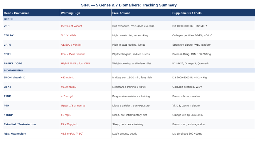
The biomarkers covered in this article — vitamin D, CTX-I, P1NP, PTH, hsCRP, sex hormones, and magnesium — are measurable, modifiable, and directly connected to the biology of bone quality. The genetic variants — VDR, COL1A1, LRP5, ESR1, and the RANKL/OPG axis — reveal structural tendencies that can be compensated for once identified. Neither list requires expensive or exotic interventions. Most of the plans described here involve food, exercise, targeted supplementation, and sleep — combined with the kind of precision that makes the difference between generic advice and an approach that actually fits your biology.
The next smart step is not a supplement order — it is a blood draw. Start with 25-OH vitamin D, CTX-I, P1NP, and hsCRP at minimum, and bring the results to a physician who understands bone metabolism. Add genetic testing if you want the deeper layer. Then build from what you find. Better information makes better decisions possible — and in bone health, that distinction can determine whether the next decade looks like progressive decline or genuine recovery.
Musculoskeletal Endocrine & Metabolic
Musculoskeletal: Bone Conditions Joint Conditions
Autoimmune: Inflammatory Conditions