This article contains AI generated content.
Subchondral Insufficiency Fracture of the Patella: 3 Genes and 7 Biomarkers to Track
If you've been told you have a subchondral insufficiency fracture of the patella, you've probably already noticed how thin the explanations tend to be. You get a diagnosis, maybe a brace, maybe "stay off it for six to eight weeks," and a vague reassurance that it should heal. What you rarely get is an answer to the question that actually matters to you: why did this happen in a bone that wasn't supposed to fail under normal load, and what, specifically, makes your case more or less likely to recur or heal well.
Generic advice exists because it has to apply to everyone, which means it applies precisely to no one. "Rest it and take vitamin D" is not wrong, but it skips past the fact that bone fragility is not one thing. It can come from a vitamin D and calcium problem, a collagen quality problem, a bone remodeling imbalance, a mechanical loading problem around the kneecap, or some combination that only shows up when you actually measure it. Two people with the same MRI report can have completely different underlying reasons their bone gave out.
This article takes the more specific route. Instead of repeating "rest, ice, see an orthopedist," it walks through the biomarkers that reveal what's actually happening inside your bone metabolism, the genes that shape your baseline fragility risk, a set of ideas from longevity-focused medicine that challenge how bone health is normally handled, and a short list of complementary approaches that have real, condition-relevant human evidence behind them, not just wellness-industry noise.
None of this promises a fix. Bone healing is a biological process with a timeline that supplements and biomarkers don't override. But better information changes decisions: what to ask your orthopedist, what to test before assuming "it's just bad luck," and what to actually do differently while the bone repairs itself and afterward, so it doesn't happen again.
Summary
Here's the short version of what follows. Subchondral insufficiency fractures happen when normal, everyday loading overwhelms bone that is weaker than it should be, and the patella's fracture pattern often traces back to a handful of measurable things: how much active vitamin D your body actually uses, how fast your bone is being broken down versus rebuilt, and whether your collagen and Wnt-signaling genetics gave you a sturdier or more fragile starting point. The genetics section below looks at three genes worth knowing about, including what the current human evidence actually shows and what realistically can be done about each one, with and without supplements. The biomarker section goes further, laying out seven specific lab and imaging markers, what each one reveals about your fracture risk, how they're measured, what they cost, and concrete corrective plans depending on whether the result comes back favorable or not. Beyond that, you'll find a distillation of what Peter Attia's Outlive gets right about treating bone fragility as a training problem rather than a disease to catch later, plus a review of the complementary approaches, tai chi, yoga, gait biofeedback, and photobiomodulation, that have genuine clinical evidence behind them for knee bone and joint health. Together, this gives you a much clearer map of where your particular fracture risk is coming from and what you can actually do about it.
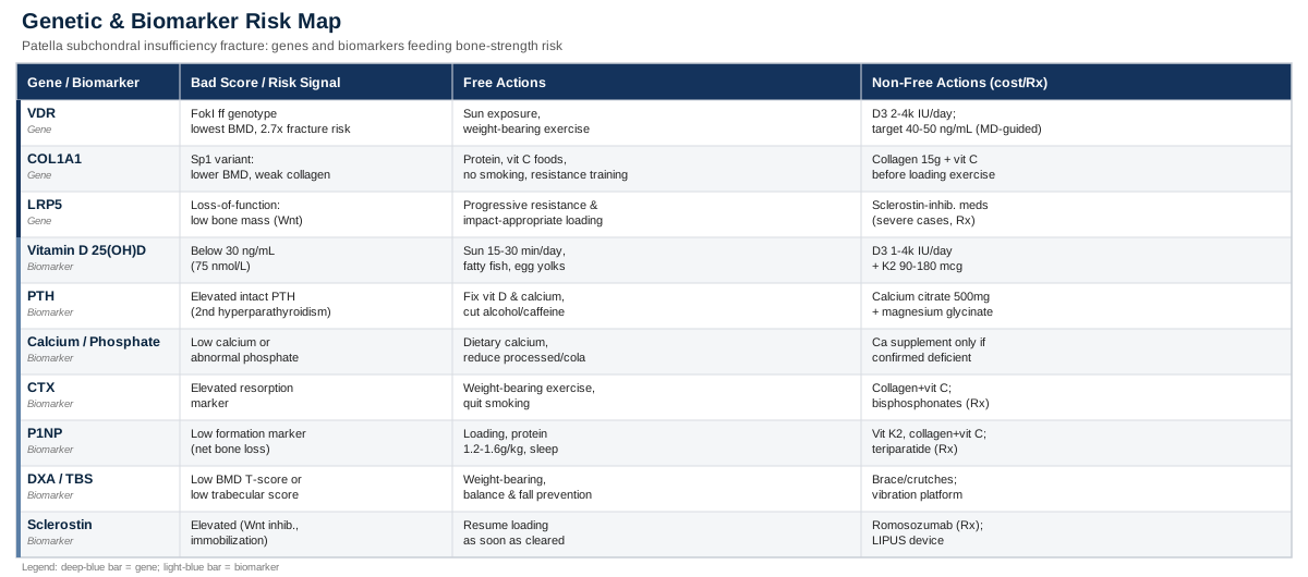
The 7 Biomarkers Worth Tracking for Patella Subchondral Insufficiency Fracture
Subchondral insufficiency fractures occur when repetitive, ordinary mechanical load exceeds what the underlying bone can absorb and repair, which is a different mechanism from a traumatic fracture caused by a single overwhelming force. That distinction matters because it points straight at bone quality and remodeling capacity as the thing to investigate, not just the joint itself. A recent review in Biomedicines on osteoporosis, osteoarthritis, and subchondral insufficiency fracture lays this out clearly: risk climbs with advancing age, female sex, meniscal damage, knee malalignment, and low vitamin D, and importantly, standard bone density scans can look deceptively normal in people whose bone is actually fragile, because osteophytes and sclerosis around an arthritic joint can inflate the reading (see the review here: Osteoporosis, Osteoarthritis, and Subchondral Insufficiency Fracture: Recent Insights). That's exactly why a single test is not enough. The seven biomarkers below, taken together, give a much more complete picture than any one of them alone.
1. Serum 25-Hydroxyvitamin D (25(OH)D)
This is the standard measure of vitamin D status, and it's the single most consistently cited risk factor for subchondral insufficiency fracture in the clinical literature. Low vitamin D impairs intestinal calcium absorption, which forces the parathyroid gland to pull calcium from bone to keep blood levels stable, quietly weakening the exact trabecular bone that supports the patellar subchondral plate.
How to measure it
A standard 25(OH)D blood draw, available through any primary care visit or direct-to-consumer lab panel. Cost typically runs from 15 to 60 USD/EUR out of pocket, less if covered by insurance. Optimal ranges for bone health are generally cited around 30 to 50 ng/mL (75 to 125 nmol/L).If the result is low: the plan without supplements
Regular, sensible sun exposure (roughly 15 to 30 minutes of midday skin exposure several times a week, adjusted for skin tone and latitude), and dietary sources like fatty fish, egg yolks, and fortified dairy. This is slower and less reliable than supplementation, especially in winter months or higher latitudes, so retesting after 8 to 12 weeks is important before assuming it worked.If the result is low: the plan with supplements or equipment
Vitamin D3, typically 1,000 to 4,000 IU daily depending on how low the baseline is and body weight, taken continuously rather than in large infrequent doses. Pairing with vitamin K2 (MK-7, roughly 90 to 180 mcg/day) is commonly recommended to help direct calcium toward bone rather than soft tissue. Retest at 3 months. Side effects are rare at these doses, but sustained intake above 10,000 IU/day without monitoring can cause hypercalcemia, so this should be dose-adjusted against actual lab values, not guessed.2. Intact Parathyroid Hormone (PTH)
PTH is the feedback signal that reveals whether your body is actively robbing bone to maintain blood calcium. A high PTH alongside low-normal vitamin D is the classic secondary hyperparathyroidism pattern seen in insufficiency fracture cases, and it's often missed because vitamin D is tested alone.
How to measure it
Intact PTH blood test, usually ordered alongside calcium and vitamin D. Cost is around 30 to 80 USD/EUR where not covered by insurance.If the result is high: the plan without supplements
Correcting vitamin D and dietary calcium intake usually normalizes PTH on its own within a few months. Reducing excess alcohol and caffeine, which both interfere with calcium handling, and confirming kidney function is normal (since impaired kidneys can independently raise PTH) are the non-supplement levers.If the result is high: the plan with supplements or equipment
Vitamin D3 correction as above, plus calcium from food first; if dietary intake is genuinely insufficient, calcium citrate at 500 to 600 mg per dose (better absorbed with food, split across the day rather than one large dose) and magnesium glycinate (200 to 400 mg/day, since magnesium is a cofactor for vitamin D metabolism). Recheck PTH after 3 months. Overshooting calcium supplementation without a documented deficiency is associated with kidney stone risk, so this is a correction, not a baseline habit.3. Corrected Calcium and Phosphate
These two, read together, distinguish a straightforward vitamin D deficiency from a more complex mineral metabolism problem, such as osteomalacia or a kidney-driven phosphate issue, either of which can independently soften bone and predispose it to insufficiency fracture.
How to measure it
Part of any basic metabolic panel, roughly 10 to 30 USD/EUR, and often already included in a standard blood panel your doctor orders.If the result is abnormal: the plan without supplements
Increase dietary calcium (dairy, leafy greens, tofu, canned fish with bones) if low, and reduce heavily processed foods and cola-type sodas if phosphate is elevated, since these are major dietary phosphate sources. If malabsorption (celiac disease, low stomach acid, prior bariatric surgery) is suspected, that needs its own workup rather than more calcium.If the result is abnormal: the plan with supplements or equipment
Supplement calcium only when dietary intake is confirmed insufficient and levels are actually low, not preemptively. Persistent phosphate abnormalities warrant referral rather than self-supplementation, since they can reflect kidney or parathyroid pathology that supplements won't touch.4. CTX (C-Terminal Telopeptide) — Bone Resorption Marker
CTX reflects how much bone is currently being broken down. In the context of an insufficiency fracture, an elevated CTX suggests your bone turnover is skewed toward resorption faster than formation can keep up, which is precisely the imbalance that produces fragile subchondral bone.
How to measure it
A specialty blood test, best drawn fasting and in the morning since it has diurnal variation. Cost ranges from 40 to 100 USD/EUR, more accessible through longevity-focused labs or functional medicine panels than routine primary care.If the result is elevated: the plan without supplements
Once cleared by your treating clinician for progressive loading (not during the acute non-weight-bearing phase), controlled resistance and weight-bearing exercise shifts the resorption/formation balance toward formation. Smoking cessation and limiting alcohol also measurably reduce resorption markers.If the result is elevated: the plan with supplements or equipment
Correcting vitamin D, calcium, and magnesium as above addresses the most common upstream driver. Collagen peptides (10 to 15 g/day) with vitamin C have some supporting data for collagen matrix synthesis. If resorption remains high despite correction, antiresorptive medication (bisphosphonates) is a physician-directed decision, not a supplement one, and should be discussed rather than self-initiated.5. P1NP (Procollagen Type I N-Terminal Propeptide) — Bone Formation Marker
P1NP is the mirror image of CTX: it reflects new bone formation. Read alongside CTX, it tells you whether your bone is in a net-building or net-losing state, which is far more informative than either marker alone. A trial testing vitamin D3 supplementation directly measured both bALP, CTX, osteocalcin, and P1NP together and found that correcting vitamin D alone, without addressing load and protein status, didn't reliably move these markers in already-replete adults (see Effects of Vitamin D Supplementation on Bone Turnover Markers: A Randomized Controlled Trial), which is a useful reminder that vitamin D correction is necessary but not sufficient on its own.
How to measure it
Blood test, similar cost range to CTX, 40 to 100 USD/EUR, ideally ordered as a pair with CTX from the same draw.If the result is low: the plan without supplements
Adequate mechanical loading appropriate to your healing stage, sufficient total calories and protein (roughly 1.2 to 1.6 g/kg bodyweight/day) to avoid an energy deficit that suppresses bone formation, and enough sleep, since much of bone remodeling activity is tied to overnight growth hormone pulses.If the result is low: the plan with supplements or equipment
Vitamin K2 and collagen/vitamin C as above support the formation side specifically. In cases of confirmed osteoporosis-driven fracture with persistently low formation markers, anabolic agents like teriparatide exist, but these are prescription, physician-supervised interventions, not something to source independently.6. DXA Bone Mineral Density and Trabecular Bone Score (TBS)
A standard DXA scan gives you a T-score/Z-score at the hip and spine, but as the Biomedicines review notes, DXA can be misleadingly reassuring around an arthritic joint due to sclerosis and osteophyte formation inflating the reading. Trabecular Bone Score, calculated from the same scan, adds information about bone microarchitecture quality that density alone misses.
How to measure it
DXA scans cost roughly 100 to 400 USD/EUR without insurance, often less or fully covered depending on age and risk factors. Not every center offers TBS software; ask specifically, since it requires additional analysis of the same scan.If the result is low: the plan without supplements
Weight-bearing and resistance exercise once cleared, balance and fall-prevention training (tai chi has real evidence here, covered later), and eliminating smoking and excess alcohol, both independently linked to lower bone density.If the result is low: the plan with supplements or equipment
Calcium and vitamin D correction as the foundation. During the active fracture phase, offloading equipment, an unloader brace or crutches as prescribed, is the standard of care, not optional. Whole-body vibration platforms have some early research interest for bone density but the evidence remains preliminary; treat it as a possible adjunct, not a replacement for loading and nutrition basics.7. Sclerostin
Sclerostin is produced by osteocytes and acts as a brake on the Wnt signaling pathway that drives new bone formation. Higher sclerostin means more inhibition of bone building, and levels rise with immobilization, aging, and mechanical unloading, all of which are directly relevant during recovery from a patella insufficiency fracture, when weight-bearing is deliberately restricted. A review of osteocyte-related biomarkers found sclerostin has the most consistent association with bone mineral density among the newer markers, though its ability to predict fracture specifically is still mixed across studies (A Review of the Potential Application of Osteocyte-Related Biomarkers, Fibroblast Growth Factor-23, Sclerostin, and Dickkopf-1 in Predicting Osteoporosis and Fractures).
How to measure it
Not yet a routine clinical test; available through specialty and research-oriented labs, typically 100 to 250 USD/EUR. Treat this as the most experimental marker on this list, useful for context, less useful for standalone decision-making.If the result is elevated: the plan without supplements
Minimizing unnecessary prolonged immobilization and returning to controlled mechanical loading as soon as your clinician clears it, since loading is the primary natural counter-signal to sclerostin's Wnt inhibition.If the result is elevated: the plan with supplements or equipment
No supplement reliably lowers sclerostin. The prescription antibody romosozumab directly inhibits sclerostin and is used in severe osteoporosis, but it is a physician-prescribed medical therapy, not a self-directed option. Low-intensity pulsed ultrasound (LIPUS) devices are sometimes used for delayed fracture healing and have research interest in modulating this same signaling pathway, discussed further in the complementary approaches section below.Taken as a set, these seven markers separate a straightforward "fix the vitamin D and calcium" case from one that needs a longer conversation about collagen quality, remodeling imbalance, or genuine bone microarchitecture fragility, which is exactly where genetics adds the missing context.
The Genetic Side: 3 Genes Linked to Bone Fragility and Insufficiency Fracture
Genetics doesn't determine your outcome, but it does set the baseline your biomarkers and habits are working against. Researchers like Ali Torkamani, whose work at Scripps Research focuses on polygenic risk scoring, have shown that bone mineral density is highly polygenic: a large genome-wide study identified over 600 loci contributing to heel bone density and built a polygenic score from them for predicting osteoporosis and fracture risk (Identification of 613 new loci associated with heel bone mineral density and a polygenic risk score for bone mineral density, osteoporosis and fracture). The three genes below are among the best-studied individual contributors, though it's worth being upfront that most single-gene studies involve smaller cohorts than the polygenic work, so treat individual gene findings as suggestive risk modifiers, not deterministic labels.
VDR — Vitamin D Receptor
The VDR gene encodes the receptor through which vitamin D actually exerts its effects on bone cells; having enough circulating vitamin D doesn't help much if the receptor doesn't respond efficiently to it. The FokI polymorphism is the most studied variant, and research in postmenopausal women found it to be the strongest single genetic determinant of bone mineral density among the genes tested, with the "ff" genotype associated with the lowest density values, and separately, a roughly 2.7-fold increase in stress fracture risk associated with the less favorable allele (Association between bone mineral density and polymorphisms of the VDR, ERα, COL1A1 and CTR genes in Spanish postmenopausal women).
If the gene is unfavorable: the plan without supplements
Since the issue is receptor sensitivity rather than vitamin D supply alone, consistent sun exposure and dietary vitamin D remain useful, but don't assume "normal" lab values mean the signal is being used efficiently. Weight-bearing exercise activates bone formation through mechanotransduction pathways that don't depend solely on VDR signaling, making it a reasonable independent lever.If the score is unfavorable: the plan with supplements or equipment
Some clinicians targeting a less-sensitive VDR variant aim for the higher end of normal serum 25(OH)D (40 to 50 ng/mL) rather than just clearing the deficiency threshold, using 2,000 to 4,000 IU/day of D3 continuously, with quarterly retesting rather than assuming the dose is working. This is a reasonable discussion to have with your clinician, not a self-directed mega-dosing plan; excess vitamin D without monitoring risks hypercalcemia regardless of genotype.COL1A1 — Collagen Type I Alpha 1
COL1A1 encodes the main structural protein of bone matrix. The Sp1 binding site polymorphism has been repeatedly linked to lower bone mineral density and higher fracture risk, including in a case report where a young man with unexplained multiple fragility fractures was found to carry the less favorable Sp1 genotype combined with a VDR FokI variant (Association of COL1A1 Sp1 and Fok-I VDR Genetic Polymorphisms in Young Male Idiopathic Osteoporosis), and separately in postmenopausal women, where the polymorphism was associated with bone mineral density differences (Collagen Type I alpha1 (COL1A1) Gene Polymorphism and Bone Mineral Density in Postmenopausal Kazakh Women). This is a structural quality issue, not just a density issue, meaning bone can look reasonably dense on a scan while still being mechanically weaker than expected.
If the gene is unfavorable: the plan without supplements
Adequate dietary protein to supply amino acid building blocks for collagen, vitamin C-rich foods (needed for collagen crosslinking enzymes), avoiding smoking (which directly impairs collagen crosslinking), and resistance training, which stimulates collagen turnover in bone and connective tissue generally.If the score is unfavorable: the plan with supplements or equipment
Collagen peptides combined with vitamin C, taken roughly 30 to 60 minutes before loading exercise, is a protocol with some supporting human data for enhancing collagen synthesis around connective tissue loading. A reasonable approach is 15 g of collagen or gelatin with 50 mg vitamin C, on training days only rather than continuously, since the synthesis benefit is tied to the loading stimulus that follows it. Side effects are minimal beyond occasional mild GI upset. If fracture healing is delayed, LIPUS devices are sometimes used under medical guidance, discussed below.LRP5 — LDL Receptor-Related Protein 5
LRP5 is a co-receptor in the Wnt signaling pathway that drives osteoblast activity and bone formation. Loss-of-function mutations cause severe low bone mass conditions, while gain-of-function variants cause unusually high bone mass, and common variants in the general population contribute meaningfully to the polygenic bone density risk scores described above. This is the same pathway that sclerostin inhibits, which is why the two are discussed together.
If the gene is unfavorable: the plan without supplements
Mechanical loading is the primary natural activator of Wnt/LRP5 signaling, meaning progressive resistance and impact-appropriate exercise (once cleared for your fracture stage) is arguably the single most direct non-pharmacological lever for this specific pathway.If the score is unfavorable: the plan with supplements or equipment
No supplement directly targets LRP5 signaling. The foundational vitamin D, K2, and calcium correction described earlier still matters, since this pathway operates within the same overall bone-mineral environment. For severe cases where this pathway is significantly impaired, sclerostin-inhibiting medication works downstream of the same signaling axis, but again, that's a physician-directed medical decision, not a supplement strategy.With the biology and the genetics covered, it's worth looking at how longevity medicine is starting to reframe bone fragility altogether, since it changes when and how aggressively these markers get acted on.
What Peter Attia's Outlive Gets Right About Bone Fragility (And Why Your Orthopedist Might Not Mention It)
Peter Attia's book Outlive built a following partly because it treats bone health as a training and monitoring problem to get ahead of decades early, rather than a diagnosis to wait for. That framing is directly useful for anyone dealing with a subchondral insufficiency fracture, because it reframes the fracture as a signal to investigate systemically, not just a local injury to splint and forget. Here are the ten most useful ideas from that framework.
1. The "Marginal Decade" Is Decided Long Before You Feel It
Attia's concept of the marginal decade, the final years of life where physical capability determines quality of life, argues that bone and muscle reserves built (or lost) in your 40s, 50s, and 60s are what determine whether that decade is spent independent or in decline. An insufficiency fracture in your 40s or 50s is an early data point, not an isolated event.2. A Hip or Femur Fracture After 65 Is a Mortality Event, Not Just an Injury
Outlive cites mortality following hip and femur fractures in people over 65 as high as 15 to 36 percent within a year, meaning roughly a third of people in the worst cohorts do not survive twelve months after the fracture. That statistic is the entire justification for treating bone fragility seriously well before an "insufficiency fracture" shows up in a weight-bearing bone.3. DEXA Should Be Routine, Not Reactive
Attia has described measuring bone mineral density via DEXA at both hips and the lumbar spine annually in his patients, rather than waiting for a fragility fracture to trigger the first scan. Applied here, that means anyone who has already had one insufficiency fracture has a strong case for annual DEXA monitoring going forward, not a one-time scan.4. Bone Density Numbers Can Mislead Without Microarchitecture Data
A DEXA T-score alone can look acceptable while trabecular quality is actually compromised, which is exactly the pattern the Biomedicines review flagged around arthritic joints. This lines up with Attia's broader point that a single biomarker rarely tells the whole story; it's the combination that matters.5. Muscle Is Bone's Insurance Policy
Strength training doesn't just build muscle; it applies the mechanical load that stimulates bone formation directly, and stronger muscles around a joint reduce the erratic loading patterns that contribute to localized bone stress. For a knee already dealing with subchondral fracture, quadriceps and hip strength are protective, not incidental.6. Stability Training Prevents the Fall That Causes the Fracture
A large share of fragility fractures follow a fall, not spontaneous failure under normal load. Balance and stability work reduces fall risk independently of bone density itself, which is why it's treated as a core pillar alongside strength and cardiovascular training rather than an optional extra.7. Zone 2 Training Supports the Metabolic Environment Bone Depends On
Sustained low-intensity aerobic training improves mitochondrial and metabolic health broadly, which supports the energy availability bone remodeling requires. It's a slower, less direct lever than strength training for bone specifically, but it supports the overall physiological environment recovery depends on.8. Protein Intake Requirements Are Higher Than Standard Guidelines Suggest
Attia advocates for protein intake well above the general population RDA, particularly for anyone recovering from an injury or actively trying to preserve muscle and bone matrix with age. This aligns directly with the protein targets mentioned earlier for supporting collagen and bone formation markers.9. Waiting for a Doctor to Flag Osteoporosis Is Reactive, Not Preventive
The conventional path is to wait for a DEXA scan ordered after age 65 or after a fracture. Attia's argument is that by the time a fragility fracture happens, meaningful bone loss has usually been underway for years; a subchondral insufficiency fracture at any age is a reasonable trigger to start monitoring now rather than waiting for an official screening age.10. Reframe Bone Health As a Training Goal, Not a Disease to Treat Later
The overarching shift Outlive argues for is treating bone density and strength the way you'd treat a fitness goal: measured, trained for, and tracked over years, rather than a diagnosis that only gets attention once it fails. For someone recovering from a patella insufficiency fracture, that means the biomarkers and genes above are a starting map, not a one-time checklist.That training-first mindset pairs naturally with a set of complementary approaches that have specific, condition-relevant human evidence, which is where the practical, hands-on part of recovery comes in.
Complementary Approaches With Real Evidence Behind Them
Not every complementary approach marketed for knee or bone health has solid evidence behind it, so this list sticks to a small number with human clinical data specific to knee osteoarthritis, knee loading, or bone healing, since subchondral insufficiency fracture of the patella sits at the intersection of both.
Tai Chi
Tai chi combines slow, weight-shifting movement with balance and proprioceptive training, which is directly relevant here because it improves lower-limb balance and control without the high impact loading that could aggravate a healing subchondral fracture. It also builds the kind of controlled joint loading that supports bone and muscle without the ballistic forces of running or jumping.A well-designed 52-week randomized trial comparing tai chi directly against standard physical therapy in 204 people with knee osteoarthritis found tai chi produced benefits comparable to physical therapy for pain and function, with additional improvements in depression and quality of life (Comparative Effectiveness of Tai Chi Versus Physical Therapy for Knee Osteoarthritis: A Randomized Trial).
Realistically, this fits best once you're cleared for weight-bearing movement, not during strict non-weight-bearing recovery. A beginner class two to three times a week, or a instructor-guided routine at home, is a reasonable starting cadence; there are no meaningful side effects beyond the usual caution around any new movement practice while a fracture is still healing.
Yoga
Yoga's relevance here comes from its combination of controlled range-of-motion work, isometric strengthening around the knee, and general stress and pain-coping benefits, all useful during a slow bone-healing process that can otherwise feel frustratingly passive.A randomized controlled trial of 250 people with knee osteoarthritis found that an integrated yoga therapy program added to standard physiotherapy produced significantly greater improvements in quality of life, including pain and physical functioning, compared to standard exercise therapy alone (Effect of an integrated approach of yoga therapy on quality of life in osteoarthritis of the knee joint: A randomized control study).
Apply this cautiously: poses that place direct compressive load on a bent, weight-bearing knee (deep kneeling positions, for instance) should be modified or avoided until your treating clinician confirms the fracture has progressed enough to tolerate it. A gentle, modified practice two to three times weekly is a reasonable starting point.
Biofeedback-Based Gait Retraining
This is one of the more directly mechanistic options on this list, since it targets the actual loading pattern crossing the knee joint during walking, which is highly relevant to a patella subchondral fracture, where abnormal patellofemoral loading is often part of the underlying problem.A randomized controlled trial protocol tested real-time biofeedback on either gait pattern or knee moment during treadmill walking over six weeks in people with knee osteoarthritis, aiming to reduce knee adduction moment, a key driver of joint loading, alongside pain and functional improvements (The effectiveness of a 6-week biofeedback gait retraining programme in people with knee osteoarthritis: protocol for a randomised controlled trial).
This requires access to a physical therapist with gait analysis or biofeedback equipment, which isn't universally available, but where it is, a structured six-week program under professional supervision is realistic and directly targets the mechanical loading problem rather than just symptoms.
Low-Level Laser Therapy / Photobiomodulation
Photobiomodulation is being explored specifically for its effects on the early cellular phases of bone healing, angiogenesis and osteoblast activity, which makes it conceptually relevant to a healing insufficiency fracture, though this is the least established option on this list.A systematic review of low-level laser therapy in bone healing found accelerated healing markers in the first two to four weeks post-fracture in some studies, but no consistent advantage over standard care by around 60 days, and the authors were explicit that dosing protocols vary too widely across studies to draw firm conclusions (The Role of Low-Level Laser Therapy in Bone Healing: Systematic Review).
Given the mixed and early-stage evidence, this is best treated as a possible adjunct discussed with your treating physician, delivered by a qualified provider with an appropriate device, rather than a primary strategy or something to pursue with consumer-grade devices expecting a guaranteed effect.
Conclusion
A subchondral insufficiency fracture of the patella is rarely just bad luck. It's usually the visible result of an invisible imbalance, somewhere between vitamin D and calcium handling, bone turnover rate, collagen quality, and inherited bone density risk, that finally showed itself under ordinary load. The seven biomarkers above give you a concrete way to find out which of those factors is actually driving your case, rather than guessing. The genetic variants add context for why your baseline risk might be higher even when your labs look reasonable, and the training-first mindset from longevity medicine, paired with the complementary approaches that have genuine evidence behind them, gives you a realistic plan for both healing well now and reducing the odds of this happening again.
The most useful next step is a practical one: ask your clinician to order the biomarker panel above if it hasn't been done already, get a DEXA scan with TBS if you haven't had one, and track how your numbers change as you recover. That combination of information and a qualified professional's judgment is a far better foundation than waiting to see what happens next.
Musculoskeletal: Bone Conditions Joint Conditions