This article contains AI generated content.
· UpdatedSubchondral Stress Reaction: 5 Genes and 6 Biomarkers to Track
Introduction
You get the MRI report back and it mentions bone marrow edema or a subchondral stress reaction. Your X-ray looked fine. The joint aches, sometimes sharply, sometimes as a dull pressure you can't shake. You are told to rest. Maybe to stop running or training for a while. The clinical picture is incomplete, and so is the advice you receive.
The standard protocol for subchondral stress reactions — relative rest, anti-inflammatory medication, gradual return to activity — works reasonably well for many people. But it does not explain why two people with near-identical scans heal at completely different rates. One athlete is back training in six weeks; another is still symptomatic six months later and is no closer to understanding why. The difference is rarely effort or compliance. It is often biology.
What happens inside the subchondral bone — the dense layer just beneath joint cartilage — depends on how your body manages bone remodeling, cartilage matrix turnover, and local inflammation. These processes are shaped both by measurable biochemical signals circulating in your blood and urine, and by genetic variants that quietly determine how your cells respond to mechanical stress and repair damage. Neither of these dimensions shows up in a standard imaging report or a routine orthopedic consultation.
This article takes a more useful approach. It introduces six blood and urine biomarkers that can give you objective, trackable insight into what is happening in your bones and joints right now, as well as five genetic factors that influence your individual predisposition to subchondral injury and repair. Understanding both opens a clearer path — not to a cure, but to better-informed decisions, smarter recovery strategies, and a real basis for progress.
Summary
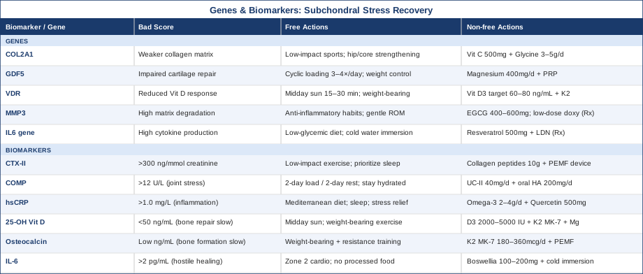
This article covers two parallel frameworks for understanding subchondral stress reactions beyond the MRI image. The first is biomarker tracking: six measurable markers — including CTX-II for cartilage breakdown, COMP for joint loading stress, hsCRP for systemic inflammation, 25-OH Vitamin D, osteocalcin, and IL-6 — that reveal what is actually happening at the tissue level and how well recovery is progressing. For each marker, you will find how to measure it, what a bad result means, and concrete plans both with and without supplementation.
The second framework is genetics: five key gene variants — COL2A1, GDF5, VDR, MMP3, and IL6 — that help explain why some people are more vulnerable to subchondral injury and slower to heal. Practical compensation strategies are included for each. Beyond these two core sections, the article also covers a paradigm-shifting podcast perspective on bone health that challenges common medical assumptions, and four complementary therapeutic approaches with genuine clinical evidence for musculoskeletal recovery.
---
6 Biomarkers to Track When You Have a Subchondral Stress Reaction
The imaging shows a lesion. What it cannot show is whether your cartilage is actively breaking down, whether bone remodeling is running at the right pace, or whether systemic inflammation is quietly prolonging healing. That is what biomarkers are for. These six markers are the most clinically relevant for tracking subchondral bone and cartilage health — together they give a functional picture no single scan can provide.
1. CTX-II — The Cartilage Breakdown Signal
What it is and why it matters
CTX-II (C-terminal cross-linking telopeptide of type II collagen) is a degradation fragment of type II collagen, the main structural protein in articular cartilage. When cartilage is under excessive mechanical stress or is breaking down, CTX-II is released into the urine. It is currently the most validated urinary biomarker for cartilage catabolism in humans, used in osteoarthritis research and increasingly in clinical monitoring.
For someone with a subchondral stress reaction, elevated CTX-II signals that the cartilage matrix overlying the stressed bone is not just under pressure — it is actively degrading. This changes the clinical picture significantly. A high CTX-II in the context of a bone marrow edema lesion suggests the joint is further along a degenerative pathway than the MRI alone might imply. Longitudinal tracking of CTX-II can also tell you whether a protocol is actually working, without waiting for a repeat MRI.
Levels correlate with disease progression in knee osteoarthritis. Research published in journals indexed on PubMed, including work from Garnero and colleagues, consistently shows that higher urinary CTX-II predicts faster joint space narrowing over two to four years.
How to measure it
CTX-II is measured in a second-void morning urine sample, typically corrected for creatinine to account for hydration. It is available through specialty labs (Quest Diagnostics, Genoptix, and some European lab networks). Cost typically ranges from $50–$150 USD depending on the lab and whether insurance covers it. Reference ranges vary by lab; a common cutoff for elevated risk is above 300 ng/mmol creatinine, though this is age- and sex-dependent.
If the score is bad, the plan without supplements
The single most effective non-supplement intervention for CTX-II is reducing high-impact mechanical loading on the affected joint while maintaining low-load movement. Pool walking, cycling, and swimming are preferred. Intermittent compression therapy (pneumatic compression boots) applied for 20–30 minutes daily appears to improve synovial fluid circulation without increasing cartilage stress.
Gait and movement mechanics matter: working with a physical therapist to identify compensatory loading patterns — excessive pronation, limb length discrepancy, or weak hip abductors — can reduce articular shear forces significantly. Studies on runners show that increasing step rate by 5–10% reduces knee joint loading and secondary cartilage stress.
Sleep quality is also relevant. Growth hormone peaks during slow-wave sleep and drives cartilage matrix synthesis. Prioritizing 7–9 hours of sleep and addressing sleep-disordered breathing directly impacts tissue regeneration capacity.
If the score is bad, the plan with supplements or equipment
Collagen peptides (10 g/day, 30–60 minutes before mechanical loading): The 2019 study by Shaw et al. (Amino Acids) showed that hydrolyzed collagen combined with vitamin C and mechanical loading increases collagen synthesis markers and lowers cartilage degradation signals. Frequency: daily, long-term (minimum 3 months). Side effects: minimal; some report mild GI discomfort at high doses.
Vitamin C (500–1000 mg/day): Required for hydroxylation of proline and lysine in collagen synthesis. Best taken around the time of collagen peptides. Cycling: daily, no specific cycling needed at these doses.
Pulsed electromagnetic field (PEMF) devices: FDA-cleared devices (brands such as Orthofix) have documented evidence for bone healing acceleration. Some studies suggest reduction in bone marrow edema on follow-up MRI. Typical protocol: 4–8 hours/day for 3–6 months. Cost: $400–$1500 for consumer-grade devices. No serious side effects documented at standard parameters.
---
2. COMP — The Joint Stress Indicator
What it is and why it matters
Cartilage Oligomeric Matrix Protein (COMP) is a large non-collagenous protein secreted by chondrocytes and synovial fibroblasts. It plays a structural role in the cartilage extracellular matrix and is acutely elevated in serum after mechanical joint loading. In the context of subchondral stress reactions, serum COMP is one of the most sensitive early indicators of acute cartilage distress — levels rise within 30 minutes of exercise and remain elevated for hours in damaged joints.
COMP is particularly useful because it responds to both acute loading and chronic degeneration. A person with a healing subchondral lesion should show post-exercise COMP spikes that decline appropriately over hours. If resting COMP remains elevated at follow-up, it suggests the joint is not coping with even baseline demands. Researchers at Lund University in Sweden and elsewhere have demonstrated COMP as a useful monitoring tool in early osteoarthritis and post-injury cartilage stress.
How to measure it
Serum COMP is typically measured via ELISA. Reference laboratories including Quest and ARUP offer it; cost is generally $80–$200. A resting morning sample is most interpretable. Levels above approximately 12 U/L (lab-dependent) in a younger adult suggest meaningful cartilage stress or damage. Ideally, measure at rest and again 2–4 hours after moderate exercise to see the loading response.
If the score is bad, the plan without supplements
Reduce loading frequency before loading intensity. COMP elevation during recovery often means the joint is being asked to absorb too many loading cycles before it has time to recover. A two-days-on, two-days-off cycle of load-bearing activity, rather than daily exercise, can allow COMP to normalize between sessions.
Joint hydration is meaningfully connected to COMP behavior. Synovial fluid viscosity declines with dehydration; ensuring adequate daily fluid intake (minimum 2.5 liters) and minimizing prolonged static positions helps maintain synovial lubrication and reduce frictional cartilage wear.
If the score is bad, the plan with supplements or equipment
Undenatured type II collagen (UC-II, 40 mg/day): Distinct from hydrolyzed collagen, UC-II works via oral tolerization. A randomized controlled trial by Lugo et al. (2016, Journal of the International Society of Sports Nutrition) showed significant reduction in joint discomfort and knee extension pain after exercise. Frequency: daily, long-term use (6+ months). Side effects: minimal.
Hyaluronic acid (oral, 200 mg/day): Improves synovial fluid viscosity. Meta-analyses support modest improvements in knee joint function in early OA. Cycling: daily for minimum 2 months. Note: evidence quality for oral HA is moderate; injectable HA has stronger evidence.
Infrared sauna or targeted thermotherapy: Heat improves synovial fluid circulation and may modulate COMP response. Sessions of 15–20 minutes at 55–65°C, three times per week. Avoid immediately post-exercise in acute flares.
---
3. High-Sensitivity CRP (hsCRP) — Systemic Inflammation
What it is and why it matters
hsCRP measures systemic low-grade inflammation with higher sensitivity than standard CRP. In subchondral stress reactions, systemic inflammation — even at subclinical levels — significantly impairs bone remodeling and cartilage repair. IL-1β and TNF-α, both downstream of elevated CRP, directly inhibit osteoblast activity and increase matrix metalloproteinase expression in chondrocytes.
Peter Attia has repeatedly emphasized hsCRP as one of the most important cardiovascular and metabolic biomarkers, but its relevance extends directly into musculoskeletal recovery. Resting hsCRP above 1.0 mg/L in a person with a joint injury should prompt investigation into its source before any sophisticated supplement protocol is started.
How to measure it
Standard blood test, widely available. Cost: $20–$60. Optimal range: below 0.5 mg/L. Below 1.0 mg/L is generally acceptable. Above 3.0 mg/L suggests meaningful systemic inflammation requiring investigation.
If the score is bad, the plan without supplements
Address dietary drivers first. Eliminate seed oils (linoleic acid burden), reduce refined carbohydrate intake, and shift toward a Mediterranean-pattern diet. A 2020 meta-analysis in Nutrients confirmed that Mediterranean dietary adherence reduces CRP by approximately 20–35% within 8–12 weeks.
Sleep optimization is the single most underused anti-inflammatory intervention. A single night of poor sleep elevates hsCRP by 12–25% in controlled studies. Targeting consistent sleep/wake timing, a cool sleep environment, and addressing sleep apnea reduces chronic CRP as reliably as many pharmacological interventions.
Chronic psychological stress is a major driver. High-cortisol states elevate NF-κB activity, which drives CRP synthesis. Structured stress reduction — even 10–15 minutes daily of diaphragmatic breathing — reduces cortisol reactivity over 8 weeks.
If the score is bad, the plan with supplements or equipment
Omega-3 fatty acids (EPA + DHA, 2–4 g/day combined): The most evidence-backed supplement for reducing hsCRP. A meta-analysis in the European Journal of Clinical Nutrition found significant CRP reduction with fish oil at doses of 2 g/day EPA+DHA or higher. Frequency: daily. Cycling: not necessary for standard doses. Side effects: GI discomfort at high doses; use enteric-coated capsules if needed.
Quercetin (500–1000 mg/day): Inhibits NF-κB. Human trials show modest CRP reduction. Prefer quercetin phytosome for improved absorption. Cycling: 8 weeks on, 2 weeks off. Side effects: mild GI effects.
Curcumin with piperine (1000 mg curcumin + 10 mg piperine/day): Robust evidence for CRP and IL-6 reduction in musculoskeletal conditions. Take with a fatty meal. Cycling: 12 weeks on, 4 weeks off. Side effects: blood thinning at very high doses; caution with anticoagulants.
---
4. 25-OH Vitamin D — Bone Repair Foundation
What it is and why it matters
Vitamin D deficiency is extraordinarily common and directly impairs subchondral bone healing. The VDR (vitamin D receptor) is expressed on osteoblasts, osteoclasts, and chondrocytes. Vitamin D regulates calcium absorption, modulates the RANK/RANKL axis governing bone remodeling, and has anti-inflammatory effects on synovial tissue.
Stress fractures and bone marrow edema lesions are significantly more common in individuals with 25-OH D below 30 ng/mL. Military studies in particular have documented a dose-response relationship: recruits with the lowest vitamin D levels have the highest rates of stress fractures during basic training. For subchondral stress reactions, this connection is direct.
How to measure it
Standard blood test, extremely affordable ($25–$60). Optimal range for bone and joint health: 50–80 ng/mL (125–200 nmol/L). Most functional medicine practitioners (Attia included) aim for above 50 ng/mL rather than the conventional threshold of 30 ng/mL.
If the score is bad, the plan without supplements
Regular midday sun exposure of 15–30 minutes on bare skin (arms and legs) increases 25-OH D production by 1,000–4,000 IU per session depending on skin type, season, and latitude. This is the most physiologically natural approach. Track levels quarterly to confirm response.
Weight-bearing exercise itself modestly elevates vitamin D metabolism and increases VDR expression in bone tissue — another reason that complete immobilization is suboptimal in mild-to-moderate subchondral stress reactions.
If the score is bad, the plan with supplements or equipment
Vitamin D3 (2,000–5,000 IU/day with K2 MK-7 at 100–200 mcg/day): The K2 is critical — it directs calcium into bone rather than soft tissue. Take with the largest fat-containing meal of the day. Retest in 90 days. Many people need 5,000 IU to reach 50 ng/mL. Cycling: not necessary at standard doses; avoid doses above 10,000 IU/day without monitoring. Side effects: hypercalcemia at very high doses (>10,000 IU/day without K2 and monitoring).
Magnesium glycinate (300–400 mg/day at night): Required for vitamin D activation and conversion to its active form (1,25-OH D). Most people who are vitamin D deficient are also magnesium insufficient. Magnesium also directly improves sleep quality — a co-benefit relevant to recovery. Side effects: loose stools if taking magnesium oxide; glycinate or malate forms are well tolerated.
---
5. Osteocalcin — Bone Formation Rate
What it is and why it matters
Osteocalcin is secreted by osteoblasts during bone matrix formation and is the primary marker of bone formation activity. In subchondral stress reactions, the clinical question is not just whether bone is resorbing too fast, but whether it is rebuilding adequately. Osteocalcin answers the formation side of the equation.
Beyond its role in bone physiology, osteocalcin has emerged — through research by Gerard Karsenty at Columbia — as a genuine metabolic hormone that influences insulin sensitivity, muscle function, and even cognition. Low osteocalcin is not merely a sign of slow bone turnover; it reflects a broader failure of bone-mediated metabolic signaling. This is relevant for recovery because muscle function and joint stability both depend on metabolic health.
How to measure it
Serum osteocalcin, measured via blood draw. Available through most reference labs at $40–$100. Normal adult range varies widely by lab and age; generally 10–46 ng/mL in adults. Low values suggest inadequate bone formation; very high values may indicate accelerated turnover (seen in conditions like Paget's disease or hyperparathyroidism).
If the score is bad, the plan without supplements
Load-bearing exercise is the most powerful stimulus for osteoblast activity. Even in the context of a stress reaction, partial loading — within pain-free limits — stimulates osteocalcin production. Zero loading (bed rest) consistently suppresses it. Aquatic exercise and cycling do not adequately stimulate osteocalcin; brief bouts of weight-bearing are needed.
Resistance training with compound movements (squats, deadlifts at low intensity) elevates osteocalcin acutely and over time. Even one or two brief sessions per week maintains the osteoblast signal.
If the score is bad, the plan with supplements or equipment
Vitamin K2 MK-7 (180–360 mcg/day): Directly carboxylates osteocalcin, converting inactive undercarboxylated osteocalcin to its active form. Studies consistently show K2 improves osteocalcin carboxylation and bone mineral density. Take with fat. Long-term use; no standard cycling needed. Side effects: theoretical interaction with warfarin — check with prescribing physician.
PEMF therapy: Stimulates osteoblast proliferation and increases osteocalcin output. Documented in multiple controlled trials at bone fracture sites. Same protocol as noted under CTX-II.
---
6. IL-6 — Local and Systemic Inflammatory Amplifier
What it is and why it matters
Interleukin-6 (IL-6) is a pleiotropic cytokine with both pro-inflammatory and anti-inflammatory roles depending on context. In joint injury and subchondral bone stress, chronically elevated IL-6 activates osteoclastogenesis (accelerating bone resorption), suppresses collagen synthesis in chondrocytes, and perpetuates synovial inflammation.
Thomas Dayspring and Peter Attia both include IL-6 in advanced cardiometabolic panels, but its relevance to musculoskeletal recovery is equally important. Elevated resting IL-6 in a person recovering from a subchondral stress reaction is a sign that the healing environment is actively hostile to tissue repair.
How to measure it
Serum IL-6 via blood draw, often part of a cytokine panel. Cost: $50–$150. Optimal fasting resting level: below 2 pg/mL. Above 5 pg/mL in a person with a joint injury warrants dietary, lifestyle, and potentially pharmacological attention.
If the score is bad, the plan without supplements
Visceral adiposity is the most powerful driver of chronically elevated IL-6. Each kilogram of visceral fat loss typically reduces IL-6 by a measurable amount. Caloric deficit of 300–500 kcal/day combined with resistance training is more effective than aerobic exercise alone for reducing IL-6. Zone 2 cardio (3–5 sessions per week of 30–45 minutes at conversational pace) specifically targets visceral fat and has documented IL-6-lowering effects.
Ultra-processed foods, especially refined sugars and trans fats, acutely elevate IL-6 within hours of consumption. A dietary elimination trial — removing processed food for four weeks — often produces significant cytokine reduction before any supplements are added.
If the score is bad, the plan with supplements or equipment
Boswellia serrata extract (AKBA fraction, 100–200 mg/day): Inhibits 5-LOX, reducing leukotriene synthesis and IL-6 downstream. A randomized trial in Phytomedicine showed significant IL-6 and CRP reduction in knee OA patients at 90 days. Cycling: 12 weeks on, 4 weeks off. Side effects: minimal; rare GI upset.
Cold water immersion (10–15°C for 5–10 minutes, 3×/week): Acutely reduces pro-inflammatory cytokines including IL-6 post-exercise. Multiple RCTs document this effect. Not a substitute for dietary change, but a meaningful adjunct with no cost beyond a cold shower or bathtub.
---
Moving from what you can measure to what you inherit, the next section explores the genetic layer beneath these biomarkers — and what can be done about it.
The Genetic Layer: 5 Genes That Shape Subchondral Bone Vulnerability
Genetic variants do not determine outcomes. They adjust probabilities. Understanding your genetic profile in relation to subchondral stress reactions allows you to personalize your recovery and prevention strategy rather than following advice designed for the average person. Work by Ali Torkamani at Scripps Research and the broader field of nutrigenomics has made this kind of personalization increasingly accessible.
Gene 1: COL2A1 — Type II Collagen Integrity
What this gene does
COL2A1 encodes the alpha-1 chain of type II collagen, the predominant structural protein in articular cartilage and subchondral bone transition zones. Variants in COL2A1 are associated with primary osteoarthritis of multiple joints, reduced collagen matrix stability, and increased vulnerability to cartilage fissuring under repetitive mechanical stress.
This is not a rare Mendelian mutation — common variants in and around COL2A1 have been identified in genome-wide association studies (GWAS) as contributors to osteoarthritis risk, with effects of modest individual size but meaningful impact when combined with high mechanical loading lifestyles.
If the gene variant is unfavorable — plan without supplements
The most important intervention is modifying loading mechanics before cumulative stress degrades an already suboptimal collagen matrix. This means: transitioning from high-impact sports (running on hard surfaces, basketball, court sports) to lower-impact alternatives for long-term joint health. Cyclic compression rather than impact loading — elliptical, cycling, swimming — provides the mechanical signaling joints need without the peak loading that degrades weaker collagen scaffolding.
Physical therapy focused on proprioception and periarticular muscle strengthening reduces articular surface stress by improving dynamic joint stabilization. Dedicated hip and core strengthening reduces knee and ankle joint loading by 10–20% in published biomechanical studies.
If the gene variant is unfavorable — plan with supplements or equipment
Vitamin C (500–1000 mg/day with collagen peptides): Essential cofactor for collagen cross-linking. For someone with COL2A1 variants, maintaining maximal collagen synthesis capacity is particularly important. Long-term daily use. Side effects: GI at very high doses (>2 g/day).
Glycine supplementation (3–5 g/day, added to food or drink): Type II collagen is approximately 33% glycine. Supplementing glycine provides building blocks for matrix synthesis, particularly relevant when COL2A1 transcription is suboptimal. No known cycling requirements. Well-tolerated.
---
Gene 2: GDF5 — Joint Morphology and Repair Potential
What this gene does
GDF5 (Growth Differentiation Factor 5) is a member of the TGF-β/BMP superfamily and plays a central role in joint development, articular cartilage maintenance, and tendon/ligament repair. A single nucleotide polymorphism (SNP) in the 5' UTR region of GDF5 — specifically the T allele at rs143384 — is among the most replicated genetic risk factors for knee and hip osteoarthritis across multiple ethnic populations.
The T allele reduces GDF5 expression in joint tissues, impairing the cartilage's ability to mount an adequate repair response to mechanical stress. This means that people carrying this variant accumulate small unrepaired insults more readily than those with the C allele — over years, this accelerates the development of subchondral bone lesions.
Evidence for this variant is strong — it has been replicated in over a dozen large human GWAS studies with consistent effect sizes.
If the gene variant is unfavorable — plan without supplements
GDF5 expression is mechanically sensitive. Moderate cyclical loading — not high-impact or sedentary — is the most reliable way to maintain GDF5 activity in joint tissue. Protocols involving short bouts (5–10 minutes) of joint loading three to four times per day, rather than single longer sessions, appear to stimulate GDF5 more consistently in laboratory models.
Body weight management is a direct lever: every kilogram of body mass reduction decreases knee joint loading force by approximately 3–4 kilograms per step. For someone with a GDF5 T allele, maintaining lean body mass and minimizing excess adipose tissue is a meaningful structural intervention.
If the gene variant is unfavorable — plan with supplements or equipment
BMP/GDF pathway support through magnesium (300–400 mg/day): BMP and GDF signaling are magnesium-dependent. In vitro, magnesium deficiency blunts BMP-pathway cartilage differentiation. Clinical evidence is indirect but the deficiency-risk ratio strongly favors supplementation. Daily, no cycling needed.
Platelet-rich plasma (PRP): GDF5 is upregulated in PRP-treated cartilage explants. For patients with persistent subchondral bone marrow edema and GDF5 risk variants, intra-articular PRP injections may provide a GDF5-independent healing signal. Evidence is moderate in quality (several RCTs show benefit in early OA and bone marrow lesions). Cost: $500–$2,000 per injection. Usually two to three injections are given. Side effects: temporary post-injection pain, rare infection.
---
Gene 3: VDR — Vitamin D Receptor Function
What this gene does
The VDR gene encodes the nuclear receptor for vitamin D. Common polymorphisms — particularly BsmI, ApaI, TaqI, and FokI variants — alter receptor binding affinity and downstream gene transcription in bone-forming cells. Even with adequate circulating 25-OH D levels, a poor-functioning VDR variant reduces the cellular response to that vitamin D.
This is clinically important because it means the standard serum 25-OH D target may not be sufficient for people with certain VDR variants. The same 50 ng/mL level that adequately stimulates osteoblast activity in one person may be functionally inadequate in another with a high-risk VDR genotype.
Evidence quality: moderate to strong. Multiple meta-analyses link VDR variants to bone mineral density and fracture risk; some also link them to osteoarthritis incidence.
If the gene variant is unfavorable — plan without supplements
Sun exposure becomes even more valuable for VDR-variant carriers because the production of local vitamin D metabolites in skin may partially bypass systemic receptor-dependent signaling. Midday UVB exposure (15–30 minutes of direct skin exposure, 4–5 days/week) is the most physiological approach.
Weight-bearing exercise upregulates VDR expression in bone tissue — a direct compensation mechanism. Resistance training, particularly exercises loading the axial skeleton and lower extremities, increases VDR mRNA expression in osteoblasts. This is one of the strongest arguments for not eliminating weight-bearing activity during subchondral stress reaction recovery.
If the gene variant is unfavorable — plan with supplements or equipment
Higher vitamin D3 targets (aim for 60–80 ng/mL rather than 50 ng/mL): For known VDR variants, saturating the receptor is a rational strategy — keeping the signal high enough that even impaired receptor function produces adequate transcriptional activation. Requires monitoring at higher doses (over 5,000 IU/day). Add K2 MK-7 (180–360 mcg/day) without exception.
Vitamin A (retinol, 2,500–5,000 IU/day from cod liver oil): Vitamin A and D act synergistically at nuclear receptors and compete at very high doses. A modest vitamin A intake from cod liver oil (rather than beta-carotene) supports the VDR-RXR complex function. Do not combine high-dose vitamin A with high-dose vitamin D without monitoring.
---
Gene 4: MMP3 — Matrix Degradation Control
What this gene does
MMP3 (Matrix Metalloproteinase 3, also called stromelysin-1) is an enzyme that degrades multiple extracellular matrix components including proteoglycans, fibronectin, and type II collagen. An insertional polymorphism in the promoter region (5A/6A) determines MMP3 transcription rate: the 5A allele produces roughly twice the MMP3 activity of the 6A allele.
Elevated MMP3 activity in joint tissue degrades cartilage matrix proteins faster than they can be replaced — a direct accelerant of subchondral bone and cartilage degradation under stress. People with the 5A/5A genotype have significantly higher synovial fluid MMP3 concentrations and show faster cartilage degradation in prospective studies. This variant is also associated with elevated serum CRP, linking genetic matrix degradation to systemic inflammation.
Evidence quality: moderate. Well-replicated in OA research; effect size is meaningful when combined with mechanical loading risk factors.
If the gene variant is unfavorable — plan without supplements
Controlling synovial inflammation is the most direct intervention, since inflammatory cytokines (especially IL-1β) are the primary inducers of MMP3 transcription. Every anti-inflammatory lifestyle measure outlined in the CRP and IL-6 sections above applies here with particular force for MMP3-variant carriers.
Avoiding prolonged joint immobility is important — synovial stasis increases inflammatory cytokine concentration. Brief, gentle joint range-of-motion exercises throughout the day (every 60–90 minutes) maintain synovial fluid circulation and dilute pro-inflammatory mediators.
If the gene variant is unfavorable — plan with supplements or equipment
EGCG from green tea extract (400–600 mg/day): EGCG has demonstrated MMP3 inhibition in human synoviocyte studies. Green tea extract standardized to 45–55% EGCG is preferable to drinking large volumes of tea. Cycling: 8 weeks on, 2–4 weeks off (hepatotoxicity risk at very high doses — stay within recommended range). Side effects: caffeine sensitivity; use decaffeinated extract if needed.
Doxycycline (low-dose, prescription only): Low-dose doxycycline (20 mg twice daily, subantimicrobial dose) is FDA-approved as an MMP inhibitor and has been studied in periodontal disease and OA. Some rheumatologists use it off-label for MMP-driven joint degradation. This requires a physician's involvement and is not a first-line recommendation. Cycling: limited to 3–6 months due to microbiome impact.
---
Gene 5: IL6 — Inflammatory Cytokine Drive
What this gene does
The IL6 gene itself (not just circulating IL-6 levels) contains functional promoter variants — most notably the -174G/C polymorphism — that determine baseline IL-6 production capacity. Carriers of the GG genotype (at rs1800795) produce significantly more IL-6 in response to inflammatory stimuli than CC carriers.
For a person with a subchondral stress reaction, the IL6 -174G/G genotype means that mechanical loading, dietary triggers, or psychological stress produce a larger inflammatory response in joint tissue, prolonging bone marrow edema and impairing bone remodeling. It also means that the IL-6 biomarker discussed earlier will be harder to normalize through generic interventions.
Evidence quality: moderate. The -174G/C variant is well-studied but effect sizes are modest; it is most meaningful in combination with environmental triggers.
If the gene variant is unfavorable — plan without supplements
Dietary glycemic load management is the most tractable IL6-modifying lifestyle factor. Postprandial blood glucose spikes directly induce NF-κB activity and IL-6 production. A lower-glycemic diet — not necessarily ketogenic, but avoiding refined starches and sugars — reduces peak IL-6 stimulus frequency throughout the day.
Cold exposure (contrast showers, cold water immersion) consistently reduces IL-6 in high-IL6 genotype individuals in controlled studies — the effect appears slightly larger for high-producer genotypes, possibly because the baseline IL-6 is higher and thus the relative reduction is more visible.
If the gene variant is unfavorable — plan with supplements or equipment
Resveratrol (500–1000 mg/day with fat): Activates SIRT1 which suppresses NF-κB. Human trials show IL-6 reduction at 90 days. Use trans-resveratrol from reputable suppliers; bioavailability is low without fat. Cycling: 12 weeks on, 4 weeks off. Side effects: theoretical estrogen-pathway interaction; caution in hormone-sensitive conditions.
Low-dose naltrexone (LDN, 1.5–4.5 mg/night, prescription required): Modulates microglial activation and reduces pro-inflammatory cytokine production including IL-6 via TLR4 antagonism. Increasing clinical use in inflammatory joint conditions; considered for high-IL6 genetic profiles by some functional medicine physicians. Requires a physician's prescription and monitoring. Side effects: vivid dreams initially (usually transient); rare GI disturbance.
---
The Huberman Lab Protocol: What Bone Biology Research Says About Recovery
The Andrew Huberman Lab podcast episode on "Building and Strengthening Bone and Cartilage" synthesizes research that challenges the passive rest model of joint injury recovery. Here are the ten most impactful concepts from that framework and related research, each of which has direct application to subchondral stress reactions.
1. Bone Is a Living Endocrine Organ
Bone does not just provide structure — it secretes hormones including osteocalcin that regulate glucose metabolism, muscle performance, and even neurotransmitter production. Treating a subchondral stress reaction as purely structural misses the systemic endocrine disruption that accompanies bone injury.
2. Brief Bouts of Impact Are Required for Bone Remodeling
Bone responds to loading through piezoelectric signaling — mechanical deformation generates electrical signals that drive osteoblast activity. Complete rest eliminates this signal. Huberman cites research showing that even brief (10–20 seconds), high-impact loading — jumping, for example — produces a disproportionately strong bone-formation stimulus. For subchondral stress reactions, this must be individualized (start with lower impact), but the principle of not completely eliminating load is well-supported.
3. Calcium Timing Matters More Than Calcium Dose
Calcium absorption and incorporation into bone matrix is time-sensitive relative to exercise. Consuming calcium-containing foods or supplements within two hours of a loading session may significantly improve bone mineral density outcomes compared to arbitrary timing.
4. Collagen Synthesis Has a Timing Window
Research by Keith Baar (UC Davis) shows that consuming collagen peptides plus vitamin C thirty to sixty minutes before mechanical loading dramatically increases the collagen synthesis response compared to post-exercise or random timing. The mechanical loading amplifies the nutritional signal.
5. Sleep Is Non-Negotiable for Bone Repair
Approximately 70% of growth hormone secretion occurs during slow-wave sleep. Growth hormone drives IGF-1, which is the primary anabolic signal for osteoblast proliferation and collagen matrix synthesis. Chronic sleep deprivation does not just slow recovery — it actively drives catabolism in bone tissue.
6. Hormonal Environment Determines Repair Rate
Estrogen and testosterone both directly stimulate osteoblast activity and inhibit osteoclast activity. Suboptimal hormone levels — including borderline-low testosterone in men or estrogen deficiency in female athletes — dramatically slow subchondral bone healing. These should be tested and optimized before assuming slow healing is inevitable.
7. Cortisol Chronically Suppresses Bone Formation
Cortisol inhibits COL1A1 and COL2A1 gene expression directly. Chronic psychological or training stress with insufficient recovery produces cortisol levels that silently suppress bone remodeling for weeks to months. This explains why overtraining is a risk factor for stress reactions independent of loading volume — the hormonal environment becomes hostile to repair.
8. Zone 2 Cardio Supports Bone Indirectly Through Metabolic Health
Zone 2 training (low-intensity aerobic exercise maintaining conversational pace) improves mitochondrial function, reduces visceral fat, and lowers systemic inflammatory cytokines. It does not directly stimulate bone formation, but it creates a metabolic and hormonal environment significantly more conducive to recovery than sedentary rest.
9. Creatine Monohydrate Has Emerging Bone Evidence
Beyond its well-known muscular effects, creatine supplementation (5 g/day) appears to support bone mineral density in several controlled trials, possibly by increasing IGF-1 locally and improving energy availability for osteoblasts. The evidence base is early-stage for joint tissue specifically, but the safety profile is excellent.
10. Mechanical Loading Must Be Progressive, Not Binary
The clinical error in most subchondral stress reaction management is treating return to activity as a binary switch — either complete rest or full activity. The research Huberman cites consistently supports progressive load introduction: start with aquatic or reduced-gravity loading, add body-weight, then external load, monitoring symptoms at each stage. The subchondral bone adapts only if challenged appropriately — never if protected indefinitely.
---
Complementary Approaches With Real Clinical Evidence
The biomarker and genetic frameworks above guide strategy, but several evidence-backed modalities can accelerate or support recovery in parallel. The four below have meaningful clinical data specifically relevant to subchondral bone and joint repair.
Low-Level Laser Therapy and Photobiomodulation
Photobiomodulation (PBM) uses specific wavelengths of red and near-infrared light (typically 630–1100 nm) to stimulate mitochondrial activity in bone and joint tissue. In the context of subchondral stress reactions, PBM increases ATP production in osteoblasts, reduces local oxidative stress, and has been shown to accelerate bone marrow edema resolution in animal models. It also reduces inflammatory cytokine production in synovial tissue, directly addressing the IL-6 and MMP3 pathways discussed above.
A systematic review published in Lasers in Medical Science (Brosseau et al.) found statistically significant reductions in knee pain and improvement in function with PBM in osteoarthritis patients. While direct trials in subchondral stress reactions are limited, the tissue-level mechanisms and adjacent evidence are robust. Wavelengths of 820–904 nm and fluences of 4–12 J/cm² per session appear most effective for bone and cartilage tissue.
Practically, home devices (red light panels from brands such as Joovv or similar) deliver 630–850 nm and can be used daily for 10–20 minutes on the affected joint. Clinical-grade devices with deeper penetrating 904 nm wavelengths are available through physiotherapy and sports medicine clinics. Start with 3 sessions per week; increase to daily if well-tolerated. PBM has an excellent safety profile; no contraindications except direct eye exposure and over suspected malignancy.
Tai Chi
Tai chi is a low-impact form of moving meditation that combines slow, deliberate joint movement with weight-shifting and proprioceptive challenges. For subchondral stress reactions, its value lies in maintaining joint loading within a controlled, pain-free envelope while improving periarticular muscle strength and neuromuscular coordination — reducing the peak articular stress that prolongs bone marrow edema.
A randomized controlled trial by Wang et al. (2016, Annals of Internal Medicine) demonstrated significant improvement in pain, function, and physical performance in knee osteoarthritis patients following 12 weeks of tai chi, with outcomes comparable to physical therapy. The study used a twice-weekly format, each session 60 minutes, and found sustained benefits at 52-week follow-up. The proprioceptive training component is particularly relevant for subchondral stress reactions involving weight-bearing joints.
For practical application, beginners should start with a guided class (in-person or video-based) focusing on Yang-style tai chi for joint health. A 20–30 minute daily practice is more effective than longer less frequent sessions. Progression should be gradual — the first four weeks should avoid postures requiring deep knee flexion or sustained single-leg stance if these provoke symptoms. Most participants can progress to standard forms within 6–8 weeks.
Biofeedback
Biofeedback trains individuals to gain awareness and voluntary control over physiological processes — including muscle tension, heart rate variability, and pain perception — by providing real-time data feedback. For subchondral stress reactions, biofeedback is particularly relevant for two reasons: it addresses the cortisol-driven suppression of bone healing by teaching the nervous system to reduce stress reactivity, and it can be used to retrain gait and loading patterns to reduce articular stress in weight-bearing joints.
A 2018 meta-analysis in Pain Medicine found that biofeedback-based interventions significantly reduced chronic musculoskeletal pain intensity and pain-related disability, with effect sizes comparable to standard physiotherapy for knee and hip conditions. HRV biofeedback (using devices such as EmWave or HeartMath) specifically reduces cortisol reactivity and improves vagal tone, creating a hormonal environment more conducive to bone repair.
The practical entry point is an HRV biofeedback device used for 5–10 minutes daily (morning or before sleep). Sessions involve paced breathing at 5–6 breaths per minute to entrain heart rate oscillations. For gait-specific biofeedback, some physiotherapy clinics offer instrumented feedback during walking and running to identify and correct asymmetric loading patterns that stress the affected joint. Both approaches can be pursued simultaneously.
Massage Therapy
Manual massage of periarticular musculature does not directly address subchondral bone pathology, but its effects on local circulation, muscle guarding, and joint proprioception are meaningfully relevant to recovery. Prolonged protective muscle splinting around an injured joint — common in subchondral stress reactions — reduces synovial fluid circulation and increases articular contact pressure during movement, slowing bone marrow edema resolution.
A systematic review in the Journal of Manual and Manipulative Therapy (Fritz and colleagues) documented significant short-term improvements in knee pain and range of motion with massage therapy in patients with knee osteoarthritis. Direct evidence in subchondral stress reactions specifically is limited, but the mechanistic rationale — releasing periarticular muscle tension, improving local circulation, and addressing pain-related movement avoidance — is well-grounded.
For practical application, deep tissue massage of the muscles directly loading the affected joint (quadriceps and IT band for knee; calf and tibialis anterior for ankle; hip rotators for hip) should be performed by a trained therapist every 1–2 weeks during recovery. Self-myofascial release using a foam roller or lacrosse ball can supplement professional sessions daily. Avoid direct pressure over the site of bone marrow edema. The goal is restoring normal tissue mobility around the joint, not treating the lesion directly.
---
Conclusion
Subchondral stress reactions sit at the intersection of bone biology, cartilage health, inflammation, and individual genetics — a complexity that standard rest-and-wait advice simply does not address. The six biomarkers covered in this article give you concrete, measurable targets: CTX-II and COMP tell you how the cartilage is coping, hsCRP and IL-6 reveal the inflammatory environment, vitamin D shows whether the bone repair foundation is solid, and osteocalcin indicates whether formation is actually occurring. Together, they transform recovery from a waiting game into an active, evidence-guided process.
The genetic layer adds another dimension of personalization. Knowing whether you carry high-risk variants in COL2A1, GDF5, VDR, MMP3, or IL6 does not change your prognosis — but it sharpens your strategy, directing you toward the interventions most likely to matter for your specific biology.
The next smart step is to choose one or two of the biomarkers most relevant to your situation and get them measured — then retest after implementing a targeted protocol for 8–12 weeks. If you have access to genetic testing (through services like Nebula Genomics, StrateGene, or a functional medicine physician using clinical panels), reviewing the five gene variants discussed is a valuable parallel investment. And whatever approach you take, doing so with the input of a qualified sports medicine physician or rheumatologist ensures the strategy is calibrated to your individual clinical picture.
Musculoskeletal: Bone Conditions Joint Conditions Sports Injuries
Autoimmune: Inflammatory Conditions Connective Tissue Conditions