This article contains AI generated content.
· UpdatedSubcutaneous Fat Necrosis — 5 Genes And 6 Biomarkers To Track
Introduction
If you are reading this, you are likely either a parent who has just received a diagnosis of subcutaneous fat necrosis of the newborn, or an adult dealing with persistent indurated lesions that no one has fully explained. Either way, you probably left your last medical appointment with more questions than answers. The condition is uncommon enough that most clinicians manage it with watchful waiting — and yet the complications, particularly the calcium dysregulation that can develop weeks after the skin clears, are serious enough that waiting without tracking can be a mistake.
What makes subcutaneous fat necrosis genuinely complex is that it sits at the intersection of several biological systems simultaneously: immune activation, vitamin D metabolism, calcium regulation, and adipose tissue inflammation. Generic anti-inflammatory advice misses most of this. Even well-intentioned dietary changes can be counterproductive if you do not know which specific pathway is most active in your case.
This article takes a different approach. It focuses on what you can actually measure — specific lab markers that map onto the mechanisms at work — and on the genetic variants that explain why some individuals experience severe or prolonged complications while others recover uneventfully. Neither of these frameworks requires a specialist referral to start exploring. They require informed conversations with your clinician and a targeted approach to testing.
Better data leads to better decisions. The two central frameworks here — biomarker tracking and genetic risk assessment — are designed to give you a sharper picture of your specific situation and a more purposeful path forward.
Summary
This article examines 6 biomarkers and 5 genes directly relevant to how subcutaneous fat necrosis develops and why some cases become complicated. The biomarker section covers serum calcium, calcitriol, PTH, high-sensitivity CRP, triglycerides, and IL-6 — each tied to a specific mechanism in the condition's progression. For each one, you will find what an abnormal result means, how to measure it and at what cost, and what concrete steps — with and without supplements — may help bring it back into range.
The genetics section explores VDR, CYP27B1, NLRP3, IL1B, and ADIPOQ — genes that govern calcium regulation, vitamin D activation, and the inflammatory machinery behind granuloma formation. If your case has been unexpectedly severe or has not resolved as expected, these variants offer a layer of explanation that standard medical workups do not provide.
Beyond labs and genes, the article includes a structured summary of Peter Attia's metabolic health framework applied to fat tissue disease, plus four evidence-informed complementary approaches — including photobiomodulation and microbiome-directed strategies. The goal throughout is not to overwhelm with options but to give you a ranked, practical set of next steps.
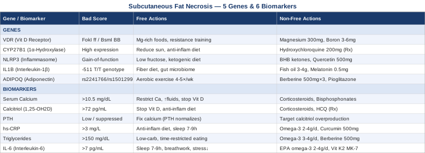
6 Biomarkers That Can Change How You Manage Subcutaneous Fat Necrosis
Subcutaneous fat necrosis does not follow a single biochemical pathway. It involves localized fat cell death, immune cell infiltration, granuloma formation, and — particularly in neonatal cases — an autonomous loop of vitamin D activation that can produce dangerous calcium elevation weeks after the visible lesions appear to be healing. The six biomarkers below map to the most clinically significant parts of that process. Tracking them gives you and your clinician something concrete to act on, rather than waiting for symptoms to declare themselves.
Biomarker 1 — Serum Calcium (Total and Ionized)
Why it matters and what it reveals: Hypercalcemia is the most dangerous complication of subcutaneous fat necrosis, particularly in neonates. It occurs because necrotic fat tissue becomes a site of ectopic enzymatic activity — macrophages infiltrating the lesions produce the active form of vitamin D, calcitriol, which drives calcium absorption from the gut and mobilization from bone. This process can continue silently well after lesions have resolved on the skin's surface. Untreated hypercalcemia causes nephrocalcinosis, cardiac arrhythmia, and in infants, potential neurodevelopmental harm. Elevated total calcium (above 10.5 mg/dL in adults, above 11 mg/dL in neonates) combined with suppressed PTH strongly suggests vitamin-D-mediated hypercalcemia — the signature pattern of subcutaneous fat necrosis complications. Ionized calcium is the more precise marker because total calcium is influenced by albumin levels.
How to measure it
A standard metabolic panel includes serum total calcium and costs $20–$50. Ionized calcium is a separate test and costs $30–$80. In neonates with confirmed subcutaneous fat necrosis, most pediatric neonatology guidelines recommend serial calcium monitoring every 1–2 weeks for at least the first 6 months of life, even after lesions have resolved. In adults, monitor at diagnosis and at 4-week intervals while lesions remain active.
If the score is bad, the plan without supplements
For mild hypercalcemia: restrict dietary calcium intake — in neonates, this may mean transitioning from breast milk to a low-calcium formula under physician guidance; in adults, avoid dairy-heavy diets and calcium supplements. Increase fluid intake significantly to promote renal calcium excretion. Limit sun exposure on skin to reduce endogenous vitamin D substrate. Discontinue all vitamin D supplementation immediately, including any formula already fortified with D3. Recheck calcium every 2–4 weeks while lesions are active.
If the score is bad, the plan with supplements or equipment
In moderate to severe hypercalcemia (total calcium above 12–13 mg/dL), pharmaceutical intervention is required and no over-the-counter supplement reliably reverses this. Corticosteroids (prednisolone at 1–2 mg/kg/day in neonates) suppress macrophage-derived enzyme activity responsible for calcitriol overproduction. Bisphosphonates (pamidronate, zoledronate) are used in refractory neonatal hypercalcemia. Ketoconazole inhibits the CYP450 enzymes responsible for calcitriol synthesis and has been used off-label in severe granulomatous hypercalcemia. All of these require physician supervision and serial calcium rechecks.
Biomarker 2 — 1,25-Dihydroxyvitamin D (Calcitriol)
Why it matters and what it reveals: In healthy physiology, calcitriol production is tightly regulated in the kidneys. In subcutaneous fat necrosis, macrophages infiltrating the necrotic fat tissue contain CYP27B1 enzyme activity and produce calcitriol autonomously — completely bypassing normal feedback controls. This is why hypercalcemia can occur even in patients who receive no vitamin D supplementation and have limited sun exposure. Elevated 1,25(OH)2D alongside suppressed PTH and elevated total calcium creates the diagnostic triad of granulomatous-mediated hypercalcemia, confirming that the source of calcium dysregulation is the lesion itself rather than a dietary or primary endocrine problem.
How to measure it
The 1,25(OH)2D test (calcitriol) should not be confused with the more common 25-OH vitamin D test, which measures inactive vitamin D storage. Calcitriol costs $80–$200 and must be specifically ordered — it is not part of standard vitamin D panels. Reference range: 18–72 pg/mL in adults; neonatal ranges are narrower. Because calcitriol has a short half-life of 6–8 hours, results can fluctuate. Testing during the active lesion phase gives the most clinically informative data.
If the score is bad, the plan without supplements
Calcitriol elevation from this mechanism does not respond to dietary restriction alone. The driving cause must be addressed: eliminate all exogenous vitamin D sources (supplements, fortified formulas, cod liver oil). Minimize direct sunlight on body skin during active disease. The most effective non-pharmacological step is reducing macrophage activation through anti-inflammatory diet and rest — as inflammation resolves, lesion-associated calcitriol production decreases. As lesions resolve, calcitriol should normalize over weeks to months.
If the score is bad, the plan with supplements or equipment
No supplement safely reduces ectopic calcitriol in this context without medical supervision. Corticosteroids remain the primary agent, suppressing macrophage CYP27B1 activity at the source. In adults with recurrent or persistent granulomatous disease driving chronic calcitriol elevation — the mechanism is identical to that seen in sarcoidosis — hydroxychloroquine has the strongest evidence for macrophage CYP27B1 suppression. Typical dosing in adults: 200–400 mg/day. Requires regular ophthalmologic monitoring due to cumulative retinal toxicity risk with long-term use. Physician prescription required.
Biomarker 3 — PTH (Parathyroid Hormone)
Why it matters and what it reveals: PTH is the body's primary calcium regulator. When calcium rises, the kidneys suppress PTH production through a tight negative feedback loop. In subcutaneous fat necrosis, this feedback system is intact — PTH is typically low or appropriately suppressed in affected patients with hypercalcemia. This distinguishes the condition from primary hyperparathyroidism, where PTH would be inappropriately elevated. Measuring PTH alongside calcium tells the clinician which mechanism is driving the calcium elevation — which directly determines the correct treatment. Measuring PTH alone without calcium can be misleading.
How to measure it
Intact PTH assay: $50–$120, widely available through standard reference labs. Reference range: 10–65 pg/mL in adults. Neonatal ranges differ by gestational age and laboratory. In some cases of malignancy-associated hypercalcemia, PTH-related protein (PTHrP) is also tested; in subcutaneous fat necrosis, PTHrP is typically normal, which helps exclude malignant causes. Both tests can be ordered simultaneously when the mechanism is unclear.
If the score is bad, the plan without supplements
Suppressed PTH in the context of elevated calcium is the expected and physiologically appropriate response in subcutaneous fat necrosis. Treating the PTH number directly would be incorrect — the priority is to correct the calcium, at which point PTH will normalize. If PTH remains low beyond 3–6 months after calcium normalization, further endocrine workup is warranted to rule out hypoparathyroidism as a separate process. No dietary or lifestyle intervention changes suppressed PTH in this context.
If the score is bad, the plan with supplements or equipment
PTH suppression in this setting is the effect, not the cause. Any attempt to raise PTH with supplements or PTH analogues (such as teriparatide) would worsen calcium elevation and is contraindicated. The appropriate medical response is always directed at calcitriol overproduction. If PTH is paradoxically high alongside elevated calcium, a different diagnosis — primary hyperparathyroidism, familial hypocalciuric hypercalcemia — should be considered urgently.
Biomarker 4 — High-Sensitivity CRP (hs-CRP)
Why it matters and what it reveals: Subcutaneous fat necrosis is fundamentally an inflammatory condition. Fat cell death triggers an immune cascade: macrophages infiltrate the lesions, form granulomas, and release pro-inflammatory cytokines that sustain the inflammatory environment locally and systemically. Systemic inflammation elevates CRP, which is produced in the liver in response to IL-6. Persistently elevated hs-CRP above 3 mg/L reflects ongoing inflammatory activity and correlates with the duration of calcitriol overproduction. Serial hs-CRP measurements give you a proxy for whether the underlying process is resolving or persisting.
How to measure it
High-sensitivity CRP: $15–$40 as a standalone test, or included in many cardiovascular risk panels. Standard interpretation: below 1 mg/L is low risk, 1–3 mg/L is moderate, above 3 mg/L is elevated. In acute inflammatory states, values can reach 50–200 mg/L. For subcutaneous fat necrosis management, measuring at diagnosis, at 4 weeks, and at 8 weeks gives the clearest trend. A rising or persistently elevated hs-CRP in a patient who appears clinically stable warrants further investigation.
If the score is bad, the plan without supplements
Anti-inflammatory dietary changes have consistent evidence for hs-CRP reduction: eliminate ultra-processed foods, refined seed oils, and added sugar from the diet — all three are independently associated with higher CRP in large observational studies. Prioritize omega-3-rich foods: fatty fish (salmon, sardines, mackerel) at least 3 times per week. Optimize sleep duration to 7–9 hours — under 6 hours per night is independently associated with elevated hs-CRP. Moderate-intensity aerobic exercise at 150 minutes per week reduces CRP by 10–30% over 8–12 weeks in most intervention studies. For nursing mothers of affected infants, maternal anti-inflammatory diet adjustments may reduce systemic inflammatory load passed through breast milk.
If the score is bad, the plan with supplements or equipment
Omega-3 fatty acids (EPA + DHA combined, 2–4 g/day) have demonstrated hs-CRP reduction in multiple randomized controlled trials, with consistent effect sizes of 15–30%. Take daily with food. No cycling required; reassess labs every 3 months. Side effects: fishy aftertaste, mild GI upset, theoretical minor bleeding risk at very high doses. Magnesium glycinate at 300–400 mg/day in adults is associated with reduced inflammatory markers in magnesium-deficient individuals; reassess at 60 days. Bioavailable curcumin with piperine: 500–1000 mg/day; evidence for CRP reduction exists in metabolic syndrome and arthritis trials. Cycle 8 weeks on, 2 weeks off. Not appropriate for neonates or young infants.
Biomarker 5 — Fasting Triglycerides and Lipid Panel
Why it matters and what it reveals: Subcutaneous fat necrosis pathology begins with adipocyte injury and death — fat cells release free fatty acids and triglycerides into surrounding tissue when they are disrupted. In pancreatic panniculitis (a form of fat necrosis driven by pancreatic lipase leaking into systemic circulation), hypertriglyceridemia is both a contributing driver and a consequence. Even in neonatal and traumatic fat necrosis, elevated triglycerides and lipid dysregulation signal broader metabolic vulnerability that may prolong inflammation and impair tissue repair. The triglyceride-to-HDL ratio (ideally below 2.0 in adults) is a particularly sensitive proxy for insulin resistance and systemic metabolic dysfunction, both of which amplify inflammatory tissue responses.
How to measure it
Standard fasting lipid panel: $30–$60 with a 12-hour fast before blood draw. For a more complete picture, advanced lipid testing via NMR lipoprotein fractionation (LDL-P particle count, sdLDL particle size) is available through LabCorp or Quest Diagnostics for $100–$300. Thomas Dayspring and Peter Attia consistently recommend ApoB measurement ($20–$60) as a single most informative lipid metric — it captures total atherogenic particle burden regardless of size and is more predictive than LDL-C alone. ApoB above 90 mg/dL warrants attention.
If the score is bad, the plan without supplements
For elevated fasting triglycerides: reducing refined carbohydrate and sugar intake is the most effective dietary lever — dietary carbohydrates drive triglyceride synthesis through de novo lipogenesis more powerfully than dietary fat in most individuals. Time-restricted eating (a 10–12 hour eating window) has demonstrated consistent triglyceride reduction in multiple intervention studies. Reduce or eliminate alcohol, which dramatically elevates triglycerides in susceptible individuals. Aerobic exercise at 150 minutes per week produces 10–20% fasting triglyceride reduction over 8–12 weeks in most studies.
If the score is bad, the plan with supplements or equipment
Omega-3 (EPA + DHA at 3–4 g/day) is the most evidence-backed supplement for triglyceride reduction, with typical reductions of 15–30% in RCTs. Prescription icosapentaenoic acid (Vascepa, 4 g/day) is FDA-approved for severe hypertriglyceridemia (above 500 mg/dL). Berberine: 500 mg taken 2–3 times daily with meals reduces triglycerides and improves insulin sensitivity through AMPK activation. Cycle 8 weeks on, 2 weeks off. Contraindicated in pregnancy and not established in neonates. Niacin: extended-release niacin at 500–1500 mg/day has strong triglyceride-lowering data but requires physician monitoring for liver function and glucose effects.
Biomarker 6 — Interleukin-6 (IL-6)
Why it matters and what it reveals: IL-6 is the cytokine most directly linked to the macrophage activation and granuloma formation that define subcutaneous fat necrosis at a cellular level. It drives hepatic CRP production and sustains the feedback loop that keeps macrophages activated and calcitriol production elevated. Persistently elevated IL-6 is a key reason why some cases of SFNN produce prolonged or severe hypercalcemia while others resolve within weeks — the intensity of macrophage activation, reflected in IL-6 levels, determines how actively the ectopic calcitriol pathway operates. Elevated IL-6 (above 7 pg/mL in fasting, non-acutely ill individuals) indicates ongoing granulomatous inflammation.
How to measure it
IL-6 serum assay: $80–$180 at specialty reference labs. Not routinely ordered but available through Quest, LabCorp, and most academic medical center labs. IL-6 is highly sensitive to acute illness — even a minor cold can spike it substantially and transiently. For the most accurate baseline reading, measure at least 2 weeks after any acute infection or significant physical stressor. Best measured in conjunction with hs-CRP to confirm whether CRP elevation is primarily IL-6-driven.
If the score is bad, the plan without supplements
Sleep quality is the most underappreciated lever for chronic IL-6 elevation: a single night of sleep below 6 hours measurably raises IL-6 the following day; consistent 7–9 hours reduces IL-6 over 4–6 weeks. Stress reduction has a direct mechanistic pathway to IL-6 reduction via the cholinergic anti-inflammatory reflex — parasympathetic activation suppresses macrophage cytokine release. Note: while cold water immersion is a documented IL-6 modulator in healthy adults, it should be avoided over affected skin areas in subcutaneous fat necrosis, where cold exposure can trigger further fat cell damage. Stress-reduction practices (covered in the complementary section below) are safer options.
If the score is bad, the plan with supplements or equipment
EPA-dominant omega-3 at 2–4 g/day has shown consistent IL-6 reduction in RCTs across several inflammatory conditions; EPA appears to have stronger IL-6-modulating properties than DHA. Vitamin K2 (MK-7 form, 100–200 mcg/day) has emerging evidence for anti-inflammatory effects through activation of gamma-carboxylation-dependent proteins; reassess at 90 days with no major side effects at standard doses. Low-dose naltrexone (LDN, 1.5–4.5 mg at night) is an off-label prescription option with a growing evidence base for chronic inflammatory suppression via glial cell modulation; requires physician prescription and periodic liver function monitoring.
5 Genes That Shape Risk and Recovery
Understanding your biomarker levels tells you the current state of the system. Knowing which genetic variants you carry explains the underlying architecture — why the system behaves the way it does under stress, why some individuals experience more severe calcium complications than others with comparable lesions, and which interventions are most likely to work for your specific biology. These five genes represent the most relevant genetic ground for subcutaneous fat necrosis.
VDR — Vitamin D Receptor
What it does: The VDR gene encodes the intracellular receptor through which calcitriol exerts its effects on hundreds of downstream genes. VDR is expressed in virtually every cell type, including macrophages, where it modulates immune activation, granuloma formation, and cytokine production. When macrophages in fat necrosis lesions produce excess calcitriol, VDR activity is the mechanism through which that calcitriol drives calcium-related gene expression. VDR polymorphisms — particularly FokI, BsmI, TaqI, and ApaI — are among the most studied functional variants in immune and inflammatory disease.
Relevant variants: The FokI ff genotype produces a longer VDR protein with higher transcriptional activity, potentially amplifying the effects of elevated calcitriol in target tissues. The BsmI BB genotype has been linked to altered macrophage differentiation and cytokine profiles in multiple autoimmune and granulomatous disease studies. High-activity VDR variants may explain why some individuals develop more severe calcium dysregulation from the same extent of fat necrosis. Evidence is primarily from sarcoidosis and autoimmune disease cohorts; direct subcutaneous fat necrosis genetic data is limited to case reports and small series.
If the gene is bad, the plan without supplements
Prioritize magnesium-rich foods — magnesium is a required cofactor for VDR activation and for CYP enzymes in the vitamin D pathway. Leafy greens, pumpkin seeds, legumes, and dark chocolate are the most concentrated dietary sources. Resistance training 2–3 times per week has been shown to upregulate VDR expression in muscle tissue. Reduce VDR-suppressing factors: chronic high-sugar diets, obesity, and chronic stress are all independently associated with reduced VDR sensitivity. In the acute phase of subcutaneous fat necrosis with hypercalcemia, avoid all vitamin D supplementation regardless of VDR status — the receptor is already being overstimulated.
If the score is bad, the plan with supplements or equipment
Magnesium glycinate or malate, 300–400 mg/day: daily without cycling; primary purpose is VDR cofactor support and CYP enzyme function. Side effects: loose stool at high doses (switch to glycinate form if this occurs). Boron, 3–6 mg/day: emerging evidence that boron reduces calcitriol catabolism and supports VDR expression in individuals with low VDR activity; reassess at 60–90 days. Avoid vitamin D supplementation during active disease — this is the most important caution for high-activity VDR variants.
CYP27B1 — 1-Alpha Hydroxylase
What it does: CYP27B1 encodes the enzyme responsible for converting inactive 25-hydroxyvitamin D into calcitriol. Under normal conditions, this conversion happens in kidney tubular cells with tight negative feedback control. In subcutaneous fat necrosis, macrophages infiltrating the lesions express CYP27B1 without any feedback regulation — they convert circulating 25-OH vitamin D into calcitriol autonomously. This is the exact step responsible for the hypercalcemia complication that makes this condition potentially dangerous. Variants that increase CYP27B1 expression or reduce its negative feedback sensitivity in immune cells could theoretically amplify this process.
Relevant variants: Gain-of-function or expression-upregulating variants in CYP27B1 are primarily studied in granulomatous disease (sarcoidosis, tuberculosis) where the same ectopic macrophage CYP27B1 mechanism operates. The genetic literature here is early-stage for fat necrosis specifically. However, high-expressing CYP27B1 haplotypes identified in sarcoidosis cohorts provide a plausible framework for why similar hypercalcemia complications occur in subcutaneous fat necrosis.
If the gene is bad, the plan without supplements
Reduce vitamin D substrate availability during active disease: minimize sun exposure on affected skin, avoid vitamin D-fortified foods in excess, and cease supplementation. An anti-inflammatory dietary pattern that reduces macrophage recruitment and activation directly decreases the cell population expressing CYP27B1 at the lesion site — this is the most impactful non-pharmacological step. Mediterranean-pattern eating with high polyphenol content and omega-3 fats has the strongest evidence for macrophage modulation.
If the score is bad, the plan with supplements or equipment
No supplement specifically inhibits CYP27B1 activity without systemic consequences. Medically, ketoconazole (a CYP450 inhibitor) has been used but carries significant hepatotoxicity concerns. Hydroxychloroquine is the most clinically supported agent for suppressing macrophage CYP27B1 in granulomatous hypercalcemia — it is the standard treatment in sarcoidosis-related calcitriol excess. Standard adult dosing: 200–400 mg/day with ophthalmologic monitoring every 6–12 months for retinal toxicity. Physician prescription required. Evidence quality: moderate, based on mechanism extrapolated from sarcoidosis.
NLRP3 — Inflammasome Sensor
What it does: NLRP3 is a cytosolic pattern recognition receptor that acts as the cell's internal danger sensor. When activated by necrotic cell debris — precisely the signal generated by dying fat cells — NLRP3 assembles into a large protein complex called the inflammasome, which activates caspase-1. Caspase-1 then cleaves pro-IL-1β and pro-IL-18 into their active, pro-inflammatory forms. This is one of the primary mechanisms of sterile inflammation — the kind driven by tissue damage rather than infection. In subcutaneous fat necrosis, NLRP3 activation is one of the first steps in the cascade that leads to macrophage infiltration, granuloma formation, and ultimately calcitriol overproduction.
Relevant variants: Rare gain-of-function mutations in NLRP3 cause cryopyrin-associated periodic syndromes (CAPS), with extreme inflammatory episodes. More common functional polymorphisms in NLRP3 and associated genes (CASP1, IL18, PYCARD) are being studied for their contribution to inflammatory response intensity. Gary Brecka's work on genetic expression and inflammation, and Ali Torkamani's research on polygenic risk profiling, both point to NLRP3 pathway variants as meaningful modifiers of inflammatory phenotype — though direct subcutaneous fat necrosis genetic studies are absent.
If the gene is bad, the plan without supplements
Reduce dietary NLRP3 activators: palmitic acid (from saturated fat overload, particularly processed meats and fried foods) and uric acid (from high fructose and purine intake) are among the most potent NLRP3 activators studied in human cells. Reduce fructose-containing beverages and ultra-processed meats. Metabolic ketosis via time-restricted eating or low-carbohydrate diet elevates beta-hydroxybutyrate, which directly inhibits NLRP3 inflammasome assembly — this is one of the more compelling mechanistic explanations for the anti-inflammatory effects of ketogenic dietary patterns.
If the score is bad, the plan with supplements or equipment
Exogenous beta-hydroxybutyrate (BHB ketone salts or esters, 10–15 g/day in adults) directly inhibits NLRP3 activation in human cell and early clinical studies; take with meals. Not established in neonates. Sulforaphane from standardized broccoli sprout extract (10–50 mg/day) activates Nrf2 pathway, which suppresses NLRP3 upstream; cycle 6 weeks on, 2 weeks off. Quercetin, 500–1000 mg/day with food: multiple RCTs demonstrate NLRP3 inhibition and general anti-inflammatory effects; widely available, low side effect profile. Cycle 8 weeks on, 2 weeks off.
IL1B — Interleukin-1 Beta
What it does: IL-1β is the primary product of NLRP3 inflammasome activation and one of the most potent pro-inflammatory cytokines in the innate immune arsenal. Once cleaved and released, it amplifies local inflammation dramatically, recruits additional macrophages and neutrophils, activates NF-κB in surrounding cells, and sustains the granuloma formation process. High sustained IL-1β keeps the calcitriol-producing macrophage machinery running. Genetic variants in IL1B that increase production in response to stimulation are associated with more intense and prolonged inflammatory responses across multiple conditions.
Relevant variants: The IL1B -511 T/T genotype is one of the most studied high-production variants, associated with significantly elevated IL-1β output in response to inflammatory stimuli. IL1B haplotypes have been studied in autoimmune, infectious, and granulomatous disease contexts. Carriers of high-production IL1B variants may experience more severe inflammatory responses to the same degree of fat cell death — a meaningful factor in whether subcutaneous fat necrosis stays localized or produces systemic complications.
If the gene is bad, the plan without supplements
Optimize gut microbiome health as a priority: gut-derived lipopolysaccharide (LPS) from dysbiotic gut bacteria is one of the most potent systemic IL-1β triggers. A diverse, fiber-rich diet directly reduces circulating LPS and downstream IL-1β production. Avoid dietary emulsifiers (carrageenan, polysorbate 80) that increase gut permeability. A traditional Mediterranean-pattern diet — high polyphenols, extra virgin olive oil, diverse plant fiber, fatty fish — has demonstrated direct IL-1β reduction in multiple intervention trials and is the most evidence-grounded dietary approach for high IL1B expressors.
If the score is bad, the plan with supplements or equipment
Anakinra (recombinant IL-1 receptor antagonist, a prescription biologic) directly blocks IL-1β signaling and has been used in refractory cases of neonatal SFNN with severe hypercalcemia as a rescue therapy. This is a specialist-level medical intervention. For non-prescription approaches: fish oil (EPA+DHA 3–4 g/day) consistently reduces IL-1β and its upstream activators in inflammatory disease RCTs. Melatonin at 0.5–1 mg at bedtime has emerging evidence for IL-1β modulation via NF-κB suppression; adults only, reassess at 8 weeks.
ADIPOQ — Adiponectin Gene
What it does: ADIPOQ encodes adiponectin, a hormone secreted by healthy adipose tissue with potent anti-inflammatory and insulin-sensitizing effects. Adiponectin suppresses NF-κB signaling in macrophages, reduces TNF-α production, and inhibits macrophage activation broadly. It is, in effect, an endogenous brake on the inflammatory process. When fat cells die — as in subcutaneous fat necrosis — local adiponectin production from that tissue drops precipitously, removing one of the key molecular checks on inflammation at exactly the moment it is most needed.
Relevant variants: SNPs in ADIPOQ, including rs2241766 (T/G) and rs1501299 (G/T), are associated with significantly lower circulating adiponectin levels in multiple human cohort studies. Carriers of low-adiponectin variants have a weaker endogenous anti-inflammatory brake, which may explain why some individuals with seemingly equivalent lesion extent experience much greater inflammatory severity and prolonged recovery.
If the gene is bad, the plan without supplements
Aerobic exercise is the most reliably documented natural adiponectin elevator available: 4–5 sessions per week at 60–70% maximum heart rate for 30–45 minutes raises circulating adiponectin by 10–20% over 8–12 weeks in most studies. Weight normalization in overweight individuals also raises adiponectin substantially. Avoiding prolonged caloric restriction is important — chronic undereating paradoxically lowers adiponectin despite reducing adiposity.
If the score is bad, the plan with supplements or equipment
Berberine at 500 mg, 2–3 times daily with meals, has demonstrated 20–30% increases in circulating adiponectin in metabolic syndrome populations across multiple RCTs. Cycle 8 weeks on, 2 weeks off; avoid in pregnancy. Thiazolidinediones (pioglitazone, rosiglitazone — prescription) are the most potent pharmacological adiponectin-raising agents but require physician oversight due to fluid retention and cardiovascular considerations. Regular cold exposure (cool showers, cold ambient temperature) has shown adiponectin-raising effects in human studies — but avoid applying cold directly to active lesion areas where it can worsen fat cell damage.
What Peter Attia's Framework in Outlive Reveals About Fat Tissue Disease
Peter Attia's Outlive (2023) is arguably the most rigorously cited contemporary framework for metabolic health and longevity medicine available to a general audience. Though not written about subcutaneous fat necrosis directly, its analysis of inflammation, lipid pathology, adipose tissue biology, and metabolic resilience maps almost precisely onto the biological mechanisms at play in this condition. The following ten points synthesize the most impactful ideas from his framework, applied to fat tissue necrosis and its downstream risks.
1. Acute Versus Chronic Inflammation — The Critical Distinction
Attia consistently distinguishes between acute inflammation — which is purposeful and drives repair — and chronic low-grade inflammation, which is self-sustaining and destructive. In subcutaneous fat necrosis, the initial immune response is appropriate. The danger is when it does not resolve. Treatment strategy should be calibrated to accelerate resolution rather than simply suppress the initial response. Blunting inflammation too aggressively in the acute phase can impair lesion clearance; failing to resolve it leads to chronic complications.
2. ApoB Over LDL-C — A More Complete Lipid Picture
Borrowing directly from Thomas Dayspring and Allan Sniderman's work, Attia places ApoB at the center of lipid-related cardiovascular and tissue inflammation risk. ApoB particles penetrate tissues, including adipose tissue, where they drive local inflammatory activity. In fat necrosis with a metabolic or pancreatic component, tracking ApoB ($20–$60) alongside the standard lipid panel gives a more complete picture of lipid-driven inflammatory load than LDL-cholesterol alone. Target ApoB below 80 mg/dL for metabolically vulnerable individuals.
3. Metabolic Flexibility as Tissue Resilience
Metabolic flexibility — the capacity to efficiently switch between glucose and fat as fuel — is Attia's marker of underlying metabolic health. Individuals with impaired metabolic flexibility have higher baseline inflammation, more reactive NF-κB signaling in adipose tissue, and slower tissue repair after injury. Improving metabolic flexibility through dietary carbohydrate reduction, zone 2 aerobic training, and intermittent fasting creates a lower-inflammation internal environment in which recovery from fat necrosis proceeds more efficiently.
4. Zone 2 Cardio as the Anti-Inflammatory Foundation
Attia's primary exercise recommendation — zone 2 training at 60–70% maximum heart rate, 45–60 minutes, 4 sessions per week — improves mitochondrial density in adipose and muscle tissue, reduces circulating inflammatory cytokines over time, raises adiponectin, and improves insulin sensitivity. For adults recovering from fat necrosis, this is the most evidence-backed exercise starting point once the acute phase has resolved.
5. Sleep as the Primary Recovery Lever
Drawing heavily on Matthew Walker's research, Attia frames sleep as the non-negotiable foundation of metabolic and immune health. REM and slow-wave sleep are the phases during which cortisol is lowest and GH is highest — the exact conditions for tissue repair. A single night below 6 hours of sleep demonstrably raises CRP, IL-6, and cortisol the following day. For parents managing an infant with SFNN, sleep deprivation compounds the metabolic stress and inflammatory load. Strategic napping and sleep consolidation deserve the same priority as dietary changes.
6. Continuous Glucose Monitoring as Real-Time Inflammation Feedback
Attia advocates for CGM use even in non-diabetic individuals to identify postprandial glucose spikes that correlate with inflammatory cytokine surges. For adults managing subcutaneous fat necrosis or its metabolic aftermath, a CGM can identify which foods or stress events are producing glucose excursions — and since glycemic spikes transiently raise IL-6 and CRP, this gives real-time insight into inflammation drivers that blood draws cannot capture. Consumer CGMs (Dexterity, Levels, Nutrisense) cost roughly $100–$200 per month without prescription.
7. Muscle as an Anti-Inflammatory Endocrine Organ
Skeletal muscle is not passive tissue — it actively secretes myokines during and after exercise. Brief IL-6 surges during exercise, for example, paradoxically reduce systemic IL-6 chronically and suppress TNF-α post-exercise. Muscle-secreted irisin suppresses inflammatory NF-κB signaling in adipose tissue. Building and maintaining lean muscle mass directly supports adipose tissue health and reduces the inflammatory burden from fat cell dysfunction. Attia recommends 2–3 resistance training sessions per week as a minimum for all adults.
8. The Insulin–Inflammation Axis
Hyperinsulinemia activates NF-κB in adipose tissue macrophages, directly amplifying inflammatory cytokine production. Reducing insulin load — primarily by reducing refined carbohydrate consumption — is the most rapid dietary lever for systemic inflammation. Attia and metabolic health researchers including Jason Fung and Satchin Panda have documented consistent CRP and IL-6 reductions within 4–8 weeks of significant carbohydrate reduction in insulin-resistant individuals.
9. Protein Adequacy for Tissue Repair and Immune Function
Attia recommends 1.6–2.2 g of protein per kilogram of body weight per day for active adults, grounded in muscle protein synthesis research. Adequate protein is equally important for immune cell production, tissue repair collagen synthesis, and the resolution of inflammatory processes. Chronic low-protein intake — common in adults who focus exclusively on calorie reduction — impairs all three and may prolong recovery from fat tissue injury.
10. Test, Intervene on One Variable, Retest — The N=1 Method
Attia's core methodological point applies directly here: subcutaneous fat necrosis is heterogeneous. Different individuals have different dominant pathways — some primarily calcitriol-driven, some predominantly inflammatory, some with underlying metabolic dysfunction. Serial biomarker testing is how you know whether what you are doing is working. Intervening on diet, sleep, and supplementation simultaneously makes it impossible to identify what is actually helping. Changing one variable at a time and rechecking labs at 8–12 weeks is slower but far more informative.
Complementary Approaches With Meaningful Clinical Evidence
Subcutaneous fat necrosis requires medical oversight — there is no equivalent of a purely lifestyle-managed protocol for its serious complications. The modalities below are evidence-informed adjuncts that can support the recovery process, reduce systemic inflammation, or address the secondary effects of the condition. They are selected for having genuine human clinical evidence relevant to the mechanisms at play, not for theoretical plausibility alone.
Low-Level Laser Therapy (Photobiomodulation)
Photobiomodulation (PBM) uses specific red and near-infrared wavelengths (typically 630–1064 nm) to stimulate mitochondrial cytochrome c oxidase activity, enhance ATP production in tissue cells, and shift macrophage polarization from the pro-inflammatory M1 phenotype toward the reparative M2 phenotype. This macrophage polarization effect is directly relevant to subcutaneous fat necrosis: lesion resolution requires the transition from inflammatory macrophages (which sustain granulomas and calcitriol production) to reparative macrophages (which clear cellular debris and promote tissue remodeling). In adult cases where indurated nodules persist long after the acute phase, PBM may support this transition in affected tissue.
A systematic review of photobiomodulation for chronic inflammatory conditions published in Photobiomodulation, Photomedicine, and Laser Surgery (2019) documented consistent anti-inflammatory effects via multiple cytokine pathways. Specific subcutaneous fat necrosis PBM studies are absent from the literature, but the mechanism of action is plausible and well-characterized. Risk at therapeutic doses and wavelengths is minimal.
Protocol: Treatment by a trained physiotherapist or dermatologist; 808 nm or 904 nm wavelength, 2–3 sessions per week for 4–8 weeks over non-inflamed, non-infected chronic lesion areas. Do not apply during the active acute inflammatory phase or over any hypercalcemia-active period. Home-use panels at 630–850 nm exist but should only be used once the acute phase has fully resolved and only after consulting a clinician.
Microbiome-Directed Therapies
The gut microbiome is a powerful regulator of systemic immune tone. Dysbiosis — disrupted microbial balance — increases intestinal permeability, which elevates circulating bacterial lipopolysaccharide (LPS). LPS is one of the most potent known triggers of macrophage IL-1β and IL-6 production via toll-like receptor 4 — the exact cytokine pathway that sustains granuloma formation and calcitriol overproduction in subcutaneous fat necrosis. A healthier, more diverse microbiome translates directly to lower systemic inflammatory baseline and potentially faster lesion resolution.
A 2021 randomized trial published in Cell by Wastyk and colleagues demonstrated that a high-fiber diet and a fermented food diet each independently reduced multiple inflammatory proteins, including IL-6 and CRP, over 10 weeks in healthy adults. The fermented food intervention showed particularly robust effects on microbiome diversity. This is among the most methodologically rigorous human dietary-microbiome-inflammation trials published to date.
Protocol: Increase dietary fiber to 35–40 g/day from diverse plant sources — prioritize variety over volume. Add 2–3 servings of fermented foods daily: kimchi, plain kefir, sauerkraut, or plain yogurt. For a more targeted approach, a clinically studied probiotic containing Lactobacillus rhamnosus GG or Bifidobacterium longum at 10–50 billion CFU/day has anti-inflammatory evidence across multiple trials. Introduce all changes gradually over 2–3 weeks to minimize digestive adjustment symptoms.
Mindfulness Meditation and MBSR
Mindfulness-Based Stress Reduction (MBSR) is a structured 8-week program combining meditation, body awareness, and yoga developed by Jon Kabat-Zinn at the University of Massachusetts Medical Center. Its anti-inflammatory relevance for subcutaneous fat necrosis — particularly for parents managing an infant's diagnosis or adults dealing with prolonged recovery — lies in its documented capacity to reduce cortisol and inflammatory cytokines. Chronic stress activates the HPA axis, which elevates cortisol, which in turn amplifies NF-κB-driven cytokine production. MBSR interrupts this loop through both neuroendocrine and behavioral pathways.
A randomized controlled trial published in Brain, Behavior, and Immunity (2016) comparing MBSR to an active relaxation control found that MBSR specifically reduced NF-κB activity and IL-6 in stressed adults — effects not seen in the relaxation control group — suggesting the meditation component rather than just stress relief drives the anti-inflammatory benefit.
Protocol: The standard MBSR program involves 8 weekly group sessions of 2.5 hours plus a full-day retreat and 45 minutes of daily home practice. Structured online versions are available through several university medical centers at reduced cost. For individuals with limited time, 10–15 minutes of daily focused breath-attention practice has demonstrated cortisol reduction in 4-week trials. No contraindications relevant to subcutaneous fat necrosis.
Breathing-Based Therapies
Controlled breathing techniques — specifically those extending the exhalation relative to inhalation — activate the vagus nerve and shift autonomic tone toward parasympathetic dominance. This has measurable downstream effects on macrophage activity: vagal nerve activation suppresses TNF-α and IL-6 release from macrophages via the cholinergic anti-inflammatory pathway, a mechanism studied extensively since Kevin Tracey's foundational work at Cold Spring Harbor Laboratory. For individuals with elevated IL-6 and hs-CRP, daily breathing practice is a low-risk, accessible tool for dampening inflammatory cytokine output.
A 2014 trial by Kox and colleagues published in PNAS found that participants trained in Wim Hof Method breathing protocols showed significantly reduced TNF-α, IL-6, and IL-1β responses when challenged with experimental endotoxemia compared to untrained controls. Mechanistically, this appears to involve voluntary sympathetic activation followed by robust parasympathetic rebound, with lasting effects on innate immune reactivity.
Protocol: For daily practice, extended-exhale breathing — 4 counts inhale, 6–8 counts exhale — practiced for 5–10 minutes daily is the most accessible and safest starting point. Box breathing (4 in, 4 hold, 4 out, 4 hold) is another well-documented variant. Wim Hof-style hyperventilation cycles should not be practiced near water or while driving due to syncope risk. Avoid any breath-hold technique with forceful intrathoracic pressure changes in individuals with active cardiac arrhythmias — a potential concern given that hypercalcemia can affect cardiac rhythm.
Conclusion
Subcutaneous fat necrosis does not fit into a single clinical box, and managing it well requires more than watchful waiting. The condition operates across interconnected systems — calcium regulation, vitamin D activation, macrophage behavior, and metabolic inflammation — and the specific pathway that is most active in any individual case is what should drive the response.
The six biomarkers covered here give you a measurable framework: not a diagnosis in themselves, but a set of data points that make the difference between knowing what is happening inside the body and simply hoping the lesions resolve without consequence. The five genes add a layer of context that explains why severity varies so much from person to person. Together, these frameworks move the conversation from reactive to informed.
The next smart step is straightforward: bring a targeted biomarker panel — calcium, calcitriol, PTH, hs-CRP, triglycerides, and IL-6 where possible — to your next clinical appointment. Ask specifically whether calcitriol has been measured if hypercalcemia has occurred. Consider a basic genetic panel if your case has been unexpectedly severe or has recurred. Match the lifestyle adjustments — dietary anti-inflammatory changes, sleep optimization, aerobic exercise — to your specific lab picture. And keep tracking. Serial measurement is how you know whether what you are doing is working.