This article contains AI generated content.
Superior Tibiofibular Joint Dislocation: 6 Genes And 6 Biomarkers To Track
If your fibular head keeps slipping, clunking, or partially dislocating against the outside of your knee, you've probably already heard some version of "strengthen the area and use a brace." That advice isn't wrong, but it's incomplete. It treats every case of proximal tibiofibular joint instability as if it came from the same place — a bad landing, a twisted knee, a one-off accident. For a lot of people, that's exactly what happened, and rehab alone gets them back to normal.
For others, the joint keeps giving way even after physical therapy, even after a brace, even after the swelling is long gone. That pattern — recurrent or bilateral instability, joint hypermobility elsewhere in the body, unusually stretchy skin, a family history of "loose joints" — points toward something more specific: an underlying difference in how connective tissue is built. That difference is often genetic, and it explains why generic strengthening protocols sometimes fall short no matter how consistently they're followed.
This article looks at the superior tibiofibular joint dislocation picture from two angles that rarely get combined in one place: the specific collagen and connective-tissue genes that make some joints inherently looser than others, and the everyday biomarkers that can flag whether your body currently has what it needs to keep ligaments strong. Neither replaces an orthopedic evaluation or imaging. Both can make the conversation with your surgeon, physiotherapist, or geneticist considerably more precise.
None of this is about miracle fixes. Structural collagen genes can't be rewritten with a supplement stack, and no biomarker panel will glue a joint back together. But better information changes decisions — whether that means asking for a hypermobility assessment, requesting a specific blood panel before your next appointment, or finally understanding why your ankles, shoulders, and fibular head all seem to move a little more than everyone else's.
Summary
Recurrent proximal tibiofibular joint dislocation is rarely just bad luck. In a meaningful subset of cases, it traces back to inherited variations in the genes that build type I, III, and V collagen, tenascin-X, or fibrillin-1 — the same genes implicated in Ehlers-Danlos syndromes and Marfan-related joint laxity. Below, you'll find the six genes most consistently tied to joint hypermobility and dislocation risk, what each one actually does, and a realistic plan for working around a "bad" variant — with and without supplements or equipment, including frequency, cycling, and side effects for each option. You'll also find six blood-and-function biomarkers worth tracking if your joints are unstable, a research-backed collagen-loading protocol borrowed from tendon physiologist Dr. Keith Baar's work, and a short review of complementary approaches with actual human evidence behind them for joint and ligament support. By the end, you should have a much clearer sense of which category you fall into — and what to actually do about it.
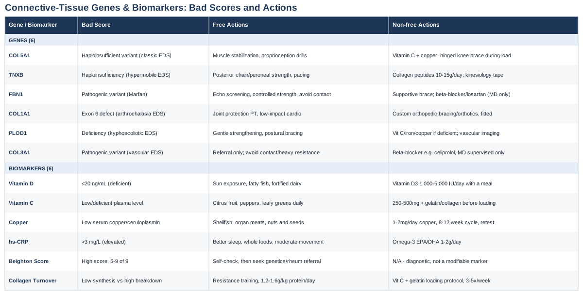
6 Genes Linked to Tibiofibular Joint Instability and Dislocation
Proximal tibiofibular joint (PTFJ) dislocation is uncommon enough that most orthopedic reviews describe it in terms of four patterns: atraumatic subluxation, posteromedial, anterolateral, and superior dislocation — the last being the rarest of the four, per a widely cited case review and literature summary (Proximal tibiofibular dislocation: a case report and review of literature). Because the joint is normally stabilized by strong ligaments and a snug capsule, most cases follow a clear traumatic mechanism — a twisting knee injury, a fall, a direct blow. But a smaller, clinically important group of cases occurs with minimal or no trauma, or recurs after the initial injury has fully healed. That pattern shows up specifically in people with Ehlers-Danlos syndrome, where joint dislocation from little to no trauma is described as "a relatively frequent complication" due to underlying ligamentous laxity (Hypermobile Ehlers-Danlos Syndrome and Hypermobility Spectrum Disorders).
This is where genetics becomes practically useful rather than academic. If your joint keeps dislocating without a clear mechanical cause, or if you notice hypermobility in your fingers, elbows, or shoulders too, the six genes below are the ones with the strongest human evidence for building the specific proteins that keep joints — including the tibiofibular joint — in place. Genomics researchers like Ali Torkamani, who studies how genetic variants translate into real-world disease risk, and health educators like Gary Brecka, who popularized direct-to-consumer genetic and biomarker testing, are a reasonable starting point if you want to see where you personally land on these genes — but the interpretation below is what actually matters for a joint-instability picture.
A note on what "compensating" for these genes really means: you cannot rewrite a collagen gene with lifestyle changes. What you can do is optimize the biochemical cofactors collagen assembly depends on, reduce unnecessary mechanical stress on already-lax ligaments, and build the surrounding musculature to serve as an active backup system for a passive structure that isn't holding up its end. That's the framework behind every plan below.
COL5A1 and COL5A2: The Classic Collagen-V Genes
COL5A1 and COL5A2 encode the two chains that combine to form type V collagen, a minor but essential collagen that regulates how thick and how tightly packed type I collagen fibrils become. Mutations here, usually causing reduced (haploinsufficient) rather than absent protein, are identified in roughly half of people with a clinical diagnosis of classic Ehlers-Danlos syndrome, a condition marked by stretchy skin, wide atrophic scars, and generalized joint hypermobility (Molecular mechanisms of classical Ehlers-Danlos syndrome; Clinical and molecular characterization of 40 patients with classic Ehlers-Danlos syndrome).
What it may affect: with thinner, poorly organized collagen fibrils, ligaments and joint capsules — including the ones stabilizing the fibular head against the tibia — have less tensile strength for the same amount of collagen mass. This shows up clinically as joints that move further than expected before anything stops them.
How it may be compensated for: because the problem is structural quality rather than a fixable enzyme deficiency, compensation means shifting the joint's stability budget away from passive ligament restraint and onto active muscular control, while making sure the collagen that is produced gets the raw materials and cofactors it needs.
The Plan Without Supplements
Build the muscles that cross the tibiofibular joint and the whole lower leg-knee complex as active stabilizers: closed-chain exercises like step-downs, single-leg balance work, and controlled calf raises, three to four sessions per week. Add proprioceptive training — single-leg stands on an unstable surface, working up to eyes-closed variations — since the joint's positional sense is often blunted along with its mechanical restraint. Avoid end-range stretching of the knee and ankle complex; if you're already hypermobile, you don't need more range, you need control within the range you have. There are no real side effects here beyond standard training fatigue, but progress slowly with balance work since a fall risk exists in more severely hypermobile individuals.
The Plan With Supplements or Equipment
Vitamin C (250-500 mg with meals) and copper (1-2 mg/day, ideally from diet like shellfish, organ meats, or nuts, or a low-dose supplement) support the two enzymatic steps — prolyl hydroxylation and lysyl oxidase cross-linking — that determine collagen fibril strength regardless of the underlying COL5A1 genotype. Take vitamin C daily; there's no need to cycle it, though doses above 2 g/day can cause GI upset or, in people prone to kidney stones, increase oxalate load. Keep copper supplementation modest and short-term (8-12 weeks at a time) since chronic excess copper interferes with zinc absorption and can, at high doses, be hepatotoxic. A hinged or figure-8 style knee-fibular brace during higher-load activities (running, cutting sports) adds passive restraint the ligament itself can't fully provide — wear it during the activity, not all day, to avoid muscle deconditioning from over-reliance.
TNXB: The Tenascin-X Gene
TNXB encodes tenascin-X, an extracellular matrix protein that helps organize collagen fibers and regulate their deposition. Haploinsufficiency of TNXB is strongly linked to hypermobile Ehlers-Danlos syndrome and benign joint hypermobility syndrome, conditions that, unlike classic EDS, often show minimal skin involvement but pronounced, widespread joint laxity (Tenascin-X: a candidate gene for benign joint hypermobility syndrome).
What it may affect: reduced tenascin-X leads to disorganized collagen fibril spacing, which weakens the tensile properties of ligaments and joint capsules more diffusely than a single-collagen-gene defect — which is part of why hypermobile EDS so often involves many joints rather than just one.
How it may be compensated for: because tenascin-X deficiency is also associated with chronic musculoskeletal pain and even nerve hypersensitivity in more severe (biallelic) cases, compensation strategies need to address pain modulation alongside mechanical stability, not just strength.
The Plan Without Supplements
Graded, low-impact strength training focused on the posterior chain and peroneal muscles (which directly influence fibular head stability) two to three times weekly, progressing load slowly to avoid flare-ups of joint pain that are common in tenascin-X-related laxity. Pair this with pacing strategies — planned rest between activity bouts — since overtraining tends to provoke pain rather than build resilience in this group. No cost, no real side effects, but expect slower strength gains than in a non-hypermobile person and don't chase soreness as a marker of progress.
The Plan With Supplements or Equipment
The same collagen-cofactor combination (vitamin C, copper, and adequate dietary protein with glycine/proline-rich sources like bone broth or collagen peptides, 10-15 g/day) supports whatever tenascin-X-organized collagen is being produced. Take collagen peptides once daily, cycling on for 8-12 weeks with a break, since long-term continuous use hasn't been well studied and the main risk is simply displacing other protein sources in the diet. Kinesiology taping around the fibular head during activity provides light proprioceptive feedback (not real mechanical restraint) and can reduce the "the joint feels like it's about to slip" sensation many people describe — reapply every 3-5 days per roll, watch for skin irritation with prolonged wear.
FBN1: The Fibrillin-1 Gene
FBN1 encodes fibrillin-1, the structural protein that forms microfibrils supporting elastic tissue throughout the body. Mutations cause Marfan syndrome, and joint hypermobility is increasingly recognized as a useful clinical indicator that helps differentiate Marfan-related joint laxity from Ehlers-Danlos-related laxity in borderline cases (Joint Hypermobility as a Potential Indicator of Marfan Syndrome and Ehlers-Danlos Syndrome; FBN1-Related Marfan Syndrome, GeneReviews).
What it may affect: microfibril disruption from FBN1 variants also increases free TGF-beta signaling, which affects long bone overgrowth and joint laxity — meaning a long, thin lower leg with a naturally looser fibular head attachment is a plausible mechanical setup for this specific dislocation pattern.
How it may be compensated for: this is the one gene on this list where the compensation conversation has to start with a safety flag, not a training plan.
The Plan Without Supplements
Before addressing the joint at all, anyone with a suspected FBN1 variant and joint hypermobility needs an echocardiogram to screen for aortic root dilation — this is standard of care for Marfan syndrome, not optional. Once cleared, joint-focused work looks similar to the COL5A1 approach: controlled strengthening of the peroneals and lateral knee stabilizers, avoiding high-impact pivoting sports that stress an already lax joint, and building single-leg balance capacity two to three times weekly. Contact and collision sports are generally discouraged given the cardiovascular risk profile, independent of the joint issue itself.
The Plan With Supplements or Equipment
There is no supplement that meaningfully alters fibrillin-1 microfibril assembly, so this category is mostly about equipment: a supportive brace for higher-risk activities, and — separately — beta-blocker or losartan therapy is often prescribed by cardiology for the aortic component, which is a medical decision, not a self-directed one. Standard vitamin C/copper collagen-support dosing (as above) is reasonable and low-risk to include, but treat it as a minor adjunct here, not the main lever.
COL1A1 and COL1A2: The Arthrochalasia Genes
COL1A1 and COL1A2 encode the two chains of type I collagen, the most abundant collagen in ligament, bone, and skin. Specific mutations causing loss of exon 6 in either gene produce arthrochalasia-type Ehlers-Danlos syndrome, a rare condition (fewer than 50 published cases) distinguished by severe generalized joint hypermobility and a very high rate of congenital hip dislocation, with minimal skin involvement compared to other EDS subtypes (Ehlers-Danlos Syndrome Type Arthrochalasia: A Systematic Review).
What it may affect: this exon-6 defect specifically impairs the site where procollagen is processed into mature collagen, leaving an abnormal N-terminal extension that disrupts fibril packing — producing joints that are recurrently, sometimes congenitally, unstable rather than instability that develops gradually.
How it may be compensated for: because this variant is rare and severe, compensation is realistically about early recognition, protective bracing, and surgical planning rather than reversing the underlying biology — treatment for arthrochalasia EDS is described in the literature as symptomatic, with no curative option currently available.
The Plan Without Supplements
Early physiotherapy focused on joint protection education (avoiding hyperextension positions, learning safe movement patterns) rather than aggressive strengthening, since connective tissue this fragile responds poorly to high mechanical loads. Low-impact activities like swimming or stationary cycling maintain conditioning without repeated joint stress. Regular orthopedic follow-up is essential since recurrent dislocation in this group often eventually requires surgical stabilization.
The Plan With Supplements or Equipment
Custom bracing or orthotics, fitted by an orthopedic specialist rather than off-the-shelf, is the most practical tool here — worn during activity and removed at rest to avoid dependency. Standard collagen-cofactor nutrition (vitamin C, copper, adequate protein) is reasonable to include but should be framed honestly to the patient or family as supportive, not corrective, given how limited the underlying collagen-processing defect is.
PLOD1: The Lysyl Hydroxylase Gene
PLOD1 encodes lysyl hydroxylase 1, the enzyme responsible for hydroxylating lysine residues in collagen so they can later form stable cross-links. Deficiency causes kyphoscoliotic Ehlers-Danlos syndrome, marked by joint hypermobility, hypotonia, early-onset kyphoscoliosis, and — importantly — a risk of arterial rupture that requires medical surveillance (PLOD1-Related Kyphoscoliotic Ehlers-Danlos Syndrome, GeneReviews).
What it may affect: without proper lysine hydroxylation, collagen fibrils form fewer and weaker cross-links, directly reducing tensile strength — this is one of the few genes on this list where the biochemistry points to a specific, testable nutrient interaction, since lysyl hydroxylase itself requires vitamin C and iron as cofactors.
How it may be compensated for: while supplementation cannot restore a deficient enzyme's function, ensuring cofactor sufficiency (vitamin C, iron, copper for the downstream lysyl oxidase step) removes any secondary nutritional bottleneck stacked on top of the genetic one.
The Plan Without Supplements
Gentle, progressive strengthening avoiding spinal loading (given the kyphoscoliosis risk) and joint hyperextension; physical therapy supervised by someone familiar with EDS is strongly preferable to generic strength programming. Postural bracing exercises for the spine, three times weekly, alongside lower-limb stabilization work for the joint itself.
The Plan With Supplements or Equipment
Vitamin C (500 mg/day, split into two doses for better absorption) ensures the cofactor for whatever residual lysyl hydroxylase activity remains isn't the limiting factor; iron should only be supplemented if bloodwork confirms deficiency, since unnecessary iron supplementation carries its own oxidative stress risk. Copper at low, cycled doses (8-week courses) supports the downstream cross-linking step. Given the arterial fragility associated with this gene, any equipment or exercise equipment involving high-impact loading should be avoided entirely, and vascular imaging surveillance should be arranged through a genetics or vascular specialist — this is not a self-managed risk.
COL3A1: The Gene That Changes the Whole Conversation
COL3A1 encodes type III collagen and is the causal gene in vascular Ehlers-Danlos syndrome (vEDS), the most severe EDS subtype. Joint hypermobility here is typically limited to the fingers, while the defining risk is spontaneous arterial or bowel rupture, reported in the majority of patients in cohort studies (Assessment of arterial damage in vascular Ehlers-Danlos syndrome).
What it may affect: this gene is included not because it's a common cause of isolated tibiofibular instability, but because unexplained joint hypermobility combined with thin, translucent skin, easy bruising, or a family history of unexplained arterial events warrants ruling this out before any exercise-based joint program begins.
How it may be compensated for: there is no fitness or supplement protocol appropriate here — this is a "the plan is a referral" situation.
The Plan Without Supplements
If this gene is suspected (based on family history, skin findings, or genetic testing), the actual plan is a referral to a geneticist and vascular specialist for surveillance imaging and blood pressure control, plus avoidance of contact sports and heavy resistance training, which are specifically discouraged in vEDS due to rupture risk. Any joint stabilization work should be low-load and cleared by that specialist team first.
The Plan With Supplements or Equipment
None recommended independently of medical supervision. Beta-blocker therapy (e.g., celiprolol) has trial evidence for reducing arterial events in vEDS but is a prescription decision made by a cardiologist or vascular geneticist, not something to self-initiate.
The genetic side of this picture answers the "why does my joint keep slipping when everyone else's doesn't" question. But genes are fixed at birth — biomarkers tell you what's happening in your body right now, which is often more actionable day to day.
6 Biomarkers Worth Tracking If Your Joints Keep Slipping
Even without a diagnosed connective tissue disorder, the biomarkers below reflect whether your body currently has the raw materials and inflammatory environment needed to keep ligaments strong. These are the kind of practical, trackable numbers that clinicians like Peter Attia and Thomas Dayspring emphasize in their broader work on measurable, modifiable health markers rather than vague symptom management.
25-Hydroxyvitamin D
Why it matters: vitamin D status is consistently associated with tendon and ligament healing outcomes — deficiency has been linked to poorer postoperative recovery, higher retear rates, and slower functional return after tendon repair procedures (The Role of Vitamin D in Postoperative Tendon Healing: A Scoping Review). Evidence in ligaments specifically is less robust than in tendons, but the biological plausibility (vitamin D reduces matrix-metalloproteinase activity, which otherwise degrades healing collagen) is strong.
How to measure it: a standard 25-OH vitamin D blood test, widely available through primary care or direct-to-consumer labs, costs roughly $30-$80 out of pocket depending on the lab and whether insurance covers it.
The plan without supplements: 15-20 minutes of midday sun exposure on exposed skin several times weekly (season and skin-tone dependent), plus dietary sources like fatty fish and fortified dairy.
The plan with supplements or equipment: vitamin D3, 1,000-2,000 IU daily for maintenance or higher (2,000-5,000 IU) under medical guidance if bloodwork shows deficiency, retesting after 8-12 weeks. Take with a fat-containing meal for absorption. Side effects are rare at these doses but excessive supplementation (>10,000 IU/day sustained) can cause hypercalcemia — this is a "test, don't guess" nutrient.
Plasma Vitamin C
Why it matters: vitamin C is a required cofactor for the prolyl hydroxylation step of collagen synthesis; even moderate changes in vitamin C status measurably affect the ratio of mature to immature collagen cross-links in connective tissue, and vitamin C-enriched gelatin taken before loading doubles a marker of new collagen synthesis in healthy adults (Vitamin C-enriched gelatin supplementation before intermittent activity augments collagen synthesis).
How to measure it: plasma or serum ascorbic acid testing is less commonly ordered than vitamin D but is available through most commercial labs, typically $40-$70.
The plan without supplements: citrus fruit, bell peppers, and leafy greens daily; deficiency is uncommon with a reasonably varied diet but worth checking if you eat a very restricted diet.
The plan with supplements or equipment: 250-500 mg vitamin C, ideally paired with 10-15 g of gelatin or collagen peptides about 60 minutes before joint-loading exercise, based on the protocol below. Daily use is fine; doses above 2 g/day risk GI upset and, in susceptible individuals, kidney stone formation.
Copper and Ceruloplasmin
Why it matters: copper is the essential cofactor for lysyl oxidase, the enzyme that forms the final cross-links stabilizing collagen and elastin fibers; copper deficiency in animal models produces connective tissue abnormalities strikingly similar to those seen in some EDS subtypes (Copper and the synthesis of elastin and collagen).
How to measure it: serum copper and ceruloplasmin (the copper-carrying protein, and often a more reliable marker of status) are available through most labs, roughly $40-$90 combined.
The plan without supplements: shellfish, organ meats, nuts, and seeds provide substantial dietary copper; frank deficiency is uncommon outside of malabsorption conditions or excessive zinc supplementation.
The plan with supplements or equipment: if levels are low, 1-2 mg/day copper for 8-12 weeks, then retest — don't run copper supplementation indefinitely, since chronic excess disrupts zinc balance and can be hepatotoxic at high doses.
High-Sensitivity C-Reactive Protein (hs-CRP)
Why it matters: chronic low-grade inflammation impairs tissue repair broadly, and elevated hs-CRP is one of the most accessible general markers of that inflammatory load — useful context when a joint isn't healing or stabilizing as expected despite appropriate rehab.
How to measure it: a standard hs-CRP blood test, around $15-$40, often bundled into broader metabolic panels.
The plan without supplements: addressing sleep quality, reducing ultra-processed food intake, and maintaining regular low-to-moderate intensity movement all lower hs-CRP over weeks to months.
The plan with supplements or equipment: omega-3 fatty acids (1-2 g EPA/DHA daily) have modest, consistent evidence for lowering inflammatory markers; cycling isn't necessary, but fish-oil supplements can cause GI upset or a mild blood-thinning effect at higher doses, worth flagging before any planned surgery.
Beighton Hypermobility Score
Why it matters: this isn't a blood test but a simple, free, five-maneuver physical exam (thumb-to-forearm, pinky hyperextension, elbow/knee hyperextension, and forward flexion with palms flat) that remains the standard clinical screening tool for generalized joint hypermobility, even though its validity outside the specific joints it tests has been questioned in more recent reviews (The Beighton Score as a measure of generalised joint hypermobility).
How to measure it: any physical therapist, sports medicine physician, or rheumatologist can perform it in under five minutes at no additional cost beyond the visit itself; self-assessment guides also exist online for a rough first pass.
The plan without supplements: if you score high (generally 5+ out of 9), that's the trigger to pursue genetic or rheumatology evaluation rather than continuing generic strengthening indefinitely.
The plan with supplements or equipment: not directly modifiable — this score reflects structural laxity, not a deficiency to correct. Its value is purely diagnostic, pointing you toward the gene-focused section above.
Collagen Turnover Markers (P1NP and CTX-I)
Why it matters: procollagen type I N-terminal propeptide (P1NP) and C-terminal telopeptide (CTX-I) reflect collagen formation and breakdown respectively; while most validated for bone turnover, they offer a rough window into whether your body's overall collagen remodeling is favoring synthesis or degradation.
How to measure it: available through specialty or endocrinology-oriented labs, typically $60-$150 for the pair; less routinely ordered than the markers above, so ask specifically if interested.
The plan without supplements: resistance training and adequate protein intake (1.2-1.6 g/kg bodyweight daily) support a favorable synthesis-to-breakdown ratio over months.
The plan with supplements or equipment: the vitamin C plus collagen peptide loading protocol described in the next section is the most direct lever here, taken 3-5 times weekly around training rather than daily indefinitely, to avoid unnecessary long-term supplementation without re-testing.
The Collagen-Loading Protocol Reshaping How We Think About Ligament Repair
Much of the recent public attention on collagen synthesis timing traces back to the work of Dr. Keith Baar, a muscle and tendon physiologist whose research has been discussed extensively on health-focused podcasts, including Andrew Huberman's, for its practical implications on ligament and tendon resilience. The central finding challenges a fairly entrenched assumption in orthopedics and physical therapy — that tendons and ligaments simply can't be meaningfully strengthened the way muscle can. Baar's research suggests that's only true if you ignore the nutrient-timing side of the equation.
1. Collagen Synthesis Responds to Mechanical Loading, Not Just Rest
Ligaments and tendons remodel in response to mechanical signals, similar to muscle and bone, but on a much slower timescale — connective tissue adaptation is measured in months, not weeks, which is the core reason generic six-week rehab programs often under-deliver for genuinely lax joints (The Effects of Mechanical Loading on Tendons).
2. Vitamin C-Enriched Gelatin Nearly Doubles a Marker of Collagen Synthesis
In a randomized, double-blind, crossover trial, healthy adults who consumed 15 g of vitamin C-enriched gelatin one hour before a bout of intermittent exercise showed roughly double the blood level of a marker indicating new type I collagen synthesis compared to placebo (Vitamin C-enriched gelatin supplementation before intermittent activity augments collagen synthesis).
3. Timing Matters More Than Total Daily Intake
The effect in that study was specifically tied to consuming the gelatin-vitamin C combination shortly before the mechanical stimulus (exercise), not simply hitting a daily collagen or vitamin C quota — the loading has to overlap with the window when fibroblasts are actively signaling for raw material.
4. Dose Matters Too — More Gelatin, More Signal
The same trial found a dose-response relationship: 15 g of gelatin produced a substantially larger rise in circulating amino acids (glycine, proline, hydroxyproline) and the collagen synthesis marker than 5 g did, peaking about an hour after ingestion.
5. This Applies Directly to Joint-Stabilizing Structures
Because the proximal tibiofibular joint depends on ligament and capsule integrity rather than bony congruence for its stability, any legitimate strategy to strengthen those specific tissues — as opposed to the surrounding muscle — has to work through this same collagen-synthesis pathway.
6. Isometric and Controlled Loading Beats High-Impact Loading Early On
For a joint with a history of instability, low-force, controlled loading (isometric holds, slow-tempo resistance work) provides the mechanical stimulus needed for adaptation without the high peak forces that risk re-injury — a middle ground between total rest and aggressive return-to-sport training.
7. Anti-Inflammatory Overuse May Blunt the Adaptation
Chronic, high-dose NSAID use around training sessions is generally discouraged in this framework, since the inflammatory signaling being dampened is part of what triggers the repair-and-remodel response in connective tissue — occasional use for pain control is reasonable, but it shouldn't become a daily habit layered on top of a loading protocol.
8. Consistency Over Months Beats Intensity Over Weeks
Because ligament collagen turnover is slow, the practical takeaway is to commit to this loading-plus-nutrient pattern several times a week for a period of months, not to expect a noticeable change in joint stability after a few sessions.
9. Genetically Lax Joints May Need a More Conservative Ceiling
For anyone whose hypermobility traces back to one of the genes discussed earlier, this protocol is a supportive tool for the muscular and secondary-ligament structures around the joint — it will not correct a haploinsufficient COL5A1 or TNXB gene, so expectations should stay realistic.
10. Sleep Is Part of the Repair Window, Not a Separate Topic
Collagen remodeling, like most tissue repair, is supported by adequate sleep, since much of the hormonal environment (including growth hormone pulses) that favors tissue synthesis over breakdown occurs during deep sleep stages — a detail easy to overlook when the focus is entirely on supplements and exercise.
A protocol like this works best alongside — not instead of — the more established rehabilitation and complementary approaches below, several of which have direct evidence in joint stability and soft-tissue injury contexts.
Complementary Approaches Worth Considering
Tai Chi
Tai chi is a slow, flowing movement practice built around continuous weight shifting, single-leg balance transitions, and controlled joint positioning — all of which directly train the proprioceptive and neuromuscular control that a lax or previously dislocated tibiofibular joint depends on to stay stable during daily movement.
A randomized controlled trial of 40 adults with knee osteoarthritis found that twice-weekly, 60-minute tai chi sessions over 12 weeks produced significantly greater improvement in pain, physical function, and balance-related measures compared to an attention-control group, with no serious adverse events reported (Tai Chi is effective in treating knee osteoarthritis: a randomized controlled trial). The evidence is specific to knee osteoarthritis rather than tibiofibular joint instability directly, so the extrapolation is reasonable but not proven for this exact condition.
Realistically, a beginner tai chi class (widely available in person or via video) twice weekly for at least 12 weeks is a low-risk way to build the balance and joint-position awareness that generic strength training often skips; avoid deep single-leg postures early on if your joint is still symptomatic, and progress toward them gradually.
Biofeedback and Neuromuscular Retraining
Biofeedback-based neuromuscular training uses visual, verbal, or sensor-based feedback to help a person consciously improve joint position sense and movement control — directly relevant when ligament laxity has degraded the passive positional feedback a joint normally relies on.
A systematic review and GRADE evidence synthesis of neuromuscular training after anterior cruciate ligament injury found genuine, if modest and inconsistent, improvements in knee proprioception across the trials reviewed, with interventions including balance training, whole-body vibration, and sport-specific proprioceptive drills (Effects of neuromuscular training on knee proprioception in individuals with anterior cruciate ligament injury). The certainty of evidence was rated low in that review, so this should be framed as a reasonable adjunct rather than a proven fix, and the research is on the knee rather than the tibiofibular joint specifically.
In practice, this looks like working with a physical therapist who incorporates balance boards, single-leg reaching drills, or simple joint-angle-matching exercises (reproducing a target knee/ankle position with eyes closed) two to three times weekly for at least six to eight weeks before expecting a meaningful change in confidence and control.
Photobiomodulation (Low-Level Laser Therapy)
Low-level laser therapy uses specific red or near-infrared wavelengths intended to stimulate mitochondrial activity, reduce inflammatory signaling, and support collagen synthesis in healing soft tissue — a plausible, if incompletely proven, adjunct for ligament recovery after a dislocation event.
Evidence is genuinely mixed: a placebo-controlled trial of soccer players with grade II ankle sprains found that an 820 nm diode laser added to standard RICE care significantly reduced swelling compared to RICE alone (Low-level laser treatment can reduce edema in second degree ankle sprains), while an earlier randomized trial concluded that neither high- nor low-dose laser therapy improved outcomes in lateral ankle sprains (Low-level laser therapy in ankle sprains: a randomized clinical trial). Neither study addressed the tibiofibular joint specifically, so this should be treated as a reasonable, low-risk option rather than an evidence-backed recommendation for this exact injury.
If pursued, it's typically administered by a physical therapist or sports medicine clinic in the acute-to-subacute phase after an injury or surgery, a handful of sessions over one to two weeks; it's non-invasive with minimal side-effect risk, but shouldn't replace formal rehabilitation or bracing.
Chinese Herbal Medicine (Topical Preparations)
Topical Chinese herbal preparations, applied as plasters or ointments over an injured joint, are traditionally used to reduce swelling and pain after sprains and soft-tissue injuries, with an intended mechanism of improving local circulation and reducing inflammatory swelling around the ligament.
A multicenter, double-blind, placebo-controlled randomized trial of Dangguixu-san, a topical herbal preparation, in patients with acute lateral ankle sprain found measurable improvements in pain and swelling compared to placebo (Effects of Dangguixu-san in patients with acute lateral ankle sprain: a randomized controlled trial). This evidence is specific to ankle sprains, a related but distinct injury from proximal tibiofibular dislocation, and the broader Chinese herbal medicine literature on soft-tissue injury varies considerably in trial quality, so results should be read as promising rather than definitive.
A realistic approach is using a topical preparation like this as an adjunct for pain and swelling in the acute phase after an injury or surgical procedure, alongside — never instead of — standard bracing, physical therapy, and medical follow-up, and checking with your treating physician about interactions if you're also using prescription anti-inflammatories.
These four approaches share a common thread: they support the joint's control and healing environment without claiming to fix the structural issue underneath it, which is exactly the honest framing this condition deserves.
Putting It Into Practice
Recurrent or unexplained superior tibiofibular joint dislocation is worth investigating past the standard "brace and strengthen" advice — not because that advice is wrong, but because it's calibrated for the average case, and your joint may not be an average case. If you have hypermobility elsewhere, a family history of loose joints or unusual skin findings, or a joint that keeps giving way despite solid rehab, the genes and biomarkers covered here give you specific, discussable next steps rather than more generic reassurance.
The most useful next move is usually the simplest one: get a Beighton score assessment, ask your physician whether a hypermobility or connective-tissue workup makes sense given your history, and if bloodwork is on the table, request vitamin D, vitamin C, and copper alongside whatever else is being ordered. Whether the answer turns out to be "isolated injury, keep rehabbing" or "let's look at the genetics," you'll be making that decision with real information instead of a guess.
Musculoskeletal: Bone Conditions Tendon & Ligament Conditions Sports Injuries
Autoimmune: Connective Tissue Conditions