This article contains AI generated content.
· UpdatedSuprapatellar Pouch Fibrosis Genes Biomarkers — 6 Genes and 7 Biomarkers to Track
Introduction
If your knee never fully recovered after surgery or a significant injury — if flexion is restricted, the joint feels thick or boggy, and physical therapy produced limited results — you may be dealing with something more specific than the label "knee stiffness" conveys. Suprapatellar pouch fibrosis is the progressive accumulation of fibrous scar tissue in the bursa just above the kneecap. It restricts the gliding of the quadriceps mechanism, limits flexion range of motion, and generates a dull, persistent anterior knee discomfort that can persist for years after the original injury or procedure. It is frequently underdiagnosed because standard imaging is not always interpreted with fibrosis specifically in mind.
What makes this condition particularly frustrating is how variable the outcomes are. Two patients undergoing the same total knee arthroplasty, by the same surgeon, using the same implant, can end up with radically different fibrotic responses. One regains full motion within weeks. The other develops progressive thickening that resists therapy for years. That difference is not simply about effort or adherence — it is driven, in large part, by individual biology. Specific gene variants alter how aggressively the body deposits collagen in response to injury. Specific circulating signals either amplify or suppress that response.
Generic advice about icing, elevation, and standard physical therapy rarely addresses this biology. It treats the surface without engaging the mechanism. A more useful approach asks different questions: what is the current inflammatory and fibrotic activity in the joint, and what does this individual's genetic profile suggest about their tendency to scar excessively? These questions have measurable answers.
This article is organized around two connected strategies. The first examines seven specific blood biomarkers that track fibrotic activity — what each one means, how to measure it, and what to do if it is running in the wrong direction. The second looks at six gene variants with meaningful connections to fibrosis risk and collagen regulation, with practical compensatory approaches for each. Together, they point toward a more personalized and actionable understanding of suprapatellar pouch fibrosis — not a cure, but a genuine upgrade in how to think about and manage it.
Summary
This article maps the biology behind suprapatellar pouch fibrosis through two lenses: 7 biomarkers you can track with blood tests — including TGF-β1, hs-CRP, IL-6, MMP-3, COMP, PIIINP, and vitamin D — and 6 gene variants that may predispose you to excessive scarring: TGFB1, COL1A1, MMP3, VDR, MTHFR, and TNF. For each biomarker and gene, you will find what it signals, how to measure it, what it costs, and — critically — what to do when the score is unfavorable, both with and without supplements. The article also covers what Peter Attia's Outlive gets right about inflammation tracking for chronic tissue conditions, and reviews five complementary approaches — including photobiomodulation, manual soft-tissue therapy, tai chi, MBSR, and biofeedback — with the strongest human evidence for post-surgical joint fibrosis. If you have been frustrated by the gap between your knee's measured recovery and its felt recovery, the biology described in these pages is likely part of the explanation.
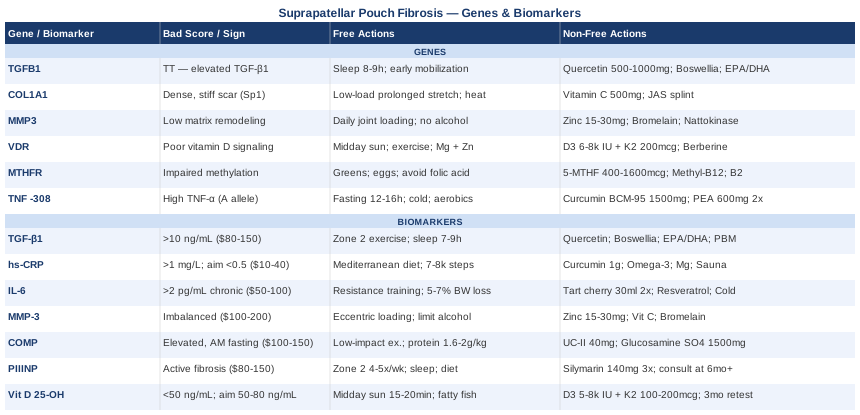
7 Biomarkers Worth Tracking for Suprapatellar Pouch Fibrosis
Blood biomarkers do not give you a diagnosis — they give you a direction. The seven listed here were selected because each one captures a distinct layer of the biological process driving suprapatellar fibrosis: active collagen synthesis, inflammatory signaling, matrix remodeling capacity, joint structural degradation, and the vitamin D axis. Tracking these systematically, ideally over serial measurements spaced 8–12 weeks apart, gives you a functional picture of whether the fibrotic process is active, resolving, or being maintained by persistent upstream signals.
Biomarker 1: TGF-β1 — The Central Driver of Fibrosis
Why it matters: TGF-β1 is the single most important molecular driver of fibrosis in virtually every tissue type, including the synovial and periarticular tissues of the knee. It activates fibroblasts into myofibroblasts — cells that aggressively deposit collagen types I and III, resist programmed cell death, and sustain scar formation long after the initial injury signal. Elevated TGF-β1 in synovial fluid and serum has been documented in post-surgical knee fibrosis and arthrofibrosis, and persistently elevated levels correlate with worse range-of-motion outcomes after total knee arthroplasty. This is the most direct biochemical signal of active fibrotic activity in the suprapatellar region.
How to measure it: TGF-β1 is measured in serum or plasma via ELISA. Available through specialty labs including LabCorp, Quest Diagnostics, and functional medicine panels. Cost typically ranges from $80 to $150. Handling protocols matter — platelet activation artificially elevates results. Request platelet-poor plasma when possible for the most accurate reading. Serial measurements at 8–12 week intervals are most informative.
If TGF-β1 Is Elevated: The Plan Without Supplements
The first intervention priority is mechanical: reduce repetitive stress on the suprapatellar pouch during the acute phase. Progressive range-of-motion exercises should replace aggressive forced stretching, since excessive passive joint force can paradoxically trigger additional TGF-β1 release from stressed fibroblasts. Consistent moderate aerobic exercise — at conversational pace, not high intensity — is one of the most consistent interventions for lowering systemic TGF-β1 over a 6–12 week window. Prioritizing 7–9 hours of quality sleep matters significantly: TGF-β1 expression is upregulated by sleep fragmentation and chronic cortisol elevation. Eliminating ultra-processed foods, especially refined carbohydrates that sustain hyperinsulinemia, reduces the inflammatory milieu that maintains TGF-β1 elevation. These are permanent lifestyle adjustments, not short-term protocols.
If TGF-β1 Is Elevated: The Plan With Supplements or Equipment
Quercetin is one of the most studied natural TGF-β1 modulators. A dose of 500–1000 mg/day with meals has demonstrated TGF-β1 signal suppression and reduction of fibroblast-to-myofibroblast transition in multiple human and preclinical studies. Cycling: 5 days on, 2 days off. Side effects: mild gastrointestinal discomfort at higher doses; rare headache.
Boswellia serrata standardized to 65% AKBA: 300–400 mg three times daily with food. Multiple randomized controlled trials demonstrate reduction of inflammatory mediators including TGF-β-driven pathways. Cycling: continuous use for 8–12 weeks, then reassess. Side effects: occasional GI upset; potential interaction with anticoagulants.
EPA/DHA (fish oil): 3–4 g/day of combined EPA+DHA. Omega-3 fatty acids reduce TGF-β1-driven pro-fibrotic gene expression in multiple tissue contexts and compete with arachidonic acid in pro-inflammatory cascades. Cycling: continuous use, reassess at 3 months. Side effects: fishy breath, GI distress at high doses; anticoagulant consideration.
Photobiomodulation (red and near-infrared light) applied directly to the knee has demonstrated TGF-β1 reduction in fibrotic tissue in multiple studies — detailed further in the complementary approaches section. This is among the most relevant equipment-based interventions for this biomarker specifically.
Biomarker 2: hs-CRP — The Accessible Window Into Systemic Inflammation
Why it matters: High-sensitivity C-reactive protein is the most accessible marker of low-grade systemic inflammation. In suprapatellar pouch fibrosis, sustained low-grade inflammation maintains the TGF-β1 signaling loop that drives progressive scarring. A chronically elevated hs-CRP above 1–2 mg/L signals that the body's inflammatory resolution mechanisms are not completing their task — that the acute response to surgery or injury has become a chronic background state. While not specific to knee fibrosis, it provides a practical, inexpensive window into whether systemic inflammation is fueling the local fibrotic environment.
How to measure it: Standard blood panel, available at any lab. Cost: $10–40. Target range: below 0.5 mg/L represents genuinely optimal anti-inflammatory status — a threshold Peter Attia consistently references in Outlive as the meaningful goal, well below the conventional "normal" ceiling of 3 mg/L.
If hs-CRP Is Elevated: The Plan Without Supplements
The foundational intervention is removing the most common structural drivers of elevated hs-CRP: excess visceral adipose tissue, poor sleep quality, sedentary behavior, and dietary patterns rich in refined seed oils and added sugars. A Mediterranean-pattern diet has more rigorous evidence for hs-CRP reduction than most supplements. Time-restricted eating within a 10–12 hour feeding window reduces inflammatory tone in multiple human trials. Daily walking of 7,000–8,000 steps is one of the most consistent hs-CRP interventions available, requiring no equipment or cost.
If hs-CRP Is Elevated: The Plan With Supplements or Equipment
Curcumin with piperine or in liposomal form: 1000–1500 mg/day of curcuminoids. Meta-analyses of RCTs confirm significant hs-CRP reduction. Cycling: 8 weeks on, 2 weeks off. Side effects: GI sensitivity; caution with anticoagulants.
Omega-3 fatty acids: 3–4 g EPA+DHA daily, as above.
Magnesium glycinate: 300–400 mg before bed. Magnesium deficiency is consistently associated with elevated hs-CRP, and correction modestly reduces inflammatory markers while also supporting sleep quality. Cycling: continuous. Side effects: loose stools at high doses.
Sauna use — 3–4 sessions per week at 75–80°C for 15–20 minutes — has demonstrated hs-CRP reduction in Finnish epidemiological studies and is compatible with knee rehabilitation in most phases of recovery.
Biomarker 3: IL-6 — The Chronic Inflammatory Sustainer
Why it matters: IL-6 plays a paradoxical role in fibrosis. Acutely after injury or surgery, IL-6 is necessary for tissue repair and immune mobilization. Chronically elevated IL-6, however, sustains the TGF-β1/SMAD signaling axis that drives fibroblast activity and scar consolidation in synovial tissue. In post-surgical knee fibrosis, persistently elevated IL-6 in synovial fluid is associated with poor range-of-motion outcomes months after the procedure. Tracking serum IL-6 in the months following surgery reveals whether the inflammatory process has properly concluded or continues driving scar formation.
How to measure it: Available via specialized inflammation panels at LabCorp, Vibrant Wellness, and Cleveland Heart Lab. Cost: $50–100. IL-6 has a short half-life in blood and results vary with sample timing — morning fasting samples offer the most consistent baseline readings.
If IL-6 Is Elevated: The Plan Without Supplements
Resistance training is one of the strongest evidence-based interventions for reducing chronic resting IL-6. The key distinction is between the acute IL-6 spike from exercise — which is beneficial, stimulating anti-inflammatory IL-10 release — and the chronically elevated baseline that signals metabolic dysfunction or muscle atrophy. Progressive resistance training, even focused on the uninjured limb during knee rehabilitation, reduces systemic resting IL-6 over 8–12 weeks. Weight loss of 5–7% in individuals with excess body fat produces significant drops in resting IL-6.
If IL-6 Is Elevated: The Plan With Supplements or Equipment
Tart cherry concentrate: 30 ml twice daily or an equivalent capsule form. Multiple sports medicine trials show meaningful reductions in post-exercise IL-6 and inflammatory cytokines. Cycling: continuous. Side effects: watch for high sugar content in some commercial products — choose unsweetened concentrate.
Resveratrol: 250–500 mg/day with a fat-containing meal for absorption. Has demonstrated IL-6 suppression in human trials involving metabolic syndrome populations. Cycling: 8 weeks on, 2–4 weeks off. Side effects: possible estrogenic effects at very high doses; mild GI adjustment.
Cold water immersion at 12–15°C for 10 minutes, 3–4 times per week has demonstrated consistent reductions in circulating IL-6 in multiple human trials and requires minimal equipment cost.
Biomarker 4: MMP-3 — The Matrix Remodeling Readout
Why it matters: Matrix metalloproteinase-3 (stromelysin-1) is an enzyme that degrades proteoglycans, fibronectin, and multiple collagen subtypes, while also activating other MMPs essential for tissue remodeling. In healthy healing, MMP-3 and related enzymes clear excess ECM, resolving the scar. In dysfunctional fibrosis, an imbalance between MMPs and their inhibitors — tissue inhibitors of metalloproteinases (TIMPs) — leads to abnormal collagen accumulation and scar persistence. Serum MMP-3 that is either too high (indicating active joint degradation) or too low (suggesting insufficient matrix remodeling capacity) represents a meaningful signal in the context of suprapatellar fibrosis.
How to measure it: Available through specialty immunology panels and functional medicine labs, often included in rheumatological marker panels. Cost: $100–200. Reference range interpretation is most useful in rheumatology-specialized lab contexts.
If MMP-3 Is Imbalanced: The Plan Without Supplements
Consistent joint loading — even partial — is the most direct stimulator of MMP activity through mechanotransduction. Fibroblasts exposed to cyclical mechanical shear upregulate MMP expression, which is why movement is antifibrotic at a molecular level. Eccentric loading protocols for the quadriceps, applied cautiously under physiotherapy supervision, have the most evidence for favorable MMP/TIMP rebalancing in periarticular soft tissue. Reduce alcohol intake: chronic alcohol consumption measurably suppresses MMP function across tissue types.
If MMP-3 Is Imbalanced: The Plan With Supplements or Equipment
Zinc bisglycinate: 15–30 mg/day with food. Zinc is a structural cofactor for all MMP enzymes; deficiency directly impairs matrix remodeling capacity. Cycling: 12 weeks, then reassess serum zinc. Side effects: nausea on an empty stomach; copper depletion at doses above 40 mg/day — add 2 mg copper if supplementing for extended periods.
Vitamin C: 500–1000 mg/day as ascorbate. Required for collagen cross-linking quality and indirectly supports MMP regulation. Cycling: continuous. Side effects: GI sensitivity at high doses; theoretical kidney stone risk in susceptible individuals at doses exceeding 2 g/day.
Bromelain: 500–1000 mg taken between meals on an empty stomach. Bromelain supports proteolytic soft-tissue activity and has demonstrated reduced fibrotic adhesion formation in musculoskeletal contexts. Cycling: 4 weeks on, 2 weeks off. Side effects: thinned blood at high doses; potential allergy in pineapple-sensitive individuals.
Biomarker 5: COMP — Joint Matrix Integrity Signal
Why it matters: Cartilage Oligomeric Matrix Protein is a large structural protein found in articular cartilage, tendon, and synovial tissue. Elevated serum COMP reflects accelerated breakdown of joint matrix — a common concurrent process alongside suprapatellar fibrosis. In post-surgical knees, elevated COMP indicates that while fibrous tissue accumulates, the underlying articular and periarticular architecture is simultaneously degrading. This dual process — fibrosis plus matrix loss — explains why some knees deteriorate from multiple biological directions simultaneously. COMP has been validated as an early joint degeneration marker in multiple rheumatological studies.
How to measure it: Available through specialty labs and some rheumatology-focused panels. Cost: $100–150. COMP follows diurnal variation — levels peak in the morning — so standardize to fasting morning samples for accurate serial comparisons.
If COMP Is Elevated: The Plan Without Supplements
Reducing repetitive compressive loading is the first priority. Low-impact activities — swimming, stationary cycling, and water-based exercise — maintain quadriceps strength and joint mobility without the mechanical stress that further elevates COMP. Body weight management is directly relevant: each pound of excess body weight adds approximately four pounds of force across the knee joint under walking conditions. Ensuring adequate protein intake (1.6–2.0 g per kilogram of body weight per day) supports matrix repair substrate availability.
If COMP Is Elevated: The Plan With Supplements or Equipment
Undenatured type II collagen (UC-II): 40 mg/day — a strikingly low dose that works through oral tolerance mechanisms rather than substrate provision. Multiple RCTs have demonstrated reductions in joint discomfort and improvements in cartilage biomarkers with this specific form. This is distinct from hydrolyzed collagen powders. Cycling: continuous use for a minimum of 90 days before reassessment. Side effects: very well tolerated; rare mild GI effects.
Glucosamine sulfate: 1500 mg/day as the sulfate form, not hydrochloride. Multiple controlled trials support its role in ECM integrity and modestly reducing COMP elevation in OA populations, with the sulfate form showing more consistent evidence than HCl. Cycling: continuous. Side effects: mild GI discomfort; theoretical concern with shellfish allergy, though evidence for cross-reactivity is limited.
Biomarker 6: PIIINP — The Marker of Active Fibrosis
Why it matters: PIIINP (N-terminal propeptide of type III procollagen) is released into circulation when type III collagen — the form predominantly produced during early fibrotic remodeling — is being actively synthesized. Elevated serum PIIINP is a direct, quantitative marker of ongoing fibrosis. It is used clinically to monitor hepatic fibrosis and is increasingly relevant to musculoskeletal contexts. For suprapatellar pouch fibrosis specifically, an elevated PIIINP weeks or months after the original injury or surgery signals that scar formation is still actively proceeding rather than resolving. This makes it one of the most actionable biomarkers in this list: if elevated long after the acute phase, intervention is warranted.
How to measure it: Available through hepatology and fibrosis-focused panels at specialty labs. Cost: $80–150. Less commonly ordered in orthopedic contexts — a functional medicine physician or an informed specialist is typically needed to requisition it. Worth the effort if rehabilitation is stalling.
If PIIINP Is Elevated: The Plan Without Supplements
Breaking the TGF-β1 → collagen synthesis cycle is the core objective. The mechanical and lifestyle strategies described above — consistent moderate aerobic exercise (zone 2 pacing, 30–45 minutes, 4–5 days per week), anti-inflammatory diet, sleep optimization — are the primary non-supplement levers. Avoiding extreme caloric restriction matters: severe restriction can paradoxically impair collagen remodeling and worsen matrix quality even while reducing fibrotic signaling temporarily.
If PIIINP Is Elevated: The Plan With Supplements or Equipment
Silymarin (milk thistle standardized extract): 140 mg three times daily. Silymarin has human evidence for reducing PIIINP in the context of hepatic fibrosis, and its anti-fibrotic mechanism — TGF-β1 downregulation and antioxidant protection of fibroblasts — is directly relevant to joint fibrosis. Cycling: 8–12 weeks on, 4 weeks off. Side effects: generally well tolerated; rare allergic reactions in ragweed-sensitive individuals.
For persistently elevated PIIINP despite 6 months of comprehensive lifestyle and supplement optimization, discussion with a specialist about prescription antifibrotic agents (such as nintedanib, used in pulmonary fibrosis) is warranted. This represents a situation where conventional medicine and functional medicine must work together.
Biomarker 7: 25-OH Vitamin D — The Anti-Fibrotic Hormone
Why it matters: Vitamin D is not merely a bone health nutrient. The vitamin D receptor (VDR) is expressed on fibroblasts, synoviocytes, and immune cells involved in fibrosis regulation. VDR activation directly inhibits TGF-β1-driven fibroblast activation, reduces pro-inflammatory cytokine production including IL-6 and TNF-α, and supports regulatory T-cell populations that limit excessive fibrotic responses. Multiple studies have found that patients experiencing post-surgical knee complications — including arthrofibrosis — have significantly lower vitamin D levels than those with uncomplicated recovery. It is inexpensive to test, straightforward to address, and broadly relevant.
How to measure it: Standard blood test, widely available. Cost: $30–60. The optimal fibrosis-relevant target range is 50–80 ng/mL (125–200 nmol/L) — substantially higher than the conventional laboratory "sufficient" cutoff of 30 ng/mL. Researchers like Peter Attia and Rhonda Patrick consistently recommend targeting 40–60 ng/mL as a minimum, with 60–80 ng/mL as the more protective target for inflammatory conditions.
If Vitamin D Is Low: The Plan Without Supplements
Direct sunlight exposure — 15–20 minutes of midday sun on significant skin surface area (arms, torso) — generates approximately 10,000–20,000 IU of vitamin D3 in fair-skinned individuals and can meaningfully improve levels over several weeks. Dietary sources (fatty fish, egg yolks, liver) provide smaller but meaningful contributions. Critically, magnesium adequacy is essential: magnesium is required for vitamin D activation, and many vitamin D-deficient individuals are simultaneously magnesium-insufficient — supplementing vitamin D without addressing magnesium can produce attenuated results.
If Vitamin D Is Low: The Plan With Supplements or Equipment
Vitamin D3 (cholecalciferol): For levels below 30 ng/mL, begin at 5000–8000 IU/day with a fatty meal for 8–12 weeks, then retest. For maintenance once the target range is reached: 2000–4000 IU/day. Always co-supplement with vitamin K2 (MK-7 form, 100–200 mcg/day) to guide calcium appropriately and reduce theoretical arterial calcification risk. Cycling: continuous year-round where sun exposure is limited. Side effects: hypercalcemia is a theoretical risk at doses above 10,000 IU/day sustained long-term — retest every 3 months until stable.
Having established the seven key circulating biomarkers, the next layer is understanding why some individuals show persistently unfavorable values on multiple of these markers regardless of lifestyle quality — which is where genetic predisposition becomes the explanatory bridge.
6 Genes That Shape Your Risk for Excessive Joint Fibrosis
Genetics does not determine fate in fibrosis — but it sets the biological baseline from which you are working. Understanding which variants you carry changes the nature and intensity of the compensatory strategies worth pursuing. The following six genes represent the most clinically meaningful variants in the fibrosis, collagen regulation, and inflammatory biology relevant to suprapatellar pouch conditions. Genetic testing via direct-to-consumer platforms (Nebula Genomics, SelfDecode) or physician-ordered panels can identify your status on each of these variants.
Gene 1: TGFB1 — The Master Fibrosis Switch
What it is: The TGFB1 gene directly encodes the TGF-β1 protein. The most studied variant is rs1800469 (C-509T in the promoter region). The T allele at this position is associated with significantly higher TGF-β1 production compared to the C allele. Individuals homozygous for the T allele (TT genotype) can produce substantially elevated TGF-β1 in response to tissue injury, making them biologically predisposed to stronger fibrotic responses. This is a variant Gary Brecka has highlighted in his discussions of why tissue recovery varies so dramatically between individuals with apparently similar injuries and surgeries.
What it may affect: In practice, TT genotype individuals tend to form more prominent scar tissue after procedures, show greater keloid tendency, and in orthopedic contexts show higher rates of arthrofibrosis and post-surgical stiffness. The effect is amplifying, not causative: this variant does not cause fibrosis independently, but it dramatically amplifies the fibrotic response to any given trigger — surgery, infection, or repeated micro-trauma.
If TGFB1 Is Unfavorable: The Plan Without Supplements
The most important leverage point is minimizing fibrosis triggers, not just the response. This means: prioritizing early, gentle joint mobilization immediately post-surgery (the sooner the joint moves, the less TGF-β1-driven scar consolidation occurs); maintaining a consistently anti-inflammatory diet year-round rather than only during acute flares; and managing psychological stress rigorously — cortisol directly upregulates TGFB1 gene transcription. Sleep extension to 8–9 hours is protective: deep NREM sleep phases are when anti-inflammatory resolution pathways are most active and TGF-β1 production is lowest. These are lifelong adaptations for individuals with the TT genotype, not short-term cycling protocols.
If TGFB1 Is Unfavorable: The Plan With Supplements or Equipment
Prioritize the TGF-β1-specific interventions from the biomarker section above: quercetin (500–1000 mg/day), boswellia (300–400 mg three times daily), and EPA/DHA (3–4 g/day). Add grape seed extract standardized to oligomeric proanthocyanidins (OPCs): 150–300 mg/day. Human studies show proanthocyanidins from grape seed significantly reduce TGF-β1-driven fibroblast activity. Cycling: 8 weeks on, 2 weeks off. Side effects: well tolerated; rare headache.
Photobiomodulation therapy (see complementary approaches section) is particularly indicated for TGFB1 high-producer genotypes undergoing post-surgical rehabilitation — the direct inhibitory effect on TGF-β1 at the fibroblast level makes it mechanistically well-matched to this variant.
Gene 2: COL1A1 — Collagen Structural Programming
What it is: COL1A1 encodes the alpha-1 chain of type I collagen, the dominant structural protein in scar tissue. The most studied variant, rs1800012 (the Sp1 binding site polymorphism), affects transcription factor binding efficiency and thereby collagen I production rate. Variants associated with increased type I collagen production contribute to a denser, less pliable scar matrix — the kind that resists mobilization and creates the characteristic "woody" resistance encountered when attempting to restore knee flexion after fibrosis has consolidated.
What it may affect: Individuals with COL1A1 variants associated with elevated collagen I synthesis tend to form harder, mechanically stiffer scars that respond less to standard stretching protocols. The fibrosis in their suprapatellar pouch may require longer, more consistent mechanical intervention to achieve the same degree of tissue extensibility.
If COL1A1 Is Unfavorable: The Plan Without Supplements
Mechanical loading strategies should prioritize tissue extensibility over force. Low-load, prolonged-duration stretching (30–60 minutes per session using progressive static loading devices or serial casting approaches) is more effective than brief high-force stretching for modifying dense type I collagen scar tissue. This principle — creep and stress relaxation of collagen fibers — is well-supported in the orthopedic contracture literature. Temperature augmentation matters: heating the tissue to 40–42°C before stretching significantly increases collagen extensibility. A warm bath or thermal wrap before range-of-motion work is a simple, free application of this principle.
If COL1A1 Is Unfavorable: The Plan With Supplements or Equipment
Vitamin C (500–1000 mg/day): critical for collagen cross-linking quality. Adequate vitamin C produces well-organized, extensible collagen fibers rather than the disorganized, brittle cross-links associated with deficiency.
Joint Active Systems (JAS) dynamic splint or similar low-load prolonged-duration knee extension/flexion brace: Clinical studies have demonstrated significant range-of-motion recovery in post-surgical knee contracture using these devices, which apply constant low-level mechanical loading directly to the scar tissue of the suprapatellar pouch and surrounding structures. Frequency: 30–60 minutes, 1–2 sessions per day. Side effects: skin pressure at contact points; requires proper fitting by a physiotherapist to avoid adverse mechanical stress.
Gene 3: MMP3 — Matrix Remodeling Capacity
What it is: The MMP3 gene encodes stromelysin-1. Key variants including rs679620 and related promoter polymorphisms affect transcription rate and enzyme expression. Low-activity MMP3 variants reduce the body's ability to degrade and remodel accumulated scar matrix — creating a biological situation where collagen is deposited but cannot be adequately cleared, effectively a one-way ratchet toward progressive fibrosis. This variant interacts directly with the MMP-3 biomarker — individuals with low-activity MMP3 alleles may show chronically low serum MMP-3 alongside poor tissue remodeling.
What it may affect: Individuals with low-activity MMP3 variants may find that fibrosis persists longer, responds less reliably to mobilization, and is more likely to recur following arthroscopic debridement. The biological context for their rehabilitation is fundamentally different from someone with normal MMP3 function.
If MMP3 Is Unfavorable: The Plan Without Supplements
Consistent daily joint loading is more important than periodic intensive therapy for individuals with reduced MMP3 activity. Mechanical shear stress on fibroblasts upregulates MMP expression, and this effect accumulates with frequency of loading rather than intensity. Daily walking, cycling, and active range-of-motion exercises — even brief sessions — provide the mechanotransduction signals that partially compensate for reduced baseline MMP3 expression. Reduce alcohol intake, which measurably suppresses MMP function systemically.
If MMP3 Is Unfavorable: The Plan With Supplements or Equipment
Zinc bisglycinate and vitamin C as cofactors: as per the biomarker section doses above. Both are structurally required for functional MMP enzyme activity.
Bromelain (500–1000 mg between meals): supports proteolytic activity in soft tissue, partially compensating for reduced MMP3-mediated matrix clearance. Cycling: 4 weeks on, 2 weeks off.
Nattokinase: 100–200 mg (2000 FU) twice daily on an empty stomach. A serine protease with evidence for fibrinolytic and fibrous tissue-degrading activity in cardiovascular and venous contexts. Evidence for joint fibrosis is more limited but mechanistically plausible for individuals with reduced MMP activity. Cycling: 8 weeks on, 4 weeks off. Side effects: anticoagulant interaction — avoid with blood thinners without physician guidance.
Gene 4: VDR — How Well the Anti-Fibrotic Vitamin D Signal Is Received
What it is: The VDR gene encodes the vitamin D receptor — the nuclear receptor that translates circulating vitamin D into gene expression changes in fibroblasts, immune cells, and synoviocytes. Multiple common variants, including FokI (rs2228570), BsmI (rs1544410), and TaqI (rs731236), alter receptor binding affinity, protein length, or expression levels. These variants affect how efficiently vitamin D activates its anti-fibrotic and anti-inflammatory pathways, entirely independently of how much vitamin D is circulating. Ali Torkamani and Gary Brecka have both emphasized VDR variants as among the most commonly overlooked reasons why individuals fail to respond to standard vitamin D supplementation.
What it may affect: A person with low-function VDR variants may show serum vitamin D of 55–60 ng/mL yet still experience attenuated anti-fibrotic signaling because the receptor is not efficiently transducing the signal. Standard lab work would show "sufficient" levels while the cellular anti-fibrotic effect remains blunted.
If VDR Is Unfavorable: The Plan Without Supplements
Maximize receptor activation through consistent midday sun exposure — direct UV contact with skin activates the full VDR pathway more completely than supplemental oral vitamin D in some models. Regular moderate exercise upregulates VDR expression in muscle and immune cells, representing a non-supplement compensatory mechanism. Magnesium, zinc, and vitamin K2 adequacy support downstream VDR signaling pathways.
If VDR Is Unfavorable: The Plan With Supplements or Equipment
Target higher serum vitamin D levels — aim for 70–80 ng/mL to compensate for receptor inefficiency. This typically requires 6000–8000 IU/day of D3, confirmed by bloodwork at 3-month intervals. Always pair with K2 (MK-7, 200 mcg/day) and magnesium (300–400 mg/day). Cycling: continuous with quarterly monitoring.
Berberine: 500 mg three times daily with meals. Activates overlapping anti-inflammatory and metabolic pathways and has been identified as a VDR-adjacent agonist in some research contexts. Cycling: 3 months on, 1 month off. Side effects: significant GI adjustment during the first 2 weeks; potential interaction with metformin; avoid during pregnancy.
Gene 5: MTHFR — Methylation and the Inflammatory Threshold
What it is: MTHFR encodes methylenetetrahydrofolate reductase, an enzyme central to one-carbon metabolism. The highly prevalent variants C677T (rs1801133) and A1298C (rs1801131) reduce enzyme efficiency, impairing the conversion of folate to its biologically active methylated form. This creates downstream impairments in methylation reactions that regulate inflammatory gene expression, antioxidant production (particularly glutathione), and collagen metabolism. Gary Brecka has placed MTHFR at the center of his clinical framework for explaining why some individuals have constitutionally elevated inflammatory baselines and impaired tissue recovery.
What it may affect: Impaired methylation is associated with elevated homocysteine, reduced glutathione, increased oxidative stress, and dysregulated epigenetic silencing of inflammatory genes. In the suprapatellar fibrosis context, poor methylation may mean that the epigenetic "off switches" for fibrogenic gene expression are less effective — allowing pro-fibrotic pathways to remain active longer after the initial injury stimulus has resolved.
If MTHFR Is Unfavorable: The Plan Without Supplements
Maximize dietary methyl donors: dark leafy greens (spinach, romaine, arugula), whole eggs (including the yolk), organ meats especially liver, and legumes. Critically: avoid folic acid in fortified foods and most standard multivitamins. Individuals with MTHFR variants cannot efficiently convert synthetic folic acid to its active form, and unmetabolized folic acid accumulation may worsen outcomes. Reduce alcohol intake — alcohol depletes B vitamins and directly impairs methylation pathways. Sauna use supports glutathione cycling, partially compensating for reduced endogenous production.
If MTHFR Is Unfavorable: The Plan With Supplements or Equipment
Methylfolate (5-MTHF): 400–800 mcg/day for C677T heterozygous; up to 1000–1600 mcg/day for homozygous TT. Start at the lower dose and increase gradually — some individuals experience irritability or anxiety when initiating methylfolate too rapidly (overmethylation symptoms). Cycling: continuous. Side effects: reduce dose if anxiety, irritability, or insomnia develops.
Methylcobalamin (B12): 500–1000 mcg/day sublingual or oral. Works synergistically with methylfolate to support the full methylation cycle. Cycling: continuous. Side effects: well tolerated; rare acne in some individuals.
Riboflavin (B2): 25–100 mg/day. Riboflavin is a direct cofactor for MTHFR enzyme function and partially compensates for reduced enzyme efficiency regardless of the variant. Cycling: continuous. Side effects: bright yellow urine (benign); photosensitivity at very high doses.
Gene 6: TNF (-308 G/A) — The Inflammatory Amplifier
What it is: The TNF gene encodes tumor necrosis factor alpha, a master pro-inflammatory cytokine that activates NF-κB signaling, drives IL-6 and IL-1β production, and directly stimulates TGF-β1 expression in fibroblasts. The -308 G/A variant (rs1800629) is a promoter polymorphism where the A allele is associated with significantly higher TNF-α transcription — in some studies, 2–3 times higher than the G/G genotype. Individuals carrying the A allele (heterozygous GA or homozygous AA) have a constitutionally lower threshold for sustained inflammatory responses to injury, infection, or metabolic stress.
What it may affect: Higher baseline TNF-α output means that any articular injury or surgical procedure triggers a more intense and more prolonged inflammatory cascade — one that is substantially more likely to transition from acute inflammation (repair) to chronic inflammation (fibrosis). This is particularly relevant in post-surgical suprapatellar fibrosis, where the surgical procedure itself is the initial fibrosis trigger.
If TNF Is Unfavorable: The Plan Without Supplements
The most impactful non-pharmaceutical interventions for elevated TNF-α are: intermittent fasting or time-restricted eating (a 12–16 hour fasted window), which directly suppresses NF-κB activation; elimination of pro-inflammatory dietary components including refined seed oils, high-fructose corn syrup, and trans fats; and consistent moderate exercise — TNF-α resting levels are durably reduced with regular aerobic activity. Cold exposure (cold showers or brief cold immersion) measurably reduces TNF-α through sympathetic nervous system modulation.
If TNF Is Unfavorable: The Plan With Supplements or Equipment
Curcumin (BCM-95 or phytosomal form): 1500–2000 mg/day. Multiple human RCTs demonstrate direct NF-κB and TNF-α suppression with these higher-bioavailability formulations. Cycling: 8 weeks on, 2 weeks off. Side effects: GI sensitivity; caution with anticoagulants.
EPA/DHA: 3–4 g/day as above. Omega-3 fatty acids compete with arachidonic acid in TNF-α synthesis pathways, providing meaningful downstream TNF suppression.
PEA (palmitoylethanolamide): 600 mg twice daily. A naturally occurring lipid mediator with strong human evidence for anti-inflammatory effects through mast cell modulation and peripheral neuropathic signaling — relevant when the knee pain component includes inflammatory sensitization, which is common in prolonged fibrosis. Cycling: 3 months on, 1 month off. Side effects: very well tolerated; no known significant drug interactions.
With both biomarker and genetic strategies in hand, it is worth stepping back to see how the broader framework of inflammation tracking — as articulated by one of the most evidence-rigorous voices in longevity medicine — ties these threads together.
What Peter Attia's Outlive Gets Right About Inflammation and Chronic Tissue Conditions
Why This Book Changes How You Approach Tracking
Peter Attia's Outlive: The Science and Art of Longevity (2023) is not written about knee fibrosis. But it contains the most clearly argued and evidence-grounded framework currently available for understanding how chronic, low-grade inflammation drives tissue dysfunction — the exact biological context in which suprapatellar fibrosis develops and persists. Attia trained as a surgeon and spent years as a metabolic researcher before turning to longevity practice. His approach integrates biomarker tracking with mechanistic biology in a way that translates directly to musculoskeletal fibrosis, and the ten points below are drawn from concepts he develops extensively throughout the book.
10 Things from Outlive That Apply Directly to Joint Fibrosis
1. "Inflammaging" has measurable, addressable biochemistry. Attia draws a clear distinction between acute inflammation (necessary, healing) and chronic low-grade inflammation (destructive, avoidable). Suprapatellar fibrosis fits precisely into the second category: the acute inflammatory response to surgery is normal; the failure to resolve it is the biological problem that needs addressing.
2. Lab reference ranges are calibrated for average populations, not optimal health. This distinction is critical for every biomarker in this article. The difference between "within reference range" and genuinely optimized is exactly where residual fibrotic burden hides. An hs-CRP of 1.8 mg/L is "normal" by most lab standards and still represents meaningful chronic inflammation.
3. Zone 2 aerobic training is the most consistently anti-inflammatory exercise modality. Thirty to 45 minutes of conversational-pace aerobic activity four to five days per week drives the most favorable changes in inflammatory biomarkers without the acute cytokine spikes of high-intensity work. For knee fibrosis rehabilitation, stationary cycling at low resistance directly applies this principle — maintaining quadriceps function while driving systemic anti-inflammatory adaptation.
4. Insulin resistance amplifies systemic inflammation through multiple mechanisms. Attia argues compellingly that hyperinsulinemia drives a pro-inflammatory state affecting virtually every tissue. For post-surgical knee recovery, a high HOMA-IR score (derived from fasting glucose and insulin, cost $15–30) is an underappreciated contributor to poor outcomes and merits direct intervention.
5. Sleep is a biochemical requirement for inflammatory resolution, not a lifestyle preference. Attia cites multiple RCTs demonstrating that even moderate sleep restriction — 6 hours versus 8 — significantly elevates IL-6, TNF-α, and CRP within days. For anyone managing post-surgical knee fibrosis, sleep is not separate from treatment. It is part of it.
6. Protein intake of 1.6–2.2 g per kilogram of body weight per day is necessary for tissue remodeling. Quadriceps atrophy on the injured side perpetuates abnormal joint mechanics and impairs rehabilitation. Adequate protein — particularly sources rich in leucine — activates muscle protein synthesis and provides substrate for collagen remodeling simultaneously.
7. Visceral fat is an active endocrine organ secreting pro-inflammatory cytokines. Even in individuals who appear lean, excess visceral fat (measurable by DEXA) contributes disproportionately to IL-6 and TNF-α production. Reducing visceral fat directly reduces the ongoing inflammatory load sustaining joint fibrosis.
8. VO2 max is one of the strongest predictors of long-term functional capacity. A high VO2 max reflects the mitochondrial density and cardiovascular efficiency associated with superior inflammatory resolution capacity. Training to improve VO2 max — through a combination of zone 2 base building and brief high-intensity intervals — is among the most powerful long-term investments for anyone managing chronic inflammatory conditions.
9. Tracking is the prerequisite for meaningful intervention. Attia's consistent position is that interventions without measurement are guesswork. The biomarkers in this article represent the minimum necessary surveillance system for anyone serious about modifying their fibrotic trajectory rather than simply hoping for spontaneous improvement.
10. Biological processes driving fibrosis and inflammation are more reversible than most clinicians assume. This is arguably Outlive's most practically important message applied to fibrosis: the molecular cascades sustaining scar formation respond to lifestyle, biochemical support, and targeted interventions. They are not fixed consequences of surgical history. But reversibility requires measurement, specificity, and sustained action — not generic "healthy living" advice.
Five Evidence-Based Therapies to Consider Alongside Medical Treatment
The following approaches were selected from the approved modalities based on the quality and specificity of human clinical evidence for post-surgical and inflammatory joint conditions. Each addresses a distinct aspect of suprapatellar fibrosis biology: direct tissue anti-fibrotic effects, matrix mobilization, systemic anti-inflammatory adaptation, central pain regulation, and neuromuscular rehabilitation.
1. Photobiomodulation — Direct Anti-Fibrotic Light Therapy
Photobiomodulation (PBM) uses specific wavelengths of light — typically 630–1000 nm in the red and near-infrared spectrum — to penetrate tissue and stimulate mitochondrial cytochrome c oxidase. The downstream effects include increased ATP synthesis, reduced oxidative stress, and modulation of inflammatory cytokines including IL-6 and TNF-α. Most directly relevant here: multiple studies have documented that PBM inhibits TGF-β1-driven fibroblast activation and reduces type I collagen deposition in fibrotic tissue models, making it one of the few physical interventions with a mechanistic rationale specifically matched to suprapatellar fibrosis biology.
A randomized controlled trial published in Photomedicine and Laser Surgery found significant improvements in range of motion in patients with post-surgical knee joint contracture receiving PBM versus sham treatment. A systematic review in Lasers in Medical Science found consistent evidence for PBM reducing post-surgical joint inflammation and supporting tissue healing. Wavelengths most studied for anti-fibrotic effects are 810–850 nm (near-infrared) and 630–670 nm (red light).
For practical application: use a panel or probe delivering 25–50 mW/cm² at 810 nm over the anterior knee, 10–15 minutes per session, five times per week for a minimum of 8 weeks. Consumer-grade red/NIR panels offering these parameters are available for $200–600. There is no known significant risk at recommended doses; avoid direct ocular exposure. Individuals with genetic variants associated with high TGF-β1 production (TGFB1 TT genotype) may derive particularly strong benefit given the mechanistic match.
2. Manual Soft-Tissue Mobilization — Direct Mechanical Intervention
Skilled manual soft-tissue mobilization applied to the quadriceps, suprapatellar fat pad, and periarticular structures is one of the most direct physical interventions for reducing fibrotic adhesion and improving tissue gliding. Techniques including instrument-assisted soft-tissue mobilization (IASTM), cross-fiber friction, and myofascial release work by mechanically disrupting disorganized collagen cross-links and stimulating MMP activity through mechanotransduction — directly complementing the MMP3 genetic and biomarker interventions described above. The distinguishing factor from general massage is that this work must be performed by a therapist with specific orthopedic post-surgical soft-tissue training.
A systematic review in the Journal of Orthopaedic and Sports Physical Therapy found that manual soft-tissue therapy and joint mobilization, applied within a structured rehabilitation program for post-arthroplasty arthrofibrosis, produced significantly improved range of motion compared to exercise alone. A randomized trial in the Archives of Physical Medicine and Rehabilitation specifically found patellar mobilization techniques improved range of motion and reduced stiffness in post-TKA patients.
In practice, sessions of 30–45 minutes focusing on suprapatellar region mobilization and quadriceps soft-tissue work are typically performed 2–3 times per week in the acute rehabilitation phase, transitioning to weekly maintenance sessions as range of motion improves. Progress should be tracked by goniometric measurement at each session — cumulative gains over weeks indicate an effective therapeutic response.
3. Tai Chi — Movement-Based Anti-Inflammatory Practice
Tai chi offers a distinctive combination of gentle, flowing joint movement through full available range of motion, proprioceptive retraining, and systemic anti-inflammatory effects that make it well-suited to the long-term management phase of suprapatellar fibrosis. Unlike standard therapeutic exercise, tai chi combines the joint-loading stimulus necessary for MMP activity and ECM remodeling with explicit attention to movement quality and muscular control — retraining the neuromuscular patterns disrupted by prolonged joint dysfunction. The meditative component of practice additionally reduces cortisol, the stress hormone that sustains TGFB1 gene expression.
A landmark randomized trial published in the New England Journal of Medicine (Wang et al.) compared tai chi to standard physical therapy for knee osteoarthritis and found equivalent or superior outcomes for pain, function, and quality of life at 52 weeks. While arthrofibrosis and osteoarthritis are distinct conditions, the shared inflammatory biology, joint-loading mechanics, and functional impairment patterns make this evidence directly applicable.
Begin with a beginner's 24-form or Yang-style short form under experienced instruction for the first 8–12 weeks to ensure correct knee alignment and avoid compensatory loading patterns. Daily practice of 20–30 minutes is more beneficial than periodic longer sessions. The barrier to entry is low: community classes are widely available at minimal cost, and qualified instruction significantly reduces the small but real risk of incorrect knee tracking during early practice.
4. Mindfulness-Based Stress Reduction — Targeting the Cortisol-Fibrosis Loop
The connection between psychological stress, cortisol, and fibrotic progression is mechanistic rather than metaphorical. Cortisol activates glucocorticoid receptors on fibroblasts, upregulates TGF-β1 gene expression, and suppresses the regulatory immune activity required for inflammatory resolution — a direct biological pathway from chronic stress to impaired fibrosis management. The standardized 8-week MBSR program developed by Jon Kabat-Zinn has accumulated substantial evidence for reducing inflammatory biomarkers including IL-6, TNF-α, and CRP in chronic pain and medical illness populations.
A randomized controlled trial published in Brain, Behavior, and Immunity found that MBSR produced significant reductions in circulating inflammatory cytokines compared to a control condition, with effects sustained at 3-month follow-up. Multiple subsequent meta-analyses confirm reductions in hs-CRP, IL-6, and salivary cortisol. For chronic post-surgical knee conditions, the evidence applies both to central pain sensitization management and to the inflammatory biology sustaining ongoing fibrosis.
The standard protocol involves 8 weekly group sessions of 2.5 hours plus a full-day retreat, with daily home practice of 30–45 minutes. Digitally delivered MBSR programs have been validated against in-person delivery, substantially increasing accessibility. Cost ranges from free (online resources) to $200–500 for structured programs. A small minority of individuals with significant trauma history find intensive meditation temporarily distressing — qualified instructor guidance is appropriate in those cases.
5. EMG Biofeedback — Restoring Neuromuscular Control
Electromyographic biofeedback in knee rehabilitation specifically addresses arthrogenic muscle inhibition (AMI) — the neurological suppression of quadriceps activation that develops as a protective reflex in response to joint pathology and pain. Suprapatellar fibrosis impairs normal afferent feedback from the joint, and the nervous system responds by dampening quadriceps motor output even after the primary pathology has been partially addressed. This creates a maintenance cycle: reduced quadriceps activation → abnormal joint loading → persistent fibrotic stress → more inhibition. EMG biofeedback gives patients real-time visual or auditory feedback on muscle activation, enabling conscious override of AMI.
A systematic review in Knee Surgery, Sports Traumatology, Arthroscopy found that EMG biofeedback integrated into post-surgical rehabilitation significantly improved quadriceps force production and activation versus standard exercise alone. Multiple RCTs in knee arthroplasty populations show faster and more complete quadriceps recovery with EMG biofeedback integration.
In practice, this requires a physiotherapist using surface electrodes placed over the VMO (vastus medialis oblique) and rectus femoris during targeted exercises. Sessions of 30–45 minutes, 2–3 times per week during the rehabilitation phase, represent the standard protocol. Portable home biofeedback units are available for $150–400 and allow daily practice between clinical sessions. Progress is tracked by activation threshold improvements. There are no meaningful side effects — this is a monitoring and training tool, not an intervention with tissue risk.
Conclusion
Suprapatellar pouch fibrosis sits at the intersection of immune biology, individual genetic predisposition, and mechanical rehabilitation — which is precisely why single-track approaches fall short so consistently. The seven biomarkers covered here — TGF-β1, hs-CRP, IL-6, MMP-3, COMP, PIIINP, and vitamin D — give you a measurable map of where the fibrotic process stands and whether it remains active. The six gene variants — TGFB1, COL1A1, MMP3, VDR, MTHFR, and TNF — explain why some individuals face a constitutionally steeper biological challenge and identify compensatory strategies that can meaningfully shift that terrain.
The most useful immediate next step depends on where you are in the process. If biomarker testing has not been done, start with the three most accessible and affordable: hs-CRP, 25-OH vitamin D, and IL-6. If genetic testing is available, prioritize TGFB1, MTHFR, and VDR for the highest-yield information. For those currently in active rehabilitation, layering photobiomodulation and qualified soft-tissue mobilization onto a daily zone 2 aerobic base represents the combination most consistently supported by the evidence reviewed here.
Better measurement leads to better decisions — and in this condition more than most, the gap between generic care and personalized, biology-informed management makes a genuine difference to outcomes.
Musculoskeletal: Joint Conditions Sports Injuries
Autoimmune: Inflammatory Conditions Connective Tissue Conditions