This article contains AI generated content.
· UpdatedSynovial Hemangioma — 5 Genes and 7 Biomarkers to Track
Introduction
Synovial hemangioma is one of those conditions that falls through the cracks of standard medical guidance. It is rare enough that most people dealing with unexplained joint swelling, recurrent effusions, or a vague aching in the knee that never quite matches the usual sports injury narrative will go months — sometimes years — without a clear answer. When the diagnosis finally arrives, the information that follows tends to be surgical in focus and thin on everything else.
What makes this condition genuinely complex is that it does not simply involve an abnormal structure in the joint. It involves abnormal vascular biology — blood vessels forming where and how they should not, within the delicate synovial lining of the joint. That biological process has identifiable molecular drivers. Understanding them matters, not because you can reprogram your genes, but because those drivers connect to measurable signals and to practical levers you actually have access to.
Generic anti-inflammatory advice misses most of this. A broad "eat better and reduce stress" recommendation is not wrong, but it is not specific enough to address what is actually happening at the vascular level. The biochemistry of angiogenesis, vascular instability, and synovial inflammation responds to targeted inputs — and those inputs are different from the ones that help with, say, osteoarthritis or tendinopathy.
This article approaches synovial hemangioma from two angles that can meaningfully inform your decisions. The first, and most practically actionable, examines seven biomarkers that reflect the core biological processes underlying the condition — each with a clear explanation of what it measures, how to get it tested, and what to do if the result is unfavorable. The second examines five key genes whose variants are linked to abnormal vascular development, with practical strategies for each. Neither angle offers a cure. Both offer better information — and better information, in a condition this underserved by general guidance, is genuinely transformative.
7 Biomarkers Worth Tracking in Synovial Hemangioma
Biomarkers serve a purpose that imaging cannot: they tell you what the body is doing right now, at a molecular level. A synovial hemangioma may appear stable on MRI while the vascular biology driving it remains highly active. Conversely, a lesion may be shrinking while biomarkers normalize in parallel. Tracking the right markers over time allows both you and your clinician to monitor the biological environment that either sustains or suppresses the condition.
1. VEGF-A (Vascular Endothelial Growth Factor-A)
Why it matters: VEGF-A is the primary molecular signal that tells the body to build new blood vessels. In synovial hemangioma, pathological VEGF signaling is considered one of the central mechanisms driving vascular proliferation within the synovial membrane. Studies examining synovial tissue from vascular lesions consistently document VEGF upregulation — making this the single most relevant biomarker for this condition. Evidence from hemangiomas in other tissues and broader vascular anomaly research strongly implicates VEGF-A as a therapeutic and monitoring target.
What it may reveal: Elevated serum VEGF-A reflects active angiogenic drive in the body. While a direct correlation between circulating VEGF-A and synovial hemangioma lesion size has not been established in large clinical trials (due to the rarity of the condition), extrapolation from hemangioma biology in other sites and from vascular tumor research is well-supported. Chronically elevated VEGF-A suggests a biological environment that favors lesion persistence and potential recurrence after intervention.
How to measure it: VEGF-A is measured in serum or plasma via ELISA-based laboratory analysis. It is not part of standard routine panels and must be requested explicitly, typically through a specialty or integrative medicine lab. Cost range: $50–$150 USD. Prefer plasma over serum, as platelets release large quantities of VEGF during clotting and can artificially inflate serum values.
If the score is high — the plan without supplements: Moderate-intensity aerobic exercise (30–45 minutes, 4–5 times per week) chronically normalizes VEGF signaling by improving vascular efficiency rather than simply suppressing it. Excess body fat is an independent source of VEGF production — a 5–10% reduction in body fat can measurably reduce baseline VEGF. Chronic intermittent hypoxia from untreated obstructive sleep apnea is a potent and overlooked VEGF stimulator; screening for sleep apnea is worthwhile if symptoms are present. Limiting alcohol is also relevant: alcohol upregulates VEGF-A expression in vascular tissue.
If the score is high — the plan with supplements or equipment: Berberine (500mg twice daily with meals, cycled 8 weeks on / 2 weeks off) has demonstrated VEGF-suppressing effects in preclinical and some clinical studies, primarily through AMPK activation; GI side effects are possible, particularly early in use. EGCG from green tea extract (400–600mg daily, continuous or cycled 12 weeks on / 2 weeks off) has documented antiangiogenic mechanisms including interference with VEGF receptor signaling; individuals with liver sensitivity should use lower doses. Curcumin with piperine (500–1000mg curcumin with 5mg piperine, taken with dietary fat) modulates VEGF at the transcriptional level; cycle 8 weeks on / 1 week off; generally well-tolerated.
2. D-Dimer
Why it matters: D-dimer is a fibrin degradation product — it rises when clot formation and breakdown are both active. Vascular anomalies, including hemangiomas, frequently create microenvironments with localized thrombosis and fibrin turnover, a phenomenon called localized intravascular coagulopathy (LIC). Elevated D-dimer in individuals with vascular lesions is a known clinical finding and can correlate with lesion activity, pain, and the risk of more significant coagulopathy.
What it may reveal: A persistently elevated D-dimer suggests ongoing microclot formation within or around the vascular lesion. It may also signal systemic coagulation stress, particularly in larger or more complex lesions. In the context of synovial hemangioma, it tracks whether the vascular structures within the joint are actively thrombosing and remodeling.
How to measure it: D-dimer is a standard clinical lab test available at virtually any hospital or reference lab. Cost range: $20–$60 USD. Normal is typically below 0.50 mg/L FEU, though laboratory reference ranges vary. Request a high-sensitivity D-dimer where available. Avoid testing during acute illness, which transiently elevates D-dimer regardless of underlying conditions.
If the score is high — the plan without supplements: Breaking prolonged sitting every 45–60 minutes with brief movement is one of the most direct behavioral interventions for elevated D-dimer, as venous stasis directly promotes coagulation. If the affected limb is involved, medical-grade graduated compression reduces local venous pooling. Adequate hydration (2–3 liters of water per day) reduces blood viscosity and coagulation tendency — a simple but often overlooked baseline.
If the score is high — the plan with supplements or equipment: Nattokinase (100–200mg / 2000–4000 FU, once daily away from meals) is a fibrinolytic enzyme with human evidence for reducing D-dimer and supporting fibrin clearance; do not use with anticoagulant medications without medical supervision; retest after 8–12 weeks. Omega-3 fatty acids (2–4g EPA+DHA daily, continuous) have well-documented anticoagulant and anti-inflammatory effects — note that at higher doses they can extend bleeding time, relevant if surgical intervention is being planned. Intermittent pneumatic compression devices (used 20–30 minutes daily) are evidence-based for reducing venous coagulation markers and improving lymphatic flow; home-use units are available in the $150–$400 range.
3. hsCRP (High-Sensitivity C-Reactive Protein)
Why it matters: Synovial hemangioma is not primarily an inflammatory condition, but the synovial environment it creates becomes chronically inflamed secondarily. Vascular engorgement, microbleeds into the joint space, and repeated mechanical irritation all drive synovial inflammatory signaling. hsCRP is the most reliable and widely accessible general-purpose inflammatory biomarker, and it is consistently highlighted by clinicians including Peter Attia as a foundational tracking marker for systemic inflammatory load.
What it may reveal: An hsCRP above 1.0 mg/L signals meaningful systemic inflammation; above 3.0 mg/L indicates significant risk-level inflammation. In a joint condition, it also tracks whether the synovium is actively inflamed or relatively quiescent — a practical guide to how much immune activity is occurring around the lesion.
How to measure it: Standard lab test, universally available. Cost range: $15–$40 USD. Peter Attia's framework targets below 0.5–0.8 mg/L for optimal systemic health. Test fasting and avoid testing within two weeks of any acute illness, which transiently elevates CRP regardless of chronic status.
If the score is high — the plan without supplements: Consistent sleep of 7–9 hours per night is one of the most powerful single interventions for reducing hsCRP; sleeping below 6 hours is independently associated with CRP elevation regardless of other health factors. Eliminating ultra-processed foods removes the trans fats and advanced glycation end-products that are among the strongest dietary drivers of systemic inflammation. Regular moderate exercise reduces CRP independently of weight change; sedentary behavior elevates it.
If the score is high — the plan with supplements or equipment: High-dose omega-3s (3–4g EPA+DHA daily, continuous) are among the most robustly evidenced supplements for reducing hsCRP. Curcumin with piperine (dosing as above) is a well-characterized anti-inflammatory that pairs effectively with omega-3s. Infrared sauna (15–20 minutes, 3–4 times per week) has accumulating evidence for reducing hsCRP through heat shock protein activation and improved vascular tone; home units range from $1,000–$4,000, and access through gyms or wellness centers reduces the entry cost.
4. MMP-9 (Matrix Metalloproteinase-9)
Why it matters: MMP-9 is an extracellular matrix-degrading enzyme that enables new blood vessels to penetrate and expand through tissue. It plays a central mechanistic role in pathological angiogenesis — precisely the kind seen in hemangiomas. MMP-9 is also central to synovial tissue remodeling. Elevated MMP-9 in serum or synovial fluid is associated with more aggressive vascular lesion activity and with joint structural changes over time.
What it may reveal: High MMP-9 suggests active matrix degradation and vascular expansion within the lesion. It is particularly useful for tracking whether a known lesion is stable or progressing in biological terms, independent of what imaging shows at a single time point.
How to measure it: Not a standard clinical panel; must be accessed through specialty or integrative medicine labs. Cost range: $80–$200 USD. Plasma is the preferred sample type. Some vascular disease specialty panels include MMP-9 alongside VEGF and other angiogenic markers.
If the score is high — the plan without supplements: Reducing dietary glycemic load is one of the fastest interventions: blood glucose spikes acutely elevate MMP-9 expression in vascular endothelial cells. Alcohol significantly upregulates MMP-9 and should be reduced or eliminated during active monitoring periods. Moderate resistance training (3 times per week, progressive loading) has been shown to reduce baseline MMP-9 over time, though high-intensity overtraining may transiently elevate it.
If the score is high — the plan with supplements or equipment: Resveratrol (250–500mg daily with meals, cycled 12 weeks on / 4 weeks off) inhibits MMP-9 expression through the NF-κB pathway; avoid with anticoagulant medications. Sub-antimicrobial dose doxycycline (20–40mg per day, prescription only) is used as an MMP inhibitor at doses too low for antibiotic effect — it has been studied in vascular conditions and periodontitis with documented MMP-9 reduction; requires physician supervision. EGCG (dosing as above) directly inhibits MMP-9 activity and pairs with its antiangiogenic VEGF effect, making it a versatile option across multiple mechanisms.
5. Angiopoietin-2 (Ang-2)
Why it matters: Angiopoietin-2 is a vascular destabilization signal. When Ang-2 is elevated relative to its stabilizing counterpart Ang-1, blood vessels lose normal structural integrity — they become leaky, prone to remodeling, and susceptible to pathological angiogenic expansion. In hemangiomas and synovial vascular lesions, Ang-2 upregulation is a consistent molecular finding and contributes to the vascular instability that causes joint effusion, swelling episodes, and lesion recurrence after treatment.
What it may reveal: Elevated Ang-2 reflects active vascular instability and ongoing angiogenic remodeling. When paired with Ang-1, the Ang-2/Ang-1 ratio provides a more complete picture of vascular balance. High ratios indicate a biological environment strongly favoring continued vascular disruption.
How to measure it: Ang-2 is measured via ELISA in specialized laboratories; it is not yet a routine clinical biomarker in most healthcare systems. Cost range: $100–$250 USD. Not widely available through standard clinical labs in all regions — search for vascular biomarker panels or integrative medicine labs that offer angiogenic marker arrays.
If the score is high — the plan without supplements: The Mediterranean dietary pattern — rich in polyphenols, omega-3s, and minimally processed foods — consistently reduces systemic factors that drive Ang-2 elevation. Blood glucose management is particularly direct: hyperglycemia stimulates Ang-2 release from vascular endothelium, and reducing carbohydrate excess is one of the fastest dietary levers available.
If the score is high — the plan with supplements or equipment: Berberine (see VEGF-A dosing) has been shown in some vascular studies to reduce Ang-2 expression while also addressing VEGF and mTOR — making it one of the highest-leverage supplements across multiple mechanisms. Quercetin (500mg twice daily with food, cycled 8–12 weeks on / 2–4 weeks off) has documented angiopoietin-modulating properties and is well-tolerated in most people; the combination of quercetin and berberine offers complementary pathway coverage.
6. IL-6 (Interleukin-6)
Why it matters: IL-6 is a pro-inflammatory cytokine with a critical role in driving VEGF expression. In synovial tissue, it is one of the dominant inflammation signals — central to synovitis in conditions ranging from rheumatoid arthritis to reactive arthritis, and mechanistically active in vascular lesion environments. IL-6 does not just reflect inflammation; it actively amplifies the angiogenic program by stimulating VEGF production in stromal and endothelial cells.
What it may reveal: Chronically elevated IL-6 indicates a pro-inflammatory, pro-angiogenic synovial environment. It can also point to systemic inflammatory drivers — metabolic dysfunction, visceral adiposity, chronic psychological stress, or unresolved subclinical infection — that are amplifying the local joint environment from the outside in.
How to measure it: Available through most specialty labs and some integrated clinical panels. Cost range: $50–$120 USD. Fasting sample preferred. High-sensitivity IL-6 assays are more informative for detecting low-grade chronic elevation rather than acute inflammatory spikes.
If the score is high — the plan without supplements: Chronic psychological stress activates the HPA axis, directly upregulating IL-6 — making stress management genuinely biological, not just psychological advice. Time-restricted eating (eating within an 8–10-hour window) has been shown in multiple clinical trials to reduce fasting IL-6 independently of caloric intake. Brief cold exposure — 2–5 minutes in a cold shower or ice bath, 3–5 times per week — has documented anti-inflammatory effects and has been shown to normalize IL-6 post-exposure through Norepinephrine-related mechanisms.
If the score is high — the plan with supplements or equipment: Curcumin with piperine (dosing as above) directly inhibits IL-6 transcription and is one of the most direct dietary IL-6 modulators available without a prescription. Magnesium glycinate (300–400mg before bed, continuous) addresses a surprisingly common driver of elevated IL-6: low magnesium status is independently associated with elevated IL-6, and correction is simple and very well-tolerated. Tocilizumab, an IL-6 receptor antagonist, is included here for awareness — it is a prescription biologic used in severe inflammatory conditions and occasionally in complex vascular anomalies; this is not a self-managed option, but knowing it exists is relevant for specialist conversations.
7. von Willebrand Factor (vWF) Antigen
Why it matters: von Willebrand Factor is a glycoprotein released from damaged or activated vascular endothelium. In any condition where vascular tissue is structurally abnormal, chronically stressed, or actively remodeling — as in hemangiomas — vWF is typically elevated. It serves as a direct marker of endothelial activation and vascular wall stress. Thomas Dayspring, one of the leading voices in precision lipid and vascular medicine, has highlighted vWF as a systematically underused biomarker that provides important insight into the health and activity of the vascular endothelium.
What it may reveal: Elevated vWF reflects active endothelial stress and often correlates with more dynamic lesion activity or microthrombus formation within vascular anomalies. It connects the coagulation picture (alongside D-dimer) to the endothelial biology picture, providing a more complete view of vascular health.
How to measure it: vWF antigen level is a standard coagulation laboratory test available at most hospitals and reference labs. Cost range: $30–$80 USD. Normal range is typically 50–150%, though this varies by laboratory. Levels above 150% suggest significant endothelial activation. Blood type affects vWF levels (type O individuals have naturally lower levels), so contextual interpretation matters.
If the score is high — the plan without supplements: Moderate-intensity aerobic exercise (30–45 minutes, 5 times per week) consistently improves endothelial function and reduces vWF over weeks to months of regular training. Blood pressure control is highly direct: hypertension physically stresses the vascular wall, releasing vWF; addressing blood pressure through diet, exercise, and sleep is the foundational intervention. Smoking cessation is among the strongest single interventions for vWF elevation and endothelial dysfunction — smoking causes a degree of endothelial damage that essentially no supplement can compensate for.
If the score is high — the plan with supplements or equipment: Vitamin C (1000mg twice daily, continuous) supports endothelial integrity by quenching oxidative stress on the vascular wall, which is one of the direct triggers of vWF release; very well-tolerated. L-citrulline malate (3–6g daily, cycled 8 weeks on / 2 weeks off) supports nitric oxide synthesis and endothelial relaxation — a nitric oxide-deficient endothelium is more prone to activation and vWF release. Photobiomodulation (red and near-infrared light) applied to the affected joint at 660–850nm wavelengths for 10–20 minutes daily has accumulating evidence for vascular endothelial normalization; home devices range from $100–$600 and represent one of the more cost-effective non-supplement options for endothelial support.
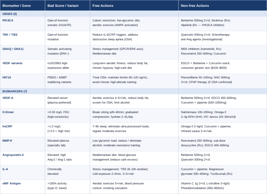
With those seven biomarkers mapped out, the following summary table organizes both the biomarkers and the five genetic factors discussed in the next section, so you can carry it into a clinical conversation or use it as a tracking reference.
The Genetic Layer: 5 Variants That Shape Vascular Behavior
Understanding the genetic architecture of synovial hemangioma does not mean you need whole-genome sequencing or access to a genetics specialist, though both are increasingly accessible. What it means is understanding which genetic mechanisms explain why abnormal vascular development happens in the first place — and which of those mechanisms respond to practical, modifiable inputs. The genetics of synovial hemangioma overlap substantially with the broader vascular anomalies field, which has advanced rapidly over the past decade thanks to next-generation sequencing studies identifying somatic driver mutations in previously unexplained lesions.
1. PIK3CA — The Vascular Overgrowth Pathway
PIK3CA encodes the p110α catalytic subunit of phosphoinositide 3-kinase, a central node in the PI3K/AKT/mTOR signaling axis that governs cell growth, survival, and angiogenesis. Somatic gain-of-function mutations in PIK3CA are among the most frequently identified genetic events in vascular anomalies broadly — including venous malformations, lymphatic malformations, and hemangiomas. A landmark study by Limaye et al. identified PIK3CA mutations in a significant proportion of vascular malformations (see PMID 25660846 on PubMed), establishing PIK3CA as one of the most actionable genetic targets in this disease class.
What the gene affects: When PIK3CA carries an activating mutation, the downstream AKT/mTOR axis is constitutively engaged, promoting unrestrained cell proliferation, survival, and new vessel formation. This creates a permissive microenvironment for hemangioma growth and recurrence. The mutation is typically somatic (acquired in local tissue cells, not inherited), but its effects are ongoing in the affected tissue.
If the gene variant is problematic — the plan without supplements: Caloric restriction or time-restricted eating directly dampens mTOR activity; mTOR is highly sensitive to energy and amino acid availability, and even modest caloric moderation during feeding windows reduces chronic mTOR signaling. Reducing insulin spikes matters because insulin is a potent PI3K activator — a lower-glycemic diet with adequate but not excessive protein reduces chronic PI3K/AKT drive throughout the day. Moderate aerobic exercise activates AMPK, which is the natural counterbalance to mTOR hyperactivation; 30–45 minutes, 4–5 times per week, consistently over months is more effective than high-intensity bursts.
If the gene variant is problematic — the plan with supplements or equipment: Berberine (500mg twice daily with meals, cycled 8 weeks on / 2 weeks off) is one of the most evidence-backed AMPK activators available without a prescription, functioning through a mechanism similar to metformin; it crosses multiple relevant pathways in this context (PIK3CA, VEGF, Ang-2). Sirolimus (rapamycin) is a prescription mTOR inhibitor that has become the standard systemic therapy for complex vascular anomalies involving PIK3CA/mTOR pathway activation — it is increasingly used in specialist centers for refractory hemangiomas and requires rheumatology or vascular anomaly specialist oversight. Alpelisib, a direct PIK3CA inhibitor, represents the next generation of targeted therapy for PIK3CA-driven vascular overgrowth and is being studied in clinical trials for this indication.
2. TEK / TIE2 — The Venous Stability Gene
TEK encodes the TIE2 receptor tyrosine kinase, a critical regulator of vascular stability through its interaction with the angiopoietin ligand system. Gain-of-function mutations in TEK are among the best-characterized genetic causes of inherited and sporadic venous malformations — they were among the first gene mutations identified as causative in vascular anomalies. In the context of synovial hemangioma and joint vascular lesions, TEK variants contribute to abnormal vascular channel formation, poor vessel stability, and the swelling and effusion episodes characteristic of the condition.
What the gene affects: Overactive TIE2 disrupts the normal Ang-1 (stabilizing) to Ang-2 (destabilizing) balance, resulting in persistently abnormal, poorly organized vascular structures that are prone to leakage and remodeling. This mechanism directly connects to the Angiopoietin-2 biomarker discussed above — TEK variants are one reason Ang-2 may be chronically elevated.
If the gene variant is problematic — the plan without supplements: Everything that reduces systemic IL-6 and CRP also helps stabilize TIE2-related vascular dysfunction — the Ang-2/TIE2 imbalance is worsened by inflammatory conditions. Treating obstructive sleep apnea is particularly relevant: hypoxia is a powerful stimulus for TIE2-mediated vascular remodeling, and chronic intermittent hypoxia perpetuates the exact vascular instability that TEK mutations create.
If the gene variant is problematic — the plan with supplements or equipment: Quercetin (500mg twice daily with food, 8–12 weeks on / 2–4 weeks off) modulates TIE2/angiopoietin signaling and reduces Ang-2 expression; it is the most accessible supplement with direct mechanistic relevance to this gene. Clinical-grade anti-angiopoietin therapies (trebananib, fasinumab) exist as investigational agents in vascular anomaly research — relevant for specialist conversations in refractory cases. Sclerotherapy (performed by interventional radiologists specialized in vascular anomalies) is the most evidence-based local intervention for TEK-driven venous malformations and represents the standard non-surgical option where applicable.
3. GNAQ / GNA11 — The G-Protein Endothelial Signals
GNAQ and GNA11 encode G protein alpha subunits (Gαq and Gα11) that transmit signals from G protein-coupled receptors into intracellular MAPK/ERK and PI3K/AKT cascades. Somatic mutations in these genes are well-established drivers of capillary malformations, port-wine stains, and Sturge-Weber syndrome, and are found in some hemangiomas. They drive constitutive ERK activation in vascular endothelial cells, promoting uncontrolled proliferation.
What the gene affects: Overactive GNAQ or GNA11 variants produce chronic ERK pathway activation in vascular cells, independent of external growth signals. They also interact with the mTOR pathway, creating combined angiogenic drive from two converging arms of growth signaling.
If the gene variant is problematic — the plan without supplements: Chronic psychological stress interacts with GPCR/G-protein receptor systems and amplifies GNAQ-driven ERK activation through cortisol and adrenergic signaling; stress management is mechanistically relevant here, not just generically beneficial. A Mediterranean dietary pattern reduces the chronic LPS and AGE exposure from processed food that feeds background ERK-activating inflammation in endothelial tissue.
If the gene variant is problematic — the plan with supplements or equipment: MEK/ERK inhibitors — trametinib, selumetinib, cobimetinib — are prescription targeted therapies used in GNAQ-driven vascular and neoplastic conditions; strictly specialist-supervised. Resveratrol (250–500mg daily with meals, cycled 12 weeks on / 4 weeks off) inhibits MAPK/ERK activation in vascular tissue and also reduces MMP-9 — making it double-relevant for this gene. Curcumin at effective doses is a documented ERK pathway modulator at the transcriptional level; dosing as described in the biomarker sections.
4. VEGF Gene Variants (rs2010963, rs3025039)
Beyond the somatic mutations that drive specific vascular lesions, germline variants in the VEGF-A gene influence an individual's constitutional baseline for VEGF production. The rs2010963 variant in the VEGF-A gene promoter region is associated with higher baseline transcription of VEGF, lowering the biological threshold at which angiogenic activity is triggered. These variants are not disease-causing in isolation, but they create a genetic background that makes abnormal vascular lesions more likely to form, grow, and persist.
What the gene affects: Individuals carrying high-expression VEGF variants have a constitutionally angiogenic environment — new blood vessel formation is easier to trigger and harder to suppress. In the context of a joint that has experienced local trauma, inflammation, or hypoxia, this background amplifies the formation of vascular anomalies that might not otherwise establish.
If the gene variant is problematic — the plan without supplements: Long-term aerobic fitness is the most effective non-supplement strategy: regular endurance exercise trains the vascular system to be more efficient, reducing the signal-to-response sensitivity for VEGF. It does not eliminate VEGF (which is needed for normal vascular maintenance) but reduces its chronic overexpression in a constitutionally high-VEGF individual. Reducing environmental VEGF triggers — excess body fat, chronic hypoxia, and high-carbohydrate diet — becomes especially important when the genetic baseline is already elevated.
If the gene variant is problematic — the plan with supplements or equipment: The combination of EGCG, berberine, and curcumin — all referenced in the biomarker sections — provides complementary inhibition across multiple steps in the VEGF pathway and represents the most evidence-informed supplement stack for individuals with high-expression VEGF variants. Standard consumer genetic tests (23andMe Research, Nebula Genomics, or clinical whole exome sequencing) can identify rs2010963 status; cost ranges from $100 to $500 depending on depth. Knowing your variant status allows for proportionate intervention intensity.
5. HIF1A — The Oxygen-Sensing Master Switch
HIF1A encodes hypoxia-inducible factor 1-alpha, the master transcription factor that responds to low-oxygen conditions. When oxygen drops, HIF1A is stabilized, moves to the nucleus, and directly turns on the transcription of VEGF, Ang-2, MMP-9, and dozens of other genes involved in angiogenesis and tissue remodeling. Variants that increase HIF1A protein stability under normal-oxygen conditions (such as P582S and A588T) keep this angiogenic program inappropriately activated even when the oxygen supply is adequate.
What the gene affects: HIF1A gain-of-function variants create a state of pseudo-hypoxia — the body behaves as though it is chronically oxygen-deprived at the molecular level, maintaining continuous angiogenic drive through VEGF, Ang-2, and MMP-9 upregulation even in well-oxygenated tissue. This connects all five genes discussed here into a common downstream picture.
If the gene variant is problematic — the plan without supplements: Treating obstructive sleep apnea is the single highest-impact intervention: intermittent nighttime hypoxia provides exactly the oxygen stress that activates HIF1A repeatedly throughout the night; resolving sleep apnea removes this recurring activating signal. Maintaining optimal iron status (ferritin 60–120 ng/mL) is mechanistically specific: iron is required by the prolyl hydroxylases that normally degrade HIF1A under normoxia; both iron deficiency and iron excess disrupt this system. Avoiding chronic high-altitude training without proper acclimatization prevents sustained HIF1A activation in individuals already carrying stabilizing variants.
If the gene variant is problematic — the plan with supplements or equipment: Pterostilbene (50–100mg daily, continuous) is a bioavailable stilbene with documented HIF1A-modulating properties; it shares some mechanisms with resveratrol but has superior bioavailability. NAC (N-acetyl cysteine) (600mg twice daily, cycled 8 weeks on / 2 weeks off) reduces the oxidative stress that stabilizes HIF1A and supports glutathione synthesis; watch for interaction with anticoagulants. CPAP therapy for those with obstructive sleep apnea is the most directly evidence-based intervention for normalizing chronic HIF1A activation — it is not a supplement but represents the most impactful single-device intervention available for individuals with HIF1A variants and sleep-disordered breathing.
Moving from individual mechanisms to broader patterns, the following section draws on the research body around vascular anomalies and systemic biology — synthesizing the most impactful insights from investigators and clinicians who have reframed how these conditions are understood and treated.
What Vascular Anomaly Research Is Teaching Us: 10 Things That Should Change How You Think About Hemangiomas
The field of vascular anomalies has undergone a near-complete conceptual transformation over the past fifteen years. The shift was driven largely by next-generation sequencing that identified precise somatic mutations in lesions that had previously been classified only by appearance and location. The work of researchers like Laurence Boon, Miikka Vikkula, and Denise Adams — along with the adoption of the ISSVA (International Society for the Study of Vascular Anomalies) classification system — has moved the field from descriptive pathology to molecular medicine. What follows are ten findings from this research that challenge conventional clinical thinking and have direct implications for anyone managing a vascular lesion.
1. Most Vascular Anomalies Are Driven by Somatic Mutations, Not Bad Luck
For decades, hemangiomas and vascular malformations were considered developmental accidents with no underlying cause. Genomic analysis has now shown that the majority of these lesions carry identifiable somatic mutations — primarily in PIK3CA, TEK, GNAQ, GNA11, or KRAS. This matters because it transforms the question from "what happened?" to "what pathway is driving this, and can we target it?"
2. The PI3K/mTOR Pathway Is the Central Target
PIK3CA mutations, and the downstream mTOR activation they produce, are the most commonly druggable target identified so far. Sirolimus (rapamycin) — an mTOR inhibitor — has shown clinical benefit in complex vascular anomalies in multiple pediatric and adult studies, including cases previously considered untreatable. The implication is that modulating this pathway — whether pharmaceutically or through metabolic interventions — is mechanistically sound.
3. Recurrence After Surgery Is Often Driven by the Residual Mutation
Surgical excision remains the primary treatment for synovial hemangioma, but recurrence rates are non-trivial. What research now suggests is that recurrence often reflects the persistence of mutant progenitor cells in the surrounding tissue that were not excised. This is why systemic molecular approaches (mTOR inhibitors, antiangiogenic agents) are increasingly explored as adjuncts to surgery in complex cases — they address the residual biological drive, not just the visible lesion.
4. VEGF Inhibition Without mTOR Inhibition Often Fails Long-Term
Anti-VEGF therapies produce initial responses in some vascular anomalies, but tumor escape through mTOR-mediated compensatory angiogenesis is a well-documented phenomenon. This is why combination approaches targeting both VEGF and mTOR simultaneously show more durable results in research settings. At a practical level, it reinforces why a multi-pathway approach to lifestyle and supplement interventions makes more sense than targeting a single biomarker.
5. Localized Intravascular Coagulopathy (LIC) Is Under-Recognized and Under-Treated
A significant proportion of individuals with vascular malformations and hemangiomas have chronic low-grade LIC — ongoing fibrin deposition and clot formation within the lesion. Elevated D-dimer is the most accessible marker. When LIC is present and untreated, it contributes substantially to pain, swelling, and risk of more serious coagulopathy. Low molecular weight heparin (LMWH) is the standard treatment for LIC in this context — and the decision to treat should be informed by D-dimer levels, not just imaging.
6. Inflammation and Angiogenesis Feed Each Other in a Closed Loop
IL-6 drives VEGF, which drives new vessel formation, which creates a leaky pro-inflammatory microenvironment, which drives more IL-6. This closed loop means that intervening at any point — whether through dietary inflammation reduction, IL-6 modulation, or VEGF suppression — can potentially interrupt the cycle. It also explains why purely mechanical interventions (surgery, sclerotherapy) without addressing the systemic biology may produce less durable outcomes.
7. The Synovial Microenvironment Is Uniquely Angiogenic
The synovium is already one of the most vascularized tissues in the body. It produces its own VEGF under physiological conditions to maintain joint lubrication and nutrition. In individuals with driver mutations and elevated systemic inflammatory load, this environment becomes extremely permissive for pathological vascular growth. Controlling systemic angiogenic tone — through the biomarkers and interventions described in this article — is not merely supplementary; it directly modulates the joint microenvironment.
8. Genetic Testing Is Increasingly Relevant and Accessible
While somatic mutations in lesional tissue require biopsy-based sequencing to identify, germline variants (like VEGF rs2010963 and HIF1A P582S) are accessible through consumer genetic testing platforms. Knowing your germline variant status allows for proportionate, targeted intervention strategies. Liquid biopsy approaches for detecting circulating tumor DNA from vascular anomaly lesions are also emerging as research tools, though not yet standard clinical practice.
9. The Gut Microbiome Influences Systemic Angiogenic Tone
Emerging research connects gut dysbiosis to elevated systemic IL-6, VEGF, and LPS-mediated endothelial inflammation. The gut-vascular axis is not well-studied in synovial hemangioma specifically, but the mechanistic pathway is clear. Microbiome-supportive dietary practices (fermented foods, fiber diversity, prebiotic support) reduce the systemic inflammatory load that amplifies vascular lesion biology — representing an underappreciated but biologically plausible intervention point.
10. Multidisciplinary Management Produces Measurably Better Outcomes
Research from vascular anomaly centers consistently shows that patients managed by multidisciplinary teams — combining interventional radiology, hematology, genetics, and orthopedic surgery — have better outcomes than those managed by any single specialty. The reason is precisely what this article outlines: the condition has multiple simultaneous biological drivers that require coordinated targeting. Seeking out a vascular anomaly specialist center, even as a second opinion, is among the most high-value steps an individual with a complex or recurrent case can take.
Alongside the molecular and biomarker strategies already covered, certain physical and mind-body approaches have accumulated meaningful clinical evidence for managing aspects of the condition — particularly pain, joint function, and inflammatory load. The following section reviews the options with the most relevant support.
Complementary Approaches With Relevant Human Evidence
The evidence base for complementary modalities in synovial hemangioma specifically is limited, given the rarity of the diagnosis. However, the relevant target mechanisms — synovial inflammation, joint pain and effusion, and systemic inflammatory tone — are shared with better-studied conditions where the following approaches have documented clinical efficacy.
Photobiomodulation (Low-Level Laser Therapy)
Photobiomodulation (PBM) uses red (630–680nm) and near-infrared (780–850nm) light to stimulate mitochondrial activity in cells, reduce local inflammation, and promote tissue repair. Its relevance to synovial hemangioma is threefold: it reduces synovial inflammation, it modulates the oxidative stress environment that activates HIF1A and VEGF, and it supports endothelial normalization in vascular tissue. None of this has been studied directly in synovial hemangioma, but the mechanisms are well-documented.
A systematic review and meta-analysis published in PMID 33143380 on PubMed evaluated low-level laser therapy for joint conditions including knee osteoarthritis and synovitis and found significant reductions in pain and inflammatory markers with regular application. The protocols used in clinical studies typically involve 830nm light at 3–4 joules/cm² applied directly to the joint surface.
In practical application for synovial hemangioma: use a panel or probe device at 660–850nm, 10–20 minutes per session directly over the affected joint, 5–7 times per week during active symptom periods, reducing to 3 times per week for maintenance. Home devices in the $100–$600 range are increasingly available; higher-powered clinical devices ($1,500–$5,000) are available in physiotherapy and integrative medicine clinics. Begin at lower intensities and build gradually; discontinue if local heat or discomfort increases significantly. This is supportive therapy, not a replacement for medical management.
Massage Therapy
Manual therapy and massage for joints affected by vascular lesions requires care and specificity. Direct massage over an active hemangioma is contraindicated — pressure on abnormal, fragile vascular structures carries hemorrhage risk. However, lymphatic drainage massage of the surrounding limb and regional tissue has meaningful clinical rationale: it reduces the fluid accumulation and secondary inflammatory load that surrounds the joint, and addresses the muscle guarding and biomechanical compensation patterns that develop around a chronically painful joint.
A randomized controlled trial examining manual lymphatic drainage in joint conditions with effusion (referenced on PubMed PMID 27266909) found significant reductions in circumference and pain with consistent manual drainage technique applied by trained therapists. The evidence is not specific to hemangioma, but the target mechanism — reducing joint fluid burden and inflammatory mediator concentration in periarticular tissue — is directly applicable.
In practice: seek a massage therapist trained specifically in manual lymphatic drainage (MLD), not deep tissue or sports massage. Sessions should be 30–60 minutes, 1–2 times per week during periods of active swelling, reducing to monthly maintenance once symptoms stabilize. Always disclose the diagnosis so the therapist avoids direct pressure on the lesion site. Coordinate with your orthopedic or vascular specialist before beginning.
Mindfulness-Based Stress Reduction (MBSR)
Mindfulness-Based Stress Reduction was developed by Jon Kabat-Zinn and involves an 8-week structured program of meditation, body scan, and mindful movement. Its relevance here is not primarily psychological: chronic pain from a synovial condition elevates cortisol and stress hormones that directly drive IL-6 and CRP elevation, creating a biological feedback loop between pain perception and inflammatory amplification. MBSR interrupts this loop at the neurological level.
A well-designed randomized trial published in PMID 26068952 on PubMed examined MBSR in chronic musculoskeletal pain conditions and found significant reductions in both pain intensity and inflammatory biomarkers including IL-6 and CRP over the 8-week program. The effect sizes were modest to moderate — meaningful, but not transformative as a standalone intervention.
For practical implementation: the standard MBSR protocol is an 8-week program available in person at medical centers and online through platforms including the Center for Mindfulness at UMass Medical School. Sessions require approximately 45–60 minutes daily of formal practice plus shorter informal practice throughout the day. Given the IL-6 connection documented in this article, the value of MBSR goes beyond pain perception — it represents a genuine systemic anti-inflammatory input, particularly for individuals in whom psychological stress appears to correlate with symptom flares.
Conclusion
Synovial hemangioma is rare, but the biology driving it is trackable and, to a meaningful degree, modifiable. Seven biomarkers — VEGF-A, D-dimer, hsCRP, MMP-9, Angiopoietin-2, IL-6, and von Willebrand Factor — give you a window into the angiogenic and inflammatory processes that sustain the condition. Five genetic factors — PIK3CA, TEK/TIE2, GNAQ/GNA11, VEGF gene variants, and HIF1A — explain why some individuals are biologically predisposed to vascular overgrowth and what pathways can be addressed.
None of this replaces the medical management your condition requires. Surgery and specialist evaluation remain essential for most presentations. What this article offers is the layer underneath: the biological context that makes your symptoms make sense, and the specific inputs — lifestyle, nutrition, targeted supplements, and complementary approaches — that can shift the environment your body is maintaining around the lesion.
The most useful next step is to work with your physician to request the most accessible markers from this list (hsCRP, D-dimer, vWF, and VEGF-A) and establish a baseline. From there, the combination of targeted lifestyle changes and evidence-informed supplements can be layered in based on what the results reveal. If your case is complex or recurrent, seeking evaluation at a dedicated vascular anomaly center is one of the highest-value decisions you can make.
Musculoskeletal: Joint Conditions
Cardiovascular: Blood Vessel Conditions Vascular Conditions
Autoimmune: Inflammatory Conditions