This article contains AI generated content.
· UpdatedSystemic Mastocytosis Genes And Biomarkers - 5 Genes And 7 Biomarkers To Track
Introduction
Living with systemic mastocytosis often means years of unexplained symptoms before a diagnosis arrives, followed by the equally difficult task of understanding a disease that looks different in every patient. Flushing, bone pain, anaphylaxis, gastrointestinal cramps, fatigue, and cognitive fog can appear in any combination, varying in intensity from week to week. What makes this especially isolating is that the symptoms are entirely real but, on the surface, seem to have no single explanation — a situation that leads many patients through years of specialist visits before anyone connects the dots.
What generic advice fails to capture is that systemic mastocytosis (SM) is, at its core, a molecularly specific disease. More than 90% of cases are driven by a single point mutation in the KIT gene, and the behavior of the disease from that point forward is shaped by a combination of measurable biomarkers and additional genetic mutations that most patients have never been told about. Treating SM as just a histamine problem — and responding with diet advice alone — misses the precision that this condition actually requires.
At the same time, monitoring SM does not have to feel opaque. There is a defined set of biomarkers that clinicians use to assess disease burden, track progression, and make treatment decisions. And there is a growing body of evidence linking specific genetic co-mutations to prognosis — with some of those genes having real, evidence-backed nutritional and lifestyle strategies that can support (not replace) medical care. Understanding these layers is not about adding complexity for its own sake; it is about having better conversations with your care team and making more informed decisions at every step.
This article covers two complementary angles for making sense of SM at a deeper level. The first is a practical framework for 7 key biomarkers — what they measure, what abnormal values mean, and what can be done when they move in the wrong direction. The second is a focused guide to 5 genes that shape how SM behaves, with concrete plans for each. Beyond those, you will find a summary of one of the most important books written on mast cell disease, plus four complementary approaches supported by real human evidence. The goal throughout is the same: better information leading to better decisions.
Summary
Here is a preview of what this article covers — and why each part is worth reading carefully.
Serum baseline tryptase is the cornerstone biomarker in SM and one of the WHO diagnostic criteria, but it tells only part of the story. KIT D816V variant allele frequency, now measurable from a standard blood draw, provides a non-invasive window into disease burden that complements tryptase. Complete blood count (CBC) with differential catches early signs of bone marrow compromise — a sign that the disease has moved beyond stable. Alkaline phosphatase and LDH together reflect bone involvement and cellular proliferation, both of which carry prognostic weight under the WHO staging system. Urinary N-methylhistamine and prostaglandin D2 metabolites reveal what mast cells are actually releasing day to day — a dimension tryptase alone does not capture. Bone marrow mast cell burden remains the gold standard for staging, and beta-2 microglobulin adds a critical prognostic layer to risk stratification. Each of these biomarkers has a specific threshold that matters for clinical decisions, and each has a plan — with and without supplements — for when values move out of range.
On the genetic side, the picture is equally specific. KIT D816V drives the disease. TET2 — the gene where vitamin C has the strongest biological justification — is the co-mutation most relevant to epigenetic strategies. SRSF2 and ASXL1 mark higher-risk disease and point toward HDAC-modulating nutritional interventions. DNMT3A connects to the methylation cycle and has practical supplementation implications. The article also covers a landmark book that has changed how thousands of patients understand their disease, four evidence-backed complementary modalities, and a complete set of action plans for every biomarker and gene discussed.
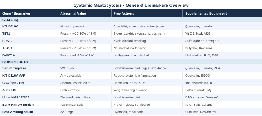
7 Biomarkers That Can Change How You Monitor Systemic Mastocytosis
Biomarkers in SM are not diagnostic checkboxes that get ticked once and forgotten. They are ongoing signals that, tracked serially over time, can reveal whether the disease is stable, progressing, or responding to treatment — often before symptoms change noticeably. The seven below represent a practical monitoring framework that goes beyond a once-yearly tryptase result and gives a far more complete picture of what is actually happening.
Biomarker 1: Serum Baseline Tryptase
Tryptase is an enzyme stored in mast cell granules and released upon activation. Baseline serum tryptase — measured outside of any allergic or anaphylactic event — reflects the total body mast cell burden more than any individual reaction. It is the most widely used biomarker in SM and serves as one of the WHO minor criteria for systemic mastocytosis when persistently elevated above 20 ng/mL.
Normal baseline tryptase is generally below 11.4 ng/mL. Values between 11.4 and 20 ng/mL raise the question of elevated mast cell mass or hereditary alpha-tryptasemia (HaT), a benign genetic variant caused by duplication of the TPSAB1 gene that raises baseline tryptase independently of SM. Values persistently above 20 ng/mL, in the appropriate clinical context, support an SM diagnosis. In aggressive or mast cell leukemia presentations, tryptase can reach several hundred or even over a thousand ng/mL.
How to Measure It
Serum tryptase is a standard blood draw ordered by allergists and hematologists. It is available through most major commercial laboratories (Quest, LabCorp, ARUP, Mayo Clinic Laboratories). Cost ranges from approximately $30 to $100; insurance typically covers it when ordered with appropriate SM or mast cell disease diagnostic codes. The test should be drawn at true baseline — ideally at least 24 hours after any reaction, in a non-acute state — and repeated at each follow-up visit. Trends over time are more informative than any single measurement.
HaT genetic testing is now available commercially and should be considered in patients with elevated tryptase but otherwise atypical presentations, to avoid misinterpretation.
If the Score Is Bad, the Plan Without Supplements
The most important non-supplement step with an elevated or rising tryptase is working with a mastocytosis-specialized hematologist or allergist to determine whether a bone marrow biopsy is indicated. On the lifestyle side, avoid established mast cell triggers: alcohol, NSAIDs (aspirin, ibuprofen, naproxen), extreme heat, vigorous exercise without antihistamine pretreatment, cold exposure, and radiocontrast media. Triggers vary between patients, making a detailed symptom-and-trigger journal a foundational tool.
A strict low-histamine diet — eliminating fermented foods, alcohol, aged cheeses, cured meats, and high-histamine vegetables such as spinach, tomatoes, and avocado — reduces the total mediator load competing with mast cell-released histamine. This does not directly lower tryptase but significantly reduces the symptom burden associated with tryptase-related mast cell activation.
If the Score Is Bad, the Plan With Supplements or Equipment
Quercetin (500 mg twice daily with meals) is the most studied natural mast cell stabilizer. It inhibits mast cell degranulation and IgE-mediated activation through multiple intracellular pathways. Cycling recommendation: 8–12 weeks on, 2–4 weeks off to reassess need. Side effects are rare at standard doses; avoid doses above 3 g/day due to potential renal effects.
Luteolin (100–200 mg daily, often formulated as palmitoylethanolamide-luteolin in products like NeuroProtek) has shown mast cell stabilizing properties in cell-based and animal studies. Cycle similarly to quercetin.
Vitamin C (1000–2000 mg daily in divided doses) supports diamine oxidase (DAO) enzyme activity and has mild antihistamine properties. Begin at 500 mg and titrate up; bowel loosening at higher doses is the practical dose ceiling.
Palmitoylethanolamide (PEA) (600–1200 mg daily, taken with food) has shown mast cell modulating and anti-neuroinflammatory effects in human clinical trials. Well-tolerated; can be taken continuously without cycling. Side effects: occasional mild GI discomfort.
Biomarker 2: KIT D816V Variant Allele Frequency in Peripheral Blood
The KIT D816V point mutation is the molecular engine of systemic mastocytosis in over 90% of cases. It causes the KIT receptor — a tyrosine kinase that regulates mast cell survival and proliferation — to remain permanently activated regardless of normal growth factor signals. The variant allele frequency (VAF), meaning the percentage of circulating cell-free DNA or mononuclear cells carrying this mutation, is now measurable from a simple blood draw using sensitive PCR or next-generation sequencing.
A detectable KIT D816V VAF in peripheral blood is diagnostically meaningful even when below 1%. Higher VAF correlates with greater mast cell burden and, in prospective studies, with increased risk of progression. Serial VAF measurement offers a non-invasive monitoring option that complements or, in some settings, reduces the frequency of repeat bone marrow biopsies.
How to Measure It
Peripheral blood KIT D816V PCR or digital droplet PCR (ddPCR — the most sensitive technique, detecting VAF as low as 0.01%) is available through specialty laboratories including Mayo Clinic Laboratories, ARUP Laboratories, and academic hematology centers. Cost ranges from $200 to $800 depending on whether a single-gene assay or a broader myeloid NGS panel is ordered. Insurance coverage is typically available when SM is established or suspected. Repeat testing every 6–12 months for stable disease; more frequently when on targeted therapy to assess response.
If the Score Is Bad, the Plan Without Supplements
A detectable or elevated KIT D816V VAF cannot be corrected through lifestyle alone — it reflects a clonal genetic event in a stem cell or mast cell precursor population. The appropriate response to a rising or high VAF is prompt hematology discussion regarding disease-modifying therapy. Midostaurin (Rydapt) was the first approved targeted agent for advanced SM; avapritinib (Ayvakit) has since demonstrated higher potency specifically against D816V-mutant KIT and has received approval for both advanced and indolent SM in the US.
While waiting for or between specialist visits, reducing systemic inflammation — through sleep optimization, stress management, and avoiding inflammatory dietary and environmental exposures — creates a less permissive environment for clonal expansion even if it cannot reverse the mutation itself.
If the Score Is Bad, the Plan With Supplements or Equipment
No supplement has demonstrated the ability to reduce KIT D816V clonal burden in human studies. However, quercetin has shown KIT kinase inhibitory activity in laboratory models — the same molecular target as avapritinib, though at far lower potency. Use as a supportive adjunct only, never as a replacement for medical evaluation.
Epigallocatechin gallate (EGCG) from standardized green tea extract (400–800 mg/day, standardized to 50% EGCG) has demonstrated tyrosine kinase inhibitory effects in vitro. Evidence in human SM is early. Cycle: 12 weeks on, 4 weeks off. Monitor liver enzymes with extended use, particularly when combining with other supplements.
Biomarker 3: Complete Blood Count With Differential
The CBC with differential measures red blood cells, white blood cells, platelets, and the specific breakdown of white cell subtypes. In SM, CBC abnormalities are among the first laboratory signs that the disease has begun to compromise the bone marrow's ability to produce normal blood cells — a transition that elevates disease risk categorization.
Anemia (low hemoglobin) is the most common abnormality, typically reflecting mast cell infiltration crowding out erythroid precursors or secondary iron or B12 deficiency from GI involvement. Thrombocytopenia (low platelets below 200,000/µL) is a formal WHO B-finding — an indicator of smoldering or more advanced disease. Eosinophilia appears in a subset, particularly SM with associated hematological neoplasm (SM-AHN). Leukopenia or leukocytosis may also occur in more advanced subtypes.
How to Measure It
A CBC with differential is one of the most affordable and widely ordered laboratory tests. Cost: $20 to $50, universally covered by insurance with a clinical indication. It should be checked at SM diagnosis and monitored at every scheduled follow-up — every 6–12 months for stable indolent SM, every 3 months or more frequently for smoldering or advanced disease or during treatment.
If the Score Is Bad, the Plan Without Supplements
The first step when the CBC is abnormal is determining the underlying cause. Iron deficiency anemia responds to dietary heme iron (lean red meat, poultry liver) combined with vitamin C-rich foods at the same meal to enhance absorption, along with reducing proton pump inhibitor use if possible (these reduce iron absorption). Anemia from bone marrow infiltration requires treatment directed at the SM itself.
Avoiding alcohol (toxic to marrow progenitor cells and a reliable mast cell trigger), NSAIDs (risk of GI bleeding), and unnecessary hematopoietic stressors preserves CBC stability between medical visits. Ensuring adequate protein intake (1.2–1.6 g/kg body weight daily) supports marrow function and red cell production.
If the Score Is Bad, the Plan With Supplements or Equipment
Iron bisglycinate (25–30 mg elemental iron daily when deficient) is gentler on the GI tract than ferrous sulfate and better tolerated in patients with mast cell-related GI reactivity. Take on an empty stomach with vitamin C; recheck ferritin and CBC after 8–12 weeks.
Methylcobalamin B12 (1000 mcg sublingual daily) and methylfolate (5-MTHF) (400–800 mcg daily) address B12 and folate deficiencies common in SM patients with intestinal mast cell involvement and impaired absorption.
Vitamin D3 plus K2 (2000–5000 IU D3 with 100–200 mcg K2 MK-7 daily) supports immune regulation and reduces marrow inflammatory signaling. Monitor 25-OH vitamin D serum levels; a target of 40–60 ng/mL is appropriate. Deficiency is common in SM patients who avoid heat and sun exposure to prevent trigger reactions.
Biomarker 4: Alkaline Phosphatase and Lactate Dehydrogenase
Alkaline phosphatase (ALP) and lactate dehydrogenase (LDH) are components of the standard comprehensive metabolic panel that carry specific prognostic weight in SM. Both are included in the WHO B-findings — clinical and laboratory features that, when present, help categorize disease as smoldering or high-risk and influence treatment decisions.
Elevated ALP most often reflects bone or liver infiltration by mast cells — two frequent sites of extramedullary involvement in SM. Elevated LDH points to accelerated cellular proliferation and high cell turnover, a sign that the mast cell clone is expanding more rapidly. When both are elevated together, urgent hematology re-evaluation is warranted. Osteoporosis and osteopenia are among the most common complications of bone mast cell infiltration and develop even when ALP is only mildly elevated.
How to Measure It
Both ALP and LDH are included in the comprehensive metabolic panel (CMP), costing $30 to $80 and covered by insurance across virtually all clinical settings. Track trends over consecutive visits rather than treating any single value as definitive.
For bone-specific monitoring, a dual-energy X-ray absorptiometry (DEXA) scan is recommended at SM diagnosis and every 1–2 years thereafter. Cost: $75 to $300. Vertebral and hip fracture risk is elevated in SM even in patients who appear otherwise healthy.
If the Score Is Bad, the Plan Without Supplements
When ALP elevation is driven by bone involvement, weight-bearing exercise — 30 minutes of low-impact activity daily (walking, resistance bands, light weights) — is among the most evidence-supported interventions for preserving and improving bone mineral density. In SM, this must be approached cautiously: vigorous exercise is a known mast cell trigger for many patients. Premedication with antihistamines before sessions, starting at low intensity, and gradually building duration is a practical protocol.
Eliminating alcohol and hepatotoxic medications protects the liver when ALP reflects hepatic infiltration. Reducing unnecessary supplementation that routes through hepatic metabolism is also prudent when liver involvement is suspected.
If the Score Is Bad, the Plan With Supplements or Equipment
Calcium citrate (500 mg twice daily with food, not calcium carbonate — poorly absorbed in the low-acid GI environment common in SM patients on proton pump inhibitors) combined with the D3/K2 protocol above is the standard bone-protective supplementation approach.
Magnesium glycinate (200–400 mg nightly) supports both bone metabolism and mast cell stability through calcium channel modulation. Well-tolerated; monitor for loose stools. Continuous use is appropriate; no cycling needed.
Whole-body vibration platforms (10–15 minutes, 3 times per week) represent an equipment-based alternative for patients unable to tolerate weight-bearing exercise due to fatigue or musculoskeletal pain. Evidence for bone density preservation is available in osteoporosis populations; SM-specific data is limited, but the mechanism is relevant.
Biomarker 5: Urinary N-Methylhistamine and Prostaglandin D2 Metabolites
While serum tryptase reflects how many mast cells are present, urinary N-methylhistamine (NMH) and prostaglandin D2 metabolites (11-beta-prostaglandin F2α) reveal how actively those mast cells are degranulating — releasing their inflammatory contents into the body. This distinction is clinically important: a patient with moderately elevated tryptase but markedly elevated urinary mediators may be experiencing significantly more active disease than tryptase alone would suggest.
N-methylhistamine is the principal urinary metabolite of histamine. Elevated levels indicate excessive histamine release over the collection period. Prostaglandin D2 (PGD2) metabolites reflect activation of the arachidonic acid pathway in mast cells — a separate mediator cascade responsible for many of the flushing, GI, and cardiovascular symptoms in SM that histamine alone does not fully explain.
How to Measure It
A 24-hour urine collection for NMH and 11-beta-prostaglandin F2α is the standard collection method. Some labs accept spot urine with creatinine correction, though 24-hour collections are more reliable for detecting true elevations versus dietary or reactive spikes. Cost: $50 to $200 depending on the panel ordered. Specialty labs including Mayo Clinic Laboratories and ARUP handle these samples reliably.
Critical collection rules: avoid histamine-rich foods (aged cheese, wine, cured meats, fermented foods) and antihistamines for at least 48 hours before and during collection if medically safe. The collection should reflect a representative symptomatic period — not during hospitalization or immediately after a severe reaction.
If the Score Is Bad, the Plan Without Supplements
The most effective non-supplement intervention is structured low-histamine dietary modification sustained for at least 4–8 weeks. This means systematically eliminating fermented products, alcohol, vinegar, aged proteins, and high-histamine vegetables. The SIGHI food list and the Mastocytosis Society dietary guidelines provide practical frameworks for implementation.
Parallel trigger identification — through a detailed food-symptom-environment diary over 2–4 weeks — typically reveals personal triggers that extend well beyond the standard list: specific fragrances, stress events, temperature transitions, certain medications (opioids, codeine, NSAIDs, some antibiotics), and even specific exercise types or intensities. Removing the 2–3 most reactive personal triggers often produces more symptom relief than broad dietary restriction alone.
If the Score Is Bad, the Plan With Supplements or Equipment
Diamine oxidase (DAO) enzyme supplements taken immediately before meals that may contain histamine support extracellular histamine degradation before absorption. Products standardized to kidney-derived DAO (such as those from Seeking Health or Umbrellux DAO) are the most studied. No cycling needed; take with meals indefinitely.
Freeze-dried stinging nettle extract (600–900 mg at onset of symptoms or twice daily) has shown H1 receptor antagonism and prostaglandin synthesis inhibition in human studies. Cycle: 12 weeks on, 2–4 weeks off.
Omega-3 fatty acids (EPA/DHA, 2–4 g daily in triglyceride or ethyl ester form, taken with food) shift arachidonic acid pathway metabolism toward less inflammatory prostaglandin species, potentially reducing PGD2 production over time. Monitor for anticoagulant interactions. Continuous use is appropriate; no cycling needed.
Biomarker 6: Bone Marrow Mast Cell Burden
The bone marrow biopsy remains the definitive staging procedure in systemic mastocytosis. It provides direct visualization of mast cell infiltration — the percentage of bone marrow cellularity occupied by mast cells, the architectural pattern (focal versus diffuse), and the immunophenotypic profile (CD117, CD25, CD2, and tryptase expression). A mast cell burden exceeding 30% of bone marrow cellularity is a WHO B-finding that places patients in the smoldering or advanced SM category and significantly influences treatment decisions.
Beyond staging, serial bone marrow biopsies during treatment with targeted therapies like avapritinib or midostaurin are currently the most direct way to assess therapeutic response — something that peripheral blood markers, while increasingly useful as surrogates, cannot yet fully replace for treatment decisions.
How to Measure It
The biopsy is performed as an outpatient procedure under local anesthesia with optional sedation, sampling the posterior superior iliac spine (hip). Procedure cost ranges from $500 to $3,000 or more depending on facility, anesthesia choice, and insurance. The specimen undergoes standard histology plus immunohistochemistry for CD117, CD25, tryptase, and CD2. At minimum, a biopsy is performed at SM diagnosis; repeat biopsies are typically scheduled every 1–3 years for smoldering SM and as clinically indicated for indolent disease or when progression is suspected.
If the Score Is Bad, the Plan Without Supplements
A high bone marrow mast cell burden requires specialist-directed medical therapy as the primary response. Targeted KIT inhibitor therapy (avapritinib, midostaurin), cladribine for cytoreduction in advanced SM, and allogenic stem cell transplant in eligible high-risk patients are the therapeutic tools. No lifestyle intervention directly reduces bone marrow mast cell infiltration.
Supporting the marrow during treatment matters, however. Adequate protein intake (1.2–1.6 g/kg daily), consistent sleep (7–9 hours), alcohol avoidance, and managing all comorbidities aggressively reduce the overall metabolic and inflammatory burden on an already stressed bone marrow environment.
If the Score Is Bad, the Plan With Supplements or Equipment
N-acetylcysteine (NAC) (600 mg twice daily) may reduce oxidative stress in the marrow microenvironment, creating conditions less permissive to clonal expansion. Evidence is indirect, drawn from oxidative stress research in hematological conditions. Cycle: 12 weeks on, 4 weeks off. Well-tolerated.
Sulforaphane from broccoli sprout extract (30–60 mg sulforaphane equivalent daily) exerts epigenetic effects via HDAC inhibition and NRF2 activation, with anti-proliferative properties demonstrated in hematological malignancy cell models. Evidence in SM specifically is early-stage. Cycle: 8 weeks on, 4 weeks off. Monitor GI tolerance; sulfurous smell is normal.
Biomarker 7: Beta-2 Microglobulin
Beta-2 microglobulin (B2M) is a small protein shed by cells with high proliferative activity. In systemic mastocytosis, elevated serum B2M reflects rapid cell turnover associated with advanced or actively progressing disease and serves as one of the prognostic markers considered in WHO B-findings and risk stratification scoring systems. Values above 2.0 mg/L are generally considered elevated; values above 3.5 mg/L are associated with higher risk of transformation and shorter survival in aggressive SM subtypes.
B2M is not SM-specific — chronic kidney disease, multiple myeloma, and lymphoma also raise it. Interpretation requires clinical context, but in a patient with known SM being followed over time, a rising B2M is a meaningful signal that warrants prompt re-evaluation.
How to Measure It
Serum B2M is a standard laboratory test available through all major commercial labs. Cost: $50 to $150, typically covered by insurance in the setting of hematological disease. Baseline measurement should be performed at SM diagnosis and monitored at 6–12 month intervals for stable disease, or more frequently during treatment or when other markers suggest progression.
If the Score Is Bad, the Plan Without Supplements
An elevated or rising B2M requires close specialist follow-up as the primary intervention. Protecting renal function independently is also important, since kidney impairment raises B2M through decreased clearance — masking whether a rise reflects SM progression or worsening renal function. Adequate hydration (2–3 liters of water daily), avoiding nephrotoxic medications (NSAIDs, certain contrast agents), and monitoring creatinine concurrently allow proper interpretation of B2M trends.
Reducing systemic inflammatory burden through sleep optimization, consistent moderate physical activity, and anti-inflammatory dietary patterns may lower the overall proliferative signaling environment, though direct effects on B2M in SM are not established by clinical trials.
If the Score Is Bad, the Plan With Supplements or Equipment
Curcumin with piperine (500–1000 mg curcumin with 5–10 mg piperine for absorption, or liposomal curcumin) inhibits NFkB signaling — a pathway activated in SM mast cells and in many hematological malignancies associated with elevated B2M. Evidence is extrapolated from broader hematological oncology research; SM-specific human data is limited. Cycle: 12 weeks on, 4 weeks off. Avoid at doses above 2 g/day in patients on anticoagulants.
Trans-resveratrol (150–500 mg daily, taken with a fatty meal) activates sirtuin pathways associated with cellular stress resistance and has shown anti-proliferative effects in hematological cancer cell studies. Evidence in SM is preliminary. Cycle: 8–12 weeks on, 4 weeks off.
With the monitoring framework established, it is worth understanding the genetic architecture that determines why different SM patients experience such different disease trajectories. The five genes below are where the molecular story behind each individual case is written.
The Genetic Blueprint of Systemic Mastocytosis
Next-generation sequencing has transformed the understanding of SM over the past decade. What was once seen as a single-mutation disease is now understood as a spectrum in which the KIT D816V mutation initiates the disease, but additional co-mutations in genes controlling epigenetics, chromatin organization, and RNA splicing determine how aggressively it behaves. Approximately 50–70% of SM patients carry at least one co-mutation alongside KIT D816V, and the specific combination shapes prognosis more reliably than any single factor.
Understanding your own genetic profile does not replace specialist management — but it informs the conversation, helps you understand why your disease may behave differently from someone else with the same diagnosis, and opens the door to specific nutritional and lifestyle interventions supported by real mechanistic evidence.
Gene 1: KIT (D816V Mutation)
KIT, also written as c-KIT, encodes the stem cell factor receptor — a tyrosine kinase that normally controls mast cell development, survival, and proliferation in response to growth factor signals. The D816V substitution (aspartic acid replaced by valine at codon 816) locks this receptor in a permanently active state, bypassing the need for stem cell factor and causing uncontrolled mast cell production regardless of what the body signals.
In many patients, KIT D816V is not confined to mature mast cells but is also detectable in other myeloid lineages — evidence that the mutation arises in an earlier, pluripotent progenitor. This multilineage involvement helps explain why SM can evolve into SM with associated hematological neoplasm (SM-AHN) or, rarely, mast cell leukemia.
If the Gene Is Bad, the Plan Without Supplements
Since KIT D816V is the primary disease driver, the most important response is ongoing engagement with a mastocytosis specialist and, when criteria are met, targeted KIT inhibitor therapy. Avapritinib (Ayvakit) was engineered specifically to overcome the resistance of the D816V mutation that limited earlier tyrosine kinase inhibitors, and is approved for both advanced and indolent SM. Midostaurin (Rydapt) was the first approved agent for advanced SM.
Beyond pharmacology: carrying a medical alert identification, maintaining an epinephrine auto-injector at all times, having a written emergency action plan, and educating family members on anaphylaxis recognition and response are non-negotiable non-pharmacological steps for all patients with confirmed KIT D816V disease.
If the Gene Is Bad, the Plan With Supplements or Equipment
No supplement replaces targeted therapy for this mutation. However, quercetin (500 mg twice daily) and luteolin (100–200 mg daily) have both demonstrated KIT signaling inhibitory activity in vitro, functioning as natural tyrosine kinase modulators at far lower potency than pharmaceutical inhibitors. These can be used as supportive adjuncts. Cycle quercetin 8–12 weeks on, 2–4 weeks off; luteolin can be taken continuously within the above dose range.
Gene 2: TET2
TET2 encodes a critical epigenetic enzyme responsible for DNA demethylation — a process that allows stem cells to differentiate normally and suppresses clonal dominance. TET2 mutations are found in approximately 20–30% of SM cases, making them the most common co-mutation after KIT D816V. When TET2 is mutated, DNA methylation patterns become dysregulated, tumor suppressor gene promoters become hypermethylated and silenced, and the affected clone gains a proliferative advantage.
TET2 mutations are also associated with clonal hematopoiesis of indeterminate potential (CHIP) and with worse prognosis in SM when present alongside KIT D816V. They also represent the gene where a specific nutritional intervention has the strongest mechanistic support.
If the Gene Is Bad, the Plan Without Supplements
TET2 enzyme activity depends on alpha-ketoglutarate (α-KG) as a metabolic cofactor and is inhibited by 2-hydroxyglutarate (accumulated in IDH-mutated cancers — rare as a co-mutation in SM, but metabolically relevant). Supporting the mitochondrial environment that maintains α-KG availability means prioritizing metabolic health: consistent sleep (7–9 hours), regular moderate aerobic exercise, low-glycemic whole-food nutrition, and stress reduction. Chronic cortisol elevation has documented epigenetic effects on hematopoietic cells, adding another dimension to the case for behavioral stress management.
If the Gene Is Bad, the Plan With Supplements or Equipment
This is where human evidence genuinely enters the picture. TET2 requires vitamin C (ascorbate) as a direct electron donor and enzymatic cofactor. Research published in Nature demonstrated that ascorbate restores TET2 enzymatic function and reduces clonal hematopoiesis in TET2-deficient mice, with follow-up human studies underway. Read the original study on PubMed. Protocol: 1000–2000 mg ascorbate daily in divided doses, ideally as liposomal vitamin C for higher bioavailability. Some researchers working in hematological applications use up to 4–6 g/day in divided doses; bowel tolerance and kidney stone risk are the practical limits at higher doses. Continuous use is appropriate. Patients with G6PD deficiency should discuss high-dose ascorbate with their physician before starting.
Alpha-ketoglutarate (AKG) as calcium AKG (1–3 g daily) directly supplies the TET2 cofactor. Evidence in hematopoietic stem cells is emerging; SM-specific human data is not yet available. Cycle: 12 weeks on, 4 weeks off. Generally well-tolerated.
Gene 3: SRSF2
SRSF2 (serine/arginine-rich splicing factor 2) regulates how pre-messenger RNA is processed into functional mRNA — a step that determines which proteins get made and in what quantities. Mutations at position P95 (the most common SRSF2 variant in SM) distort this regulation, altering splicing of genes involved in apoptosis, cell differentiation, and immune signaling. SRSF2 mutations occur in approximately 10–15% of SM cases and are strongly associated with the SM-AHN category and aggressive disease behavior.
SRSF2 mutation is one of the independent prognostic markers in modern SM risk scoring (including the MARS score), and its presence should prompt discussion of earlier therapeutic intervention even when clinical parameters appear relatively stable.
If the Gene Is Bad, the Plan Without Supplements
SRSF2 mutations have no direct nutritional correction, and the priority response is specialist care at a center with dedicated mastocytosis expertise — including more frequent bone marrow monitoring, proactive discussions about clinical trial eligibility, and early consideration of cytoreductive or targeted therapies. SRSF2 mutation in SM warrants particular vigilance for signs of transformation to AHN.
Lifestyle-wise: avoid all hematopoietic stressors rigorously — alcohol, smoking, unnecessary medications with marrow toxicity. Anti-inflammatory lifestyle practices (sleep, exercise, dietary discipline) support marrow resilience as a complement to medical management.
If the Gene Is Bad, the Plan With Supplements or Equipment
Sulforaphane (30–60 mg sulforaphane equivalent daily from broccoli sprout standardized extract) has demonstrated splicing regulation and epigenetic modulation via HDAC inhibition and NRF2 pathway activation in cancer cell models. Evidence specific to SRSF2-mutated disease is limited; this is an extrapolated general anti-proliferative approach. Cycle: 8–10 weeks on, 4 weeks off.
Omega-3 fatty acids (EPA/DHA, 3–4 g daily in triglyceride form) reduce inflammatory cytokine signaling that amplifies the downstream consequences of aberrant splicing in immune progenitor cells. Continuous use; monitor for anticoagulant interactions.
Gene 4: ASXL1
ASXL1 (additional sex combs like 1) maintains the polycomb repressive complex — a chromatin-organizing system that silences developmental genes in adult stem cells. Mutations in ASXL1 disrupt this silencing, allowing growth-promoting genes to reactivate in stem cell populations. ASXL1 mutations are present in approximately 10–15% of SM patients and, when combined with KIT D816V, are associated with a substantially worse prognosis and higher risk of transformation to SM-AHN or acute mast cell leukemia.
ASXL1 is one of the negative prognostic markers included in the MARS mutation-adjusted risk score for SM, a validated clinical stratification tool.
If the Gene Is Bad, the Plan Without Supplements
ASXL1 mutations mandate specialist hematological oversight at a high-frequency interval. Patients with this co-mutation should discuss the full range of therapeutic options — including cytoreductive therapy, targeted agents, and, in eligible patients with advanced disease, allogenic stem cell transplant — with centers that have volume experience in SM.
Avoiding alcohol and tobacco is especially important here: both have direct epigenetic effects on histone modification in hematopoietic cells, compounding the chromatin dysregulation caused by ASXL1 haploinsufficiency.
If the Gene Is Bad, the Plan With Supplements or Equipment
ASXL1 operates within the histone modification machinery, and supporting histone acetylation balance through natural HDAC inhibitors is the principal supplement-based approach:
Sodium butyrate (600–1500 mg daily) or dietary butyrate from cooked-and-cooled resistant starch foods (if low-histamine variants are tolerated) acts as a natural HDAC inhibitor. Evidence is primarily from colorectal and hematological cancer models. Cycle: 8 weeks on, 4 weeks off. GI side effects are possible in sensitive patients.
Sulforaphane (as above) compounds the HDAC inhibitory approach and can be used alongside butyrate.
Berberine (500 mg twice daily with meals) has shown AMPK-activating and epigenetic modulatory properties in hematological cell studies. Use with caution in patients on multiple medications due to its strong CYP enzyme interaction profile; discuss with your prescribing physician. Cycle: 8 weeks on, 4 weeks off.
Gene 5: DNMT3A
DNMT3A encodes DNA methyltransferase 3 alpha, the enzyme responsible for establishing de novo methylation patterns across the genome during cell differentiation. When DNMT3A is mutated, key regulatory gene promoters fail to become properly methylated, leaving growth-promoting genes inappropriately expressed. DNMT3A mutations appear in approximately 5–10% of SM cases and are associated with both clonal hematopoiesis and — when combined with other co-mutations — aggressive disease phenotypes.
Like TET2 and ASXL1, DNMT3A mutations often predate SM in the clonal history, suggesting they create a permissive epigenetic environment in which the KIT D816V clone then expands preferentially.
If the Gene Is Bad, the Plan Without Supplements
DNMT3A mutation in SM calls for close specialist monitoring with awareness of elevated progression risk. On the lifestyle side, dietary methyl donor optimization provides the methyl groups that DNMT3A — even when partially functional — needs to methylate DNA. Methyl-donor-rich foods include leafy greens (folate), eggs (choline), beets (betaine), and lean animal proteins (methionine). Equally important: avoiding methyl-depleting exposures — alcohol disrupts one-carbon metabolism, and smoking alters DNMT3A function through direct genomic effects.
If the Gene Is Bad, the Plan With Supplements or Equipment
Supporting the one-carbon cycle — the interconnected metabolic network that provides methyl groups for DNA methylation — is the primary supplement-based strategy:
Methylfolate (5-MTHF) (400–800 mcg daily), methylcobalamin B12 (1000 mcg sublingual daily), and betaine (trimethylglycine, TMG) (500–1000 mg daily) together optimize the remethylation pathway and supply methyl groups that partially compensate for impaired DNMT3A activity. These are widely available and well-tolerated. Monitor homocysteine levels (ideally below 8 µmol/L) to gauge methylation sufficiency. Caution: excessive methyl donors in genetically predisposed individuals can occasionally trigger anxiety or overmethylation symptoms — reduce dose if this occurs.
Choline (400–600 mg daily as alpha-GPC or choline bitartrate) is a betaine precursor and key methylation support nutrient — particularly important in patients who do not consume eggs regularly (the primary dietary choline source). Continuous use is appropriate.
The Book That Reframes Mast Cell Disease
Never Bet Against Occam: Mast Cell Activation Disease and the Modern Epidemics of Chronic Illness and Medical Complexity was written by Dr. Lawrence Afrin, a hematologist-oncologist who spent decades treating patients with mast cell disease at the University of Minnesota and later at Mount Sinai. The book documents what mast cell disease actually looks like across thousands of patients — and makes a detailed, evidence-grounded case for why medicine systematically misses it.
Its central argument is both simple and disruptive: mast cells are distributed throughout virtually every tissue in the body, and one dysfunctional mast cell population can produce symptoms across seemingly unrelated organ systems simultaneously. Rather than violating Occam's razor — the medical principle favoring the simplest explanation — a mast cell explanation is the simplest explanation for patients presenting with multisystem, seemingly unconnected complaints.
1. Mast Cells Are Everywhere — and So Are Their Symptoms
Mast cells reside in highest density at barrier tissues: skin, gut mucosa, airways, and connective tissue. A single dysfunctional mast cell population can produce GI symptoms, neurological symptoms, dermatological reactions, and cardiovascular instability simultaneously — not because four different systems are independently failing, but because one cell type is distributed throughout all of them.
2. MCAS and SM Are Related but Not Identical
Dr. Afrin makes a careful distinction between systemic mastocytosis (clonal mast cell expansion meeting WHO criteria) and mast cell activation syndrome (MCAS — normal or near-normal mast cell numbers, but aberrant behavior). Both produce similar symptom profiles and often overlap. Many SM patients simultaneously meet MCAS criteria; many MCAS patients have low-level clonal features without meeting full SM diagnostic thresholds.
3. The Diagnostic Odyssey Is Real and Documented
The average patient in Dr. Afrin's clinical experience sees between 7 and 10 specialists before a mast cell diagnosis is considered. This is not attributed to incompetent physicians — it reflects a systematic gap in medical education, where mast cell biology is taught in the context of allergy and anaphylaxis rather than as a multisystem regulatory network.
4. Symptoms Are Not Psychosomatic — They Are Mediator-Driven
One of the book's most important contributions is its documented pattern of patients being diagnosed with anxiety, depression, or conversion disorder before mast cell disease is found. Dr. Afrin argues that each symptom — the cognitive fog, the palpitations, the GI spasms — is mechanistically traceable to specific mast cell mediators acting on specific receptors in specific tissues. These are measurable events, not psychological constructs.
5. Baseline Tryptase Is Not Enough
Normal baseline tryptase does not rule out mast cell disease. Many MCAS patients have entirely normal tryptase but markedly elevated urinary N-methylhistamine, prostaglandins, or heparin. Dr. Afrin advocates for comprehensive mediator panel testing rather than relying on tryptase as a gate for further workup.
6. Mediator Collection Must Be Done Correctly
The book describes, with clinical precision, how frequently mediator tests are done incorrectly — wrong timing, inadequate dietary preparation, improper sample handling — producing false negatives that delay diagnosis. The 24-hour urine collection must be performed during a representative symptomatic period, with appropriate dietary restrictions, and sent to a laboratory experienced in handling these unstable analytes.
7. The Low-Histamine Diet Is a Tool, Not a Treatment
Dr. Afrin discusses the low-histamine diet as a valuable but incomplete intervention. It reduces the total mediator load the mast cell system is managing, which can reduce reaction frequency and severity — but it does not address the underlying mast cell dysfunction and should always be part of a broader, medically supervised treatment plan.
8. Mast Cells and the Nervous System Are in Constant Dialogue
Mast cells are located within microns of nerve endings and release mediators that directly modulate nerve firing. Conversely, stress-related neuropeptides — particularly corticotropin-releasing hormone (CRH) and substance P — directly trigger mast cell degranulation. This bidirectional neuroimmune axis is why stress reliably worsens symptoms and why behavioral stress management is not optional but mechanistically justified.
9. Treatment Is Deeply Individual
There is no standard mast cell treatment protocol that works across patients. Finding effective symptom control typically requires a systematic, patient-specific trial of H1 antihistamines, H2 antihistamines, mast cell stabilizers (cromolyn sodium, ketotifen), leukotriene antagonists (montelukast), and, where appropriate, targeted KIT inhibitors. This process can take months to years and requires a knowledgeable, persistent clinical partner.
10. The Scale of Undiagnosed Mast Cell Disease Is Vastly Underestimated
Dr. Afrin estimates that mast cell activation disease in some form may affect a surprisingly high percentage of the population — a figure that remains debated but reflects the growing consensus that MCAS exists on a spectrum and that diagnosed cases represent a small fraction of those actually affected. This reframes SM and related conditions not as rare curiosities but as underrecognized contributors to the epidemic of chronic multisystem illness.
Evidence-Based Complementary Approaches Worth Exploring
The five approaches below were selected for having meaningful human clinical evidence relevant to systemic mastocytosis, mast cell biology, or closely related immune and inflammatory conditions. Each is described honestly — including where the evidence is direct versus extrapolated. None replaces medical treatment or specialist oversight.
Mindfulness Meditation and MBSR
Mindfulness-Based Stress Reduction (MBSR) is an 8-week structured program integrating body-scan meditation, mindful movement, and breath awareness practice. Its relevance to SM is mechanistically direct: psychological stress activates the hypothalamic-pituitary-adrenal (HPA) axis, releasing corticotropin-releasing hormone (CRH) and substance P — neuropeptides with documented direct mast cell degranulation-triggering effects. Reducing the frequency and magnitude of this stress-mast cell signaling loop is a meaningful, evidence-grounded therapeutic target.
The standard MBSR format involves approximately 2.5 hours of weekly group practice plus 45 minutes of daily home practice over 8 weeks. A systematic review published in Brain, Behavior, and Immunity found MBSR significantly reduced inflammatory cytokine markers across multiple chronic inflammatory conditions. While SM-specific MBSR randomized trials are not yet available, the neuroimmune pathway is directly applicable and the intervention carries minimal risk.
SM patients can access MBSR through in-person hospital-based programs, online structured courses (Sounds True, Mindful.org), or apps like Insight Timer with MBSR-specific content. Starting with 10–15 minutes of daily breath-focused practice is realistic and avoids the overwhelm that can accompany full-length body-scan sessions in patients with high somatic symptom burden. Consistency over months matters more than session duration in early practice.
Microbiome-Directed Therapies
The intestinal mucosa contains the highest concentration of mast cells in the body outside the bone marrow, and gut bacteria directly regulate mast cell behavior through short-chain fatty acid production, toll-like receptor signaling, and IgE pathway modulation. Dysbiosis — an imbalance in gut microbial composition — increases intestinal permeability and drives chronic mast cell activation in ways that amplify every other symptom in SM. Addressing the gut microbiome is both mechanistically sound and practically important for patients with GI involvement, which includes the majority of SM patients.
Lactobacillus rhamnosus GG is the most studied probiotic strain in human GI immune trials and has demonstrated reductions in intestinal permeability and IgE-mediated mast cell activation in randomized controlled trials in allergic and GI inflammatory populations. Dietary support through prebiotic fiber from tolerated vegetables (cooked carrots, zucchini, asparagus, sweet potato) provides substrate for anti-inflammatory short-chain fatty acid-producing bacteria.
SM patients with histamine intolerance face a specific challenge: many fermented foods and probiotic strains (including Lactobacillus casei, L. bulgaricus, L. delbrueckii) produce histamine and worsen symptoms. Safer alternatives include L. rhamnosus GG, Bifidobacterium infantis, and Bifidobacterium longum — strains with low histamine-producing potential. Introduce at 1 billion CFU and increase gradually over 4–6 weeks, monitoring for initial GI adjustment reactions that are usually transient.
Breathing-Based Therapies
Controlled breathing techniques — particularly resonance frequency breathing at approximately 5.5 breaths per minute (inhale 5.5 seconds, exhale 5.5 seconds) — reliably increase vagal tone, measured as heart rate variability (HRV), and shift autonomic balance from sympathetic toward parasympathetic dominance. This shift has direct mechanistic relevance to SM: sympathetic nervous system activation promotes CRH release, which triggers mast cell degranulation. Chronically low vagal tone creates a baseline of heightened mast cell reactivity; improving it systematically can reduce this reactive threshold over time.
A 2021 randomized controlled trial published in Applied Psychophysiology and Biofeedback demonstrated significant HRV improvements from 20 minutes of resonance frequency breathing daily over 8 weeks, with accompanying reductions in inflammatory markers in the intervention group. Higher HRV is independently associated with reduced inflammatory cytokine levels and better immune regulation across multiple populations.
Guided resonance breathing sessions are available through apps including Elite HRV, Coherent Breathing, and HeartMath Inner Balance (which also includes a biofeedback sensor to visualize HRV in real time). Practice 10–20 minutes daily at a consistent time — morning and pre-sleep sessions are most practical for SM patients. Avoid practicing in warm rooms or environments with fragrances that may act as mast cell triggers. If lightheadedness occurs, reduce session pace and duration rather than stopping abruptly.
Low-Level Laser Therapy and Photobiomodulation
Low-level laser therapy (LLLT), now more broadly called photobiomodulation, delivers specific wavelengths of light — typically 630–850 nm (red to near-infrared) at low power densities — to stimulate mitochondrial energy production and reduce inflammatory signaling. In the context of SM, its relevance is twofold: photobiomodulation has shown anti-inflammatory effects through reduced NFkB activation and pro-inflammatory cytokine production, and studies in animal models of allergic inflammation have found reductions in mast cell degranulation in treated tissue areas.
A 2019 systematic review in Lasers in Medical Science found that photobiomodulation reduced inflammatory cytokines including IL-6, IL-1β, and TNF-α across multiple clinical populations. Near-infrared protocols using 810–850 nm wavelengths at 4–10 J/cm² energy densities are the most widely studied parameters. Evidence specific to human SM is extrapolated from adjacent inflammatory and allergic conditions rather than direct SM clinical trials.
Consumer-grade near-infrared panels from established manufacturers deliver appropriate wavelengths and can be used for systemic anti-inflammatory purposes at home. Begin with 5-minute sessions at maximum distance from the panel (12–18 inches), 2–3 times per week, gradually increasing to 10–20 minutes as heat tolerance is established. SM patients who are heat-sensitive — a common trigger pattern — should treat this approach with particular caution: the mild warmth generated by devices can provoke mast cell reactions in reactive patients. Start slow, track any post-session reactions in your symptom diary, and do not increase session length faster than your tolerance allows.
The Autoimmune Protocol (AIP) From Sarah Ballantyne
The Autoimmune Protocol (AIP), developed by Dr. Sarah Ballantyne (PhD, medical biophysics), is a structured elimination-and-reintroduction dietary intervention designed to reduce intestinal permeability, dampen immune overactivation, and identify personal food triggers through systematic challenge. While SM is not a classical autoimmune disease, it shares critical features of immune dysregulation — particularly at the gut-immune interface — that the AIP approach specifically addresses. Intestinal permeability allows food antigens and microbial components to cross the gut barrier and activate the dense submucosal mast cell population, amplifying the systemic mediator burden.
A pilot study in Inflammatory Bowel Diseases found clinically meaningful remission rates in IBD patients — a condition with strong gut mast cell involvement — following the AIP elimination protocol. The anti-inflammatory dietary framework overlaps substantially with low-histamine principles, making it particularly relevant for SM patients with GI involvement who need a systematic approach to identifying both autoimmune-type triggers and histamine-specific triggers.
SM patients implementing AIP must cross-reference the AIP food list with a validated low-histamine food list, as some AIP-included foods (bone broth, fermented vegetables, organ meats) are high in histamine and may provoke rather than resolve symptoms. A modified AIP-low-histamine approach focusing on fresh single-ingredient animal proteins, well-cooked low-histamine vegetables (zucchini, carrots, asparagus, sweet potato, beets, broccoli), and fruit in moderation is a practical starting point. Reintroduction should be done one food at a time over several days per food, tracking symptoms carefully. Work with a registered dietitian experienced in both elimination diets and mast cell conditions before starting, particularly if nutritional deficiencies or significant GI dysfunction are already present.
Conclusion
Systemic mastocytosis is a disease where the details carry disproportionate weight. The difference between an accurate prognosis and an incomplete one, between a treatment plan that works and one that misses the mark, often comes down to which biomarkers are being tracked, how often, and whether the genetic co-mutation profile has been characterized. The seven biomarkers covered in this article — serum tryptase, KIT D816V variant allele frequency, complete blood count, alkaline phosphatase and LDH, urinary histamine metabolites, bone marrow burden, and beta-2 microglobulin — together offer a monitoring framework that provides far more actionable information than any single number in isolation.
The genetic layer adds a dimension that changes the trajectory conversation. Knowing whether TET2, SRSF2, ASXL1, or DNMT3A mutations are present alongside KIT D816V shifts risk stratification and opens the door to specific, evidence-grounded nutritional strategies — particularly for TET2, where the ascorbate evidence is mechanistically rigorous and clinically actionable. For the other genes, the data is more preliminary, but the framework of supporting epigenetic function through targeted nutritional and lifestyle approaches is biologically coherent and practically low-risk.
The smartest next step is to bring this framework directly into your next specialist appointment. Ask which of these seven biomarkers are being tracked and how frequently. Ask whether comprehensive mast cell genetic profiling has been done. Ask what the trend in your key values looks like over your last three or four visits — not just whether individual numbers are in or out of range, but which direction they are moving. Those are the questions that lead to better-informed decisions, and being equipped to ask them puts you in a meaningfully stronger position in managing this disease over time.
Digestive Endocrine & Metabolic
Musculoskeletal: Bone Conditions
Autoimmune: Inflammatory Conditions
Cancer & Oncology: Blood Cancer