This article contains AI generated content.
· UpdatedTakayasu Arteritis Genes And Biomarkers - 5 Genes And 6 Biomarkers To Track
Introduction
Takayasu arteritis is one of the more elusive conditions in modern medicine. It hides in plain sight — mimicking fatigue, flu, or vague musculoskeletal pain before revealing itself as a serious large-vessel vasculitis that can silently narrow arteries supplying the brain, kidneys, and limbs. For most people living with it, getting a diagnosis can take years. And once that diagnosis arrives, the harder question begins: how do you monitor something that standard tests often miss?
That is the frustration many people encounter. Up to half of Takayasu arteritis patients show normal or near-normal ESR and CRP levels even during periods of confirmed active disease, a fact that leaves patients and their physicians uncertain about what is actually happening inside their vessel walls. Generic inflammatory markers were not designed for this condition. They were built for a different kind of inflammation, and using them as the sole guide in TA can lead to under-treatment, over-treatment, or simply false reassurance.
What makes this condition harder to navigate is its rarity. There are few large-scale trials, fewer dedicated biomarker guidelines, and almost no personalized management frameworks available to the average patient. Most people are simply told to monitor symptoms and repeat imaging every few years. That is not nothing — but it is not enough for someone trying to understand what their body is doing between appointments.
This article takes a more specific approach. It examines six biomarkers that are more informative than standard inflammatory panels for tracking Takayasu arteritis activity, including both affordable first-line tests and more specialized options that experienced specialists are beginning to rely on. It also covers the five genes most consistently associated with TA susceptibility and disease expression — because understanding your biological risk profile can meaningfully shape how you and your doctor monitor and manage the condition. Beyond that, it touches on lifestyle-level interventions with genuine scientific support, a landmark protocol that is quietly reshaping how autoimmune disease is managed, and a handful of complementary approaches that have real evidence behind them.
Summary
This article covers six biomarkers and five genes with real clinical relevance in Takayasu arteritis — not the generic inflammatory panel your lab usually reports, but the deeper signals that reflect vessel wall activity, immune dysfunction, and vascular remodeling. You will find out why PTX3 and MMP-3 outperform CRP as markers of vascular inflammation, why IL-6 matters so much that it became a treatment target, and how VEGF levels tie into disease progression in ways most physicians do not routinely check. On the genetics side, the article breaks down HLA-B*52:01 and four other gene variants linked to TA — and for each, outlines what you can actually do about it, with or without supplements. You will also find a condensed version of Terry Wahls' mitochondrial-autoimmune framework, which challenges conventional thinking on immune regulation. Finally, the article presents evidence-based complementary approaches — including the Autoimmune Protocol — that have meaningful human evidence for autoimmune vasculitis management.
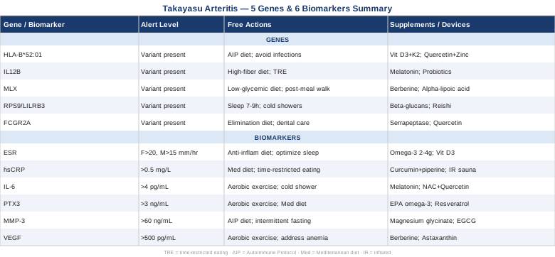
6 Biomarkers to Track in Takayasu Arteritis
Monitoring Takayasu arteritis with standard labs is like trying to see a fire through frosted glass. The tools work — just not well enough. The biomarkers below represent a more complete picture, chosen for their clinical relevance to this specific condition, their measurability in real-world settings, and the quality of evidence supporting their use. They range from affordable first-line tests to more specialized panels that forward-thinking rheumatologists increasingly rely on.
1. Erythrocyte Sedimentation Rate (ESR)
ESR is the most commonly ordered inflammatory marker in Takayasu arteritis, and it has been for decades. It measures how quickly red blood cells settle in a tube — a proxy for circulating inflammatory proteins like fibrinogen. In active TA, ESR is often elevated. But its most important limitation is its unreliability: a normal ESR does not rule out active disease. Multiple studies and clinical registries have documented that a significant minority of TA patients with confirmed vascular inflammation on imaging have completely normal ESR values. Anemia, which is common in chronic TA, can also artificially elevate ESR and distort interpretation.
How to measure it: Ordered through any standard laboratory as part of a basic inflammatory panel. Cost typically ranges from $10 to $25. Results are available within hours.
What it may reveal: Elevated ESR (above 20 mm/hr in women, 15 mm/hr in men) can indicate active systemic inflammation. Sustained normalization alongside clinical improvement is a useful reassurance signal. But it should never be used alone to assess disease activity in TA.
If the score is elevated, the plan without supplements: ESR responds to underlying inflammation control. The most effective lifestyle approaches are consistent low-inflammatory diet (Mediterranean or Autoimmune Protocol), sleep optimization (7–9 hours), stress reduction through HRV-based breathing practices, and eliminating pro-inflammatory exposures (processed foods, smoking, excessive alcohol). Daily 20–30 minute walks at moderate intensity reduce systemic inflammatory tone over 8–12 weeks without triggering TA flares.
If the score is elevated, the plan with supplements or equipment: Omega-3 fatty acids (EPA+DHA, 2–4 g/day) have consistent evidence for lowering ESR in inflammatory and autoimmune conditions. Use high-quality triglyceride-form fish oil. Cycling: continuous use is appropriate; monitor LDL and bleeding time if on anticoagulants. Vitamin D3 (2000–5000 IU/day with K2 100–200 mcg) normalizes immune regulation and is frequently deficient in TA patients. Side effects: hypercalcemia at very high doses — test serum 25-OH-D before starting.
2. High-Sensitivity C-Reactive Protein (hsCRP)
CRP is synthesized by the liver in response to IL-6 and other cytokines released during inflammation. The high-sensitivity version (hsCRP) detects lower concentrations and is more useful for chronic, low-grade inflammatory states. In TA, hsCRP has the same limitation as ESR — it can be normal during active vascular inflammation — but it rises and falls more quickly than ESR, making it a better short-term signal of flare activity. Research published on PubMed consistently shows that hsCRP normalization alone is insufficient to confirm disease remission.
How to measure it: Available at any standard lab. Cost ranges from $15 to $40. Optimal target in general cardiovascular health (per Peter Attia and Thomas Dayspring) is below 0.5 mg/L. In TA context, trending the value over time matters more than any single measurement.
What it may reveal: Values above 3 mg/L during a monitoring period suggest active systemic inflammation. Sharp rises above baseline often precede or accompany clinical flares. CRP is also useful for assessing response to corticosteroid dose adjustments.
If the score is elevated, the plan without supplements: Anti-inflammatory dietary adjustments — specifically eliminating refined carbohydrates, trans fats, and industrial seed oils — can reduce hsCRP meaningfully over 6–12 weeks. Evidence from the PREDIMED trial showed Mediterranean diet adherence reduced CRP by 20–30% over one year. In TA specifically, this forms a useful adjunct to medical management. Time-restricted eating (12–16 hour fasts) also reduces postprandial inflammatory spikes.
If the score is elevated, the plan with supplements or equipment: Curcumin with piperine (500–1000 mg curcuminoids, 3–5 mg piperine, twice daily) has consistent evidence for CRP reduction. Cycling: 8 weeks on, 2 weeks off is reasonable. Side effects: may affect drug metabolism via CYP3A4 — discuss with prescribing physician if on immunosuppressants. Infrared sauna (3–4 sessions/week, 20 minutes at 60–70°C) has emerging evidence for reducing systemic inflammatory markers through heat shock protein activation.
3. Interleukin-6 (IL-6)
IL-6 is arguably the most important biomarker in Takayasu arteritis, and not just because it reflects inflammation. It is mechanistically central to the disease: IL-6 drives the differentiation of T helper 17 cells, which are directly implicated in the granulomatous inflammation that damages vessel walls in TA. This is why tocilizumab — an IL-6 receptor antagonist — has become an important therapy for TA, shown in the TAKT trial to reduce relapse rates compared to placebo. Measuring IL-6 gives you a window into the upstream driver of the inflammatory cascade, not just its downstream effects.
How to measure it: Serum IL-6 is available at specialty labs and academic medical centers. Cost ranges from $50 to $150. It is not part of standard inflammatory panels and must be specifically ordered. Optimal values in healthy individuals are typically below 2–4 pg/mL.
What it may reveal: Elevated IL-6 in TA correlates with disease activity more reliably than ESR alone in several cohort studies. It also reflects active macrophage and dendritic cell activity in the vessel wall. Notably, IL-6 can remain elevated even when ESR and CRP appear normal, making it a valuable add-on test in ambiguous clinical situations.
If the score is elevated, the plan without supplements: IL-6 is powerfully suppressed by consistent aerobic exercise. Studies in autoimmune cohorts show that 30–45 minutes of moderate-intensity aerobic activity 4–5 times per week reduces circulating IL-6 over 8–12 weeks. In TA, this must be done carefully — avoid high-intensity training that dramatically increases blood pressure in patients with arterial stenosis. Walking, gentle cycling, and water-based exercise are first choices. Cold shower exposure (2–5 minutes, 3–4×/week) also activates anti-inflammatory pathways that attenuate IL-6 production.
[BOLD]If the score is elevated, the plan with supplements or equipment:[/TITLE] Melatonin (1–3 mg at bedtime) has documented IL-6 suppression activity in immune-mediated conditions. Use physiological doses; avoid 10+ mg doses without medical supervision. Cycling: nightly use is generally safe long-term. N-acetylcysteine (NAC, 600 mg twice daily) reduces oxidative stress upstream of IL-6 production. Side effects: mild GI discomfort at higher doses. Quercetin (500 mg twice daily) inhibits NF-κB, a major transcriptional driver of IL-6 — pair with bromelain for improved absorption. 8-week cycles are reasonable.
4. Pentraxin-3 (PTX3)
Pentraxin-3 is where the biomarker picture gets genuinely more specific. Unlike CRP, which is produced primarily in the liver in response to systemic signals, PTX3 is synthesized locally in the vessel wall — by endothelial cells, smooth muscle cells, and macrophages. This makes it a much more direct reporter of what is actually happening inside the arteries affected by TA. A landmark study by Dagna et al. in Annals of Internal Medicine demonstrated that PTX3 reflected active vascular inflammation in TA even when CRP was normal — a finding that has since been replicated and that underscores why this marker deserves more attention than it currently receives in routine clinical practice. See related studies on PubMed.
How to measure it: PTX3 is available at specialized reference labs and academic rheumatology centers. Cost ranges from $100 to $300 depending on the laboratory. It is not yet widely available at standard community labs in all countries. In healthy individuals, PTX3 is typically below 2–3 ng/mL.
What it may reveal: PTX3 elevation specifically signals local endothelial and vascular wall inflammation, making it particularly useful for distinguishing true vessel wall activity from systemic inflammatory noise. It may also help identify subclinical disease activity between imaging studies.
If the score is elevated, the plan without supplements: PTX3 responds to the same anti-inflammatory lifestyle foundations as IL-6 and CRP, but its vascular specificity means that endothelial-targeted interventions matter especially. Consistent aerobic movement (low-to-moderate intensity, daily), mediterranean diet adherence, and avoiding smoking or vaping are the highest-leverage behavioral tools. Eliminating highly processed foods and refined oils reduces endothelial oxidative stress. Adequate sleep (7–9 hours, consistent schedule) restores endothelial function through growth hormone and repair signaling.
If the score is elevated, the plan with supplements or equipment: High-dose EPA-rich omega-3s (3–4 g EPA specifically) are the most evidence-backed supplement for endothelial inflammation. Resveratrol (250–500 mg/day with a fat-containing meal) activates SIRT1, which suppresses NF-κB signaling in vascular endothelium. Cycling: 12 weeks on, 4 weeks off. Side effects: mild blood-thinning effect — caution with anticoagulants. Photobiomodulation (low-level red/near-infrared light, 630–850 nm, 10–15 minutes 4×/week over affected vascular regions) has emerging evidence for endothelial NO production and local anti-inflammatory effects.
5. Matrix Metalloproteinase-3 (MMP-3)
MMP-3, also known as stromelysin-1, is an enzyme released by activated macrophages and fibroblasts during tissue remodeling. In Takayasu arteritis, the granulomatous inflammation that attacks vessel walls also triggers MMP activity as the body attempts to repair and remodel damaged arterial tissue. Elevated MMP-3 levels reflect the degree of active vascular wall remodeling and may predict structural progression — including progression toward stenosis or aneurysm formation — even when clinical symptoms are minimal. Studies on MMP-3 in vasculitis on PubMed show it as a complementary marker alongside PTX3 for assessing tissue-level disease activity.
How to measure it: Serum MMP-3 can be ordered through reference laboratories. Cost ranges from $80 to $200. Normal values in healthy adults are typically below 30–60 ng/mL depending on the assay. Age and sex affect reference ranges, and lab-specific normal ranges should be confirmed.
What it may reveal: Persistently elevated MMP-3 despite clinical remission suggests ongoing subclinical vessel wall activity. It is particularly useful for predicting whether a patient who appears stable clinically and on standard labs is actually experiencing silent progression.
If the score is elevated, the plan without supplements: Reducing MMP activity requires addressing the upstream macrophage activation that drives it. Anti-inflammatory diet (AIP or Mediterranean), elimination of ultra-processed foods, and maintaining a healthy body weight all reduce macrophage activation state. Intermittent fasting (14–16 hours) promotes autophagy, which clears inflammatory cellular debris that activates macrophages. Heat shock protein upregulation through sauna use (3–4×/week) also appears to modulate MMP expression in preclinical models.
If the score is elevated, the plan with supplements or equipment: Magnesium (300–400 mg/day as glycinate or malate) reduces NF-κB activation upstream of MMP-3 production. Cycling: continuous use is generally appropriate. EGCG from green tea extract (400–600 mg/day) inhibits MMP expression in endothelial tissue in human cell studies. Side effects: EGCG may affect iron absorption — take away from iron-rich meals. Collagen peptides (10–20 g/day) support vascular matrix integrity and may buffer MMP-driven degradation, though evidence in TA specifically is limited — this is extrapolated from general vascular health research.
6. Vascular Endothelial Growth Factor (VEGF)
VEGF is a signaling protein that drives the formation of new blood vessels — a process called angiogenesis. In healthy tissue, VEGF supports repair. In Takayasu arteritis, elevated VEGF reflects pathological neovascularization occurring in inflamed vessel walls as the body tries to compensate for ischemia. Multiple studies have documented elevated serum VEGF in active TA, and levels correlate with disease activity scores and imaging findings. VEGF is also elevated in response to tissue hypoxia — meaning its elevation can reflect both active inflammation and downstream ischemia caused by vessel stenosis, making it a dual-signal marker with particular relevance in TA. Related research on PubMed.
How to measure it: Serum VEGF is available through specialty and academic labs. Cost ranges from $100 to $300. Normal serum VEGF values are typically below 500 pg/mL, though ranges vary by assay and laboratory. Like all TA biomarkers, trends over time are more informative than isolated values.
What it may reveal: Elevated VEGF during a period of clinical stability may indicate ongoing subclinical disease or worsening ischemia due to silent arterial progression. A sharp rise in VEGF alongside rising IL-6 or PTX3 is a signal worth acting on — it may precede imaging-detected progression.
If the score is elevated, the plan without supplements: VEGF is strongly upregulated by hypoxia and systemic inflammation. Optimizing tissue oxygen delivery through regular moderate aerobic exercise (which improves arterial collateralization in a controlled way) and reducing metabolic inflammation are the core approaches. Avoiding prolonged sitting, which worsens peripheral ischemia, and ensuring hemoglobin levels are optimal (addressing iron-deficiency anemia if present) reduces hypoxia-driven VEGF signaling.
If the score is elevated, the plan with supplements or equipment: Berberine (500 mg twice daily with meals) has documented effects on VEGF modulation through AMPK activation and NF-κB inhibition in inflammatory conditions. Cycling: 8–12 weeks on, 4 weeks off. Side effects: GI discomfort is common, especially initially — start with lower doses and titrate up. Do not combine with metformin without physician supervision. Astaxanthin (12 mg/day) has antioxidant and anti-angiogenic properties in preclinical and small clinical studies. Side effects: generally mild; skin may take on a slightly orange hue at high doses.
Beyond these measurable signals, the genetic landscape of Takayasu arteritis adds another layer of understanding — one that helps explain why some people are far more vulnerable to this condition, why disease expression varies so dramatically between individuals, and how the inflammatory response is wired at the DNA level.
5 Genes That Shape Takayasu Arteritis Risk and Response
Genetics does not determine destiny in Takayasu arteritis, but it shapes the terrain considerably. Understanding the major genetic variants associated with TA helps explain individual differences in susceptibility, disease severity, and response to treatment. It also opens the door to targeted compensatory strategies — because while you cannot change your genes, you can often modify the downstream biological processes they influence. The evidence for most TA-associated genes comes from genome-wide association studies (GWAS), and the strength of evidence varies by gene.
Gene 1: HLA-B*52:01
HLA-B*52:01 is by far the most established genetic risk factor for Takayasu arteritis. Located within the major histocompatibility complex (MHC) on chromosome 6, this specific allele of the HLA-B gene is found in 40 to 70% of TA patients in Japanese and Korean populations, compared to about 3–10% in the general population. It affects how immune cells present peptides to T lymphocytes, and it appears to drive an abnormal immune response against arterial wall antigens. Clinically, HLA-B*52:01 carriers may have more severe aortic involvement and higher rates of aortic regurgitation. Research on HLA-B52 in TA on PubMed.
If the gene variant is present, the plan without supplements: You cannot modify the MHC complex, but you can modulate what triggers it. The most powerful non-supplement intervention is rigorous infection prevention — HLA-B*52:01-associated TA activity is frequently triggered or worsened by infectious episodes. Handwashing, dental hygiene, sleep optimization, and avoidance of immune-depleting stressors all reduce trigger exposure. Anti-inflammatory diet (Autoimmune Protocol or Mediterranean) reduces the inflammatory background against which HLA-B*52:01 presents antigens more aggressively. Physical stress avoidance — particularly overtraining — is important, as exercise-induced immune perturbation can trigger flares in susceptible individuals.
If the variant is present, the plan with supplements or equipment: Vitamin D3 (3000–5000 IU/day with K2 100–200 mcg) powerfully modulates MHC class I antigen presentation and T cell differentiation. Low vitamin D is extremely common in TA patients and strongly associated with autoimmune activity. Test serum 25-OH-D and target 50–70 ng/mL. Cycling: continuous, year-round. Side effects: reassess levels every 6 months; hypercalcemia is rare at these doses but monitor if taking high-calcium supplements. Quercetin (500–1000 mg/day) reduces NLRP3 inflammasome activation downstream of HLA-mediated immune activation. Pair with zinc (15–30 mg/day) for synergistic immunomodulatory effect.
Gene 2: IL12B
The IL12B gene encodes the p40 subunit shared by both interleukin-12 (IL-12) and interleukin-23 (IL-23) — two cytokines that play central roles in directing the immune system toward Th1 and Th17 responses respectively. GWAS studies, including a landmark 2015 analysis published on PubMed, identified variants in IL12B as significantly associated with Takayasu arteritis across multiple ancestral populations. The Th17 cell pathway is particularly implicated in the granulomatous inflammation that characterizes TA vessel wall damage. Variants in IL12B that increase IL-23 signaling amplify Th17 cell differentiation, creating a pro-inflammatory bias in the immune system.
If the variant is present, the plan without supplements: Th17 activity is potently regulated by the gut microbiome. A high-fiber, diverse plant-based diet (within AIP guidelines) promotes short-chain fatty acid production by gut bacteria, which suppresses Th17 polarization through epigenetic mechanisms. Eliminating gluten and processed grains reduces intestinal barrier permeability, which is a major trigger for Th17 activation via lipopolysaccharide (LPS) translocation. Time-restricted eating reduces the metabolic stress that activates the Th17 pathway via insulin and mTOR signaling.
If the variant is present, the plan with supplements or equipment: Melatonin (3–5 mg at night) suppresses IL-23/Th17 axis activity in autoimmune research models and has an excellent safety profile. Continuous use is appropriate. Probiotic supplementation with strains shown to reduce Th17 responses — particularly Lactobacillus rhamnosus and Bifidobacterium longum — taken daily (20–50 billion CFU) helps regulate IL12B downstream effects via the gut-immune axis. Cycling: 3 months on, 1 month off. Side effects: generally minimal; temporary bloating during the first 1–2 weeks.
Gene 3: MLX
MLX encodes a transcription factor involved in metabolic sensing and glucose homeostasis, and its connection to Takayasu arteritis is more indirect but no less interesting. Variants in the MLX gene region have been identified in GWAS studies of TA, suggesting that metabolic-inflammatory crosstalk plays a role in disease susceptibility. MLX partners with other transcription factors (including ChREBP) to regulate genes that respond to carbohydrate metabolites — meaning that dietary carbohydrate load and metabolic stress can interact with this genetic variant to amplify or dampen inflammatory tone. Evidence for MLX in TA is preliminary and based primarily on GWAS signal rather than deep mechanistic studies in humans.
If the variant is present, the plan without supplements: The most direct intervention is carbohydrate management. Reducing refined carbohydrate and sugar intake lowers the metabolic substrate that activates ChREBP/MLX-driven gene expression. Low-glycemic eating patterns — not necessarily ketogenic, but avoiding processed starches and sugary foods — appear most relevant. Time-restricted eating (14–16 hours fasting window) reduces carbohydrate-sensitive transcription activity and lowers fasting insulin. Regular post-meal walking (10–15 minutes) blunts postprandial glucose excursions that would otherwise activate MLX-associated pathways.
If the variant is present, the plan with supplements or equipment: Berberine (500 mg before meals, twice daily) activates AMPK — a nutrient sensor that counteracts the ChREBP/MLX pathway — and has documented glucose-lowering and anti-inflammatory effects. Cycling: 8–12 weeks on, 4 weeks off. Side effects: GI discomfort, especially at first. Alpha-lipoic acid (300–600 mg/day) modulates glucose metabolism and has NF-κB-inhibiting properties relevant to the inflammatory dimension of MLX-related pathways. Side effects: generally mild at standard doses; may lower blood sugar — caution if on diabetes medications.
Gene 4: RPS9/LILRB3 Region
The LILRB3 (leukocyte immunoglobulin-like receptor B3) gene encodes an inhibitory receptor expressed on NK cells and monocytes. Its function is essentially to put a brake on immune activation — it signals cells to stand down. Variants in the RPS9/LILRB3 region identified in TA GWAS suggest that impaired immune inhibitory signaling may contribute to the uncontrolled immune activation characteristic of Takayasu arteritis. When LILRB3 function is reduced, NK cells and monocytes may remain activated longer and with less restraint, contributing to sustained vascular inflammation. Evidence at this point is associative from GWAS data rather than mechanistically confirmed in human TA studies.
If the variant is present, the plan without supplements: NK cell and monocyte regulation is powerfully influenced by sleep quality. Deep, consistent sleep (7–9 hours, with prioritized slow-wave sleep) is one of the most potent regulators of NK cell activity and innate immune balance. Sleep restriction activates monocytes and reduces LILRB3-mediated inhibitory signaling. Cold thermogenesis (cool showers 2–4 minutes, 3–5×/week) has evidence for NK cell modulation. Reducing psychological stress through HRV biofeedback or mindfulness practice also attenuates monocyte hyperactivation via the HPA and sympathetic nervous system axes.
If the variant is present, the plan with supplements or equipment: Beta-1,3/1,6-glucans (250–500 mg/day from certified sources such as Baker's yeast or oats) have documented NK cell modulatory effects — importantly, they appear to help normalize NK activity in both directions (suppressing excess, enhancing deficiency). Cycling: 12 weeks on, 4 weeks off. Side effects: well-tolerated; not recommended in transplant patients on immunosuppression without physician consultation. Medicinal mushroom extracts — particularly reishi (Ganoderma lucidum, 1–2 g/day standardized extract) — have documented effects on NK and monocyte regulation via beta-glucan and triterpene content.
Gene 5: FCGR2A
FCGR2A encodes the Fc gamma receptor IIA, a surface receptor on immune cells (particularly macrophages and neutrophils) that binds the tail region of IgG antibodies. When antibody-antigen complexes form — as happens during autoimmune responses — FCGR2A determines how efficiently macrophages and neutrophils clear those complexes and how intensely they activate in response. Variants in FCGR2A that alter binding affinity or signaling efficiency have been associated with multiple autoimmune vasculitides, including TA. A lower-efficiency FCGR2A variant may impair immune complex clearance, leading to sustained macrophage activation in vessel walls. The association with TA specifically has been identified in GWAS analyses of Asian cohorts.
If the variant is present, the plan without supplements: The most relevant non-supplement approach is reducing the total antigen load that generates immune complexes in the first place. An elimination-style diet that reduces common food antigens (gluten, dairy, soy, processed food additives) lowers the burden on FCGR2A-dependent clearance pathways. Environmental toxin reduction (filter drinking water, reduce chemical cleaning product exposure, use air filtration at home) similarly reduces total antigen and allergen load. Dental health maintenance is particularly important — chronic oral infections generate immune complexes that directly stress FCGR2A pathways.
If the variant is present, the plan with supplements or equipment: Serrapeptase (60,000–120,000 IU enteric-coated, between meals) is a proteolytic enzyme with evidence for breaking down immune complexes and reducing fibrin deposition — processes directly relevant to FCGR2A-impaired clearance. Cycling: 6 weeks on, 2 weeks off. Side effects: avoid in patients on anticoagulants without medical oversight. Quercetin (500–1000 mg/day) stabilizes mast cells and reduces the immune complex-driven activation of macrophages expressing FCGR2A. Pair with nettle leaf extract for additive mast cell stabilization. Side effects: generally mild; mild blood-thinning effect at high doses.
With a clear picture of the key biomarkers and genes in Takayasu arteritis, the next step is understanding the broader framework that can move the needle on autoimmune disease at a systemic level — and few approaches challenge conventional thinking as directly as what Terry Wahls has spent years researching and documenting.
The Wahls Protocol: What This Book May Change About How You See Autoimmune Disease
The Wahls Protocol by Dr. Terry Wahls is not a fringe self-help book. Wahls is a clinical professor of medicine at the University of Iowa who reversed her own secondary-progressive multiple sclerosis — a condition considered irreversible — through a rigorously researched dietary and lifestyle protocol. She later conducted clinical trials on the protocol and published results in peer-reviewed journals. Related research on PubMed. While TA is not MS, both are autoimmune conditions driven by dysregulated T cell activity and systemic inflammation — and the mechanistic principles Wahls outlines apply broadly to immune-mediated disease.
The book draws on hundreds of studies from nutrition science, mitochondrial biology, and immunology. Here are the ten most impactful ideas for someone managing Takayasu arteritis:
1. Mitochondrial dysfunction drives immune dysregulation
Wahls argues that autoimmune disease is inseparable from mitochondrial underperformance. Immune cells — particularly T cells and macrophages — are extraordinarily energy-intensive. When mitochondria are not generating ATP efficiently, immune cell behavior becomes dysregulated. Fixing cellular energy production is therefore not peripheral to autoimmune management — it is foundational to it. This reframes TA management beyond simple inflammation suppression.
2. Nine cups of vegetables per day is not optional
The Wahls Protocol's dietary centerpiece is nine cups of specific vegetables daily: three cups of dark leafy greens, three cups of sulfur-rich vegetables (cabbage, onions, garlic, mushrooms), and three cups of deeply colored vegetables or fruits. This is not a general wellness recommendation — each category provides specific micronutrients required for mitochondrial function, methylation, and myelin repair. For TA patients, this nutrient density approach directly feeds immune cell metabolic needs.
3. Sulfur-rich foods are a specific therapeutic tool
Wahls draws specific attention to sulfur-containing compounds in vegetables — sulforaphane (from broccoli), allicin (from garlic), and N-acetylcysteine precursors. These compounds upregulate Nrf2, the master antioxidant transcription factor, which reduces vascular oxidative stress directly relevant to TA. This is not a general observation — it is a mechanism-specific dietary recommendation.
4. Animal proteins are necessary, not optional, for autoimmune patients
Wahls challenges the idea that plant-based diet is universally superior for autoimmune conditions. She argues that complete proteins from quality animal sources (grass-fed meat, wild fish, organ meats) provide irreplaceable substrates for cell membrane synthesis, immune signaling molecule construction, and mitochondrial enzyme production. Organ meats — particularly liver — are uniquely dense in co-enzyme Q10, riboflavin, and folate, all of which are mitochondrial cofactors.
5. Omega-3 to omega-6 ratio is a controllable immune switch
Wahls emphasizes that the ratio of omega-6 to omega-3 fatty acids in cell membranes directly determines the inflammatory potential of every immune cell in the body. The modern diet runs at 15:1 or higher omega-6:omega-3. Wahls recommends targeting 4:1 or better by eliminating industrial seed oils and supplementing EPA/DHA. In TA, this ratio change affects every immune cell involved in vessel wall inflammation.
6. Gluten and dairy elimination removes two major immune triggers
Wahls argues — with considerable evidence from celiac, gluten sensitivity, and intestinal permeability research — that gluten and casein are among the most common food-derived drivers of intestinal barrier disruption and subsequent systemic immune activation. For autoimmune conditions including vasculitis, removing these foods for a minimum of 90 days often reveals clinical benefits that standard inflammatory markers miss.
7. Electrical muscle stimulation (E-stim) and neuromuscular movement are essential tools
Wahls personally used functional electrical stimulation as part of her recovery and studied it clinically. For TA patients who have reduced exercise tolerance due to claudication or blood pressure limitations, E-stim devices (medical-grade neuromuscular stimulators) offer a way to maintain muscle mass, improve circulation, and reduce metabolic inflammation without imposing cardiovascular stress.
8. Stress is a direct immune activator, not a side variable
Wahls documents the direct pathway from psychological stress through cortisol dysregulation to Th17 amplification. She recommends HRV training, meditation, and nature exposure not as wellness extras but as immune management tools. For TA patients, chronic stress is not just emotionally uncomfortable — it is biologically pro-inflammatory through documented neuroimmune pathways.
9. Sleep is the most important immune regulation tool you have
The Wahls Protocol dedicates significant attention to sleep as the primary immune "reset" mechanism. During deep sleep, the glymphatic system clears neuroimmune debris, anti-inflammatory cytokines peak, and regulatory T cells are replenished. Poor sleep directly elevates IL-6 and TNF-alpha — the same cytokines driving TA inflammation. Wahls recommends sleep as a prescription, not a lifestyle suggestion.
10. The gut microbiome is a modifiable immune organ
Wahls frames the gut as the primary site of immune programming. The composition of gut bacteria directly regulates Th1/Th2/Th17 balance, regulatory T cell development, and intestinal barrier integrity. She recommends fermented foods (sauerkraut, kimchi, kefir if tolerated), prebiotic fiber, and probiotic supplementation as core tools for shifting the immune system toward tolerance rather than reactivity — a shift that is directly relevant to the autoimmune mechanisms at play in TA.
Complementary Approaches With Evidence for Autoimmune Vasculitis
Takayasu arteritis is a condition where conventional medicine is essential — but where evidence-based complementary approaches can meaningfully support outcomes, reduce inflammation, and improve quality of life. Each of the following has human clinical evidence worth knowing about.
The Autoimmune Protocol (AIP) — Sarah Ballantyne
The Autoimmune Protocol, developed by Dr. Sarah Ballantyne (a medical biophysicist), is a structured elimination and reintroduction diet specifically designed for autoimmune conditions. It removes foods that research links to intestinal permeability, immune activation, and molecular mimicry — including grains, legumes, dairy, eggs, nightshades, nuts, seeds, refined oils, and alcohol — while emphasizing nutrient density through organ meats, colorful vegetables, bone broth, and fermented foods. For autoimmune conditions like Takayasu arteritis, AIP addresses the gut-immune axis — the most consistently implicated upstream driver of autoimmune activity.
A randomized trial published in Inflammatory Bowel Diseases found that AIP produced significant clinical remission in inflammatory bowel disease — another inflammatory condition with shared immunological pathways. See related studies on PubMed. For TA specifically, peer-reviewed studies are lacking, but the mechanistic rationale is directly applicable: reducing intestinal antigen exposure and repairing gut barrier integrity reduces the immune complex burden and macrophage activation that drives vessel wall inflammation.
For practical application: commit to the elimination phase for a minimum of 60–90 days before attempting reintroductions. Work with a registered dietitian familiar with AIP to ensure nutritional completeness, especially for patients on corticosteroids who have increased micronutrient needs. Reintroductions should be done one food at a time, with 3–5 days between each trial and close monitoring of any inflammatory biomarker trends.
Mindfulness-Based Stress Reduction (MBSR)
MBSR, developed by Jon Kabat-Zinn at the University of Massachusetts, is an 8-week structured program combining mindfulness meditation, body scan practices, and gentle yoga. Its relevance to Takayasu arteritis lies in its documented capacity to reduce systemic inflammatory markers — specifically CRP and IL-6 — through neuroimmune pathways involving the vagus nerve, HPA axis, and sympathetic nervous system. Chronic psychological stress directly amplifies the Th17 response implicated in TA vessel wall damage through sustained cortisol-NF-κB activation.
A 2019 meta-analysis on PubMed of randomized controlled trials found that MBSR significantly reduced CRP and IL-6 in participants with chronic inflammatory conditions. The effect sizes were modest but consistent across populations. For autoimmune conditions, a separate analysis found that mindfulness practice was associated with improvements in measures of immune regulation.
For TA patients: the standard 8-week MBSR program (available in-person through certified centers and online through programs like the UMass Medical School model) is the most evidence-backed format. 40–45 minutes of daily formal practice is recommended during the 8-week program. After completion, maintaining 20–30 minutes of daily practice sustains the neuroimmune benefits. A key practical note: MBSR should be framed as a medical intervention alongside rheumatologic care, not as an alternative to it.
Tai Chi
Tai chi is a low-intensity mind-body movement practice with origins in Chinese martial arts. It involves slow, controlled, continuous movements paired with breath awareness and meditative focus. For Takayasu arteritis patients — many of whom have exercise limitations due to claudication, stenosis, or blood pressure instability — tai chi offers a uniquely accessible form of movement that improves cardiovascular function, reduces systemic inflammation, and enhances balance without demanding high-intensity vascular stress.
A systematic review published in PLOS ONE found that tai chi practice significantly reduced CRP, IL-6, and TNF-alpha in participants with chronic inflammatory conditions and cardiovascular disease. Related research on PubMed. Another review found improvements in autonomic nervous system regulation — specifically increased parasympathetic tone — which reduces sympathetically driven immune activation.
For TA patients: begin with 20–30 minute sessions, 3–5 days per week, under supervision of a qualified instructor. The Yang style, short form (24 movements) is the most widely studied and most accessible for beginners. Blood pressure should be monitored before and after sessions, especially in patients with renal artery involvement or significant hypertension. Tai chi is best done in the morning or early afternoon. Avoid heated environments during practice, as vasodilation in the context of arterial stenosis can cause dizziness.
Breathing-Based Therapies
Slow, controlled breathing — particularly techniques that extend exhalation and reduce breathing rate to 4–6 breaths per minute — has documented effects on autonomic nervous system balance, blood pressure, and inflammatory cytokine production. For Takayasu arteritis patients who frequently develop renovascular hypertension, device-guided slow breathing (using tools like RESPeRATE) or guided coherent breathing practice offers a drug-free complement to blood pressure management that works through vagal activation and endothelial NO upregulation.
A randomized controlled trial published in Hypertension found that device-guided slow breathing (15 minutes/day, 6 breaths/minute) reduced systolic blood pressure by 7–10 mmHg in patients with essential hypertension after 8 weeks. Related hypertension breathing research. For TA patients, who may have complex, medication-resistant hypertension due to renal artery stenosis, this approach is unlikely to replace pharmacological management but may reduce the dose required and improve overall vascular tone.
Practically: begin with 5–10 minutes of 5-6 breaths per minute breathing daily, using an app (Breathwrk, Othership) or device (RESPeRATE) for pacing. Extend to 15–20 minutes as tolerated. HRV monitoring (via wearable such as Garmin or Polar H10) provides real-time feedback on autonomic regulation. Practice before sleep to reduce nighttime inflammatory signaling. Note that this should not replace antihypertensive medications without physician supervision — it is additive, not substitutive.
Chinese Herbal Medicine
Several traditional Chinese medicine herbal formulations have been studied in the context of large vessel vasculitis and autoimmune arterial inflammation, with moderate evidence from Chinese medical literature. The most relevant compounds are those containing Tripterygium wilfordii (thunder god vine), astragalus, and salvia miltiorrhiza (dan shen), which have documented immunosuppressive and anti-fibrotic properties in peer-reviewed research. Tripterygium wilfordii extract has been used as a corticosteroid-sparing agent in some Chinese studies of autoimmune vasculitis and rheumatoid arthritis. Evidence in Takayasu arteritis specifically is limited to case series and small trials, predominantly published in Chinese journals.
A systematic review available through PubMed's Chinese medical literature index found that combined Western-Chinese medicine approaches using dan shen and astragalus showed improved inflammatory marker control versus Western medicine alone in small vasculitis cohorts. See related studies on PubMed. The evidence is preliminary and the risk of herb-drug interaction is real — particularly with immunosuppressants.
For practical application: only pursue Chinese herbal medicine under the supervision of a licensed practitioner (L.Ac. or DAOM) with specific experience in autoimmune conditions. Never combine herbal preparations with corticosteroids, methotrexate, or tocilizumab without full physician disclosure. Tripterygium wilfordii in particular has significant toxicity risk and must be used under professional supervision only. Astragalus and salvia miltiorrhiza in standardized extract forms have better safety profiles and can be a starting point for a supervised Chinese medicine approach to immune modulation.
Conclusion
Takayasu arteritis is a condition where the standard monitoring toolkit often falls short, and where patients who ask deeper questions tend to do better than those who wait for symptoms to declare themselves. The six biomarkers covered in this article — ESR, hsCRP, IL-6, PTX3, MMP-3, and VEGF — give a progressively more granular picture of what is happening at the vessel wall level, well beyond what a standard inflammatory panel can reveal. The five genes discussed help explain individual differences in risk, disease expression, and immune response — and each comes with actionable, evidence-grounded compensatory strategies. None of this replaces rheumatologic care, and none of the lifestyle or supplement approaches discussed should displace proven immunosuppressive treatment. But better information leads to better questions, and better questions lead to better decisions.
The smart next step is straightforward: discuss the more specific biomarkers (IL-6, PTX3, MMP-3, VEGF) with your rheumatologist and ask whether they are appropriate additions to your monitoring panel. If you have not had a basic genetic panel or HLA typing, consider asking about it. Pick one lifestyle lever — diet quality, sleep, slow breathing, or a gentle movement practice — and give it 8–12 weeks of consistent application while tracking how your biomarkers respond. Small, measurable, consistent changes accumulate into meaningful shifts over time.
Cardiovascular: Blood Pressure Conditions Blood Vessel Conditions Vascular Conditions
Autoimmune: Inflammatory Conditions