This article contains AI generated content.
· UpdatedThalassemia Arthropathy — 6 Genes and 7 Biomarkers to Track
Introduction
If you are living with thalassemia and dealing with joint pain, the standard clinical conversation can feel incomplete. Ferritin is measured, chelation is adjusted, and the joint discomfort is acknowledged — but rarely explained in enough depth to be actionable. Arthropathy affects a significant and underappreciated proportion of people with thalassemia major and intermedia, quietly progressing while the clinical focus stays on hemoglobin counts and transfusion schedules. The joints hurt, mobility decreases, and the cause gets compressed into a phrase — "iron overload" — that explains everything and nothing at the same time.
The reality of thalassemia arthropathy is layered. It develops at the intersection of chronic iron toxicity, impaired bone remodeling, micronutrient depletion, and sustained systemic inflammation — all of which are shaped by both genetic predispositions and measurable biological markers. Two patients with the same transfusion history and the same ferritin level can have very different joint trajectories, and that difference usually traces back to factors that standard protocols do not routinely capture.
This article is not about replacing your hematologist or your chelation regimen. It is about going one layer deeper — identifying the biological signals that precede and accelerate joint deterioration in thalassemia, so that you can track what matters, make smarter lifestyle decisions, and have more informed conversations with your care team. The gap between patients who maintain reasonable joint function and those who experience rapid deterioration often comes down to a small set of overlooked biomarkers and genetic factors.
Two complementary frameworks are explored here. The first focuses on seven biomarkers you can measure regularly — markers that reveal the state of your iron burden, bone remodeling balance, inflammation levels, and micronutrient status. The second examines six genes that shape your underlying risk and modify how strongly any given intervention will work for you. Neither is a cure. Together, they form a more accurate map — and a more accurate map leads to better decisions.
7 Biomarkers That Reveal the State of Your Joints
Biomarkers in thalassemia are usually tracked for anemia management — hemoglobin, reticulocytes, lactate dehydrogenase. When joint health is the primary concern, a different panel becomes essential. The seven markers below target the core mechanisms behind thalassemia arthropathy: iron accumulation in synovial tissue, bone remodeling failure, persistent systemic inflammation, and micronutrient depletion. Each is measurable, most are affordable, and all of them point toward something you can act on.
1. Serum Ferritin
Why it matters: Ferritin is the most accessible clinical marker of body iron stores, and in thalassemia major, chronically elevated ferritin reflects the transfusional iron accumulation that the body has no natural mechanism to excrete. When storage capacity is exceeded, excess iron deposits in synovial membranes, articular cartilage, and subchondral bone — triggering Fenton-mediated oxidative reactions and driving local inflammatory cascades. Synovial iron deposition is directly observed on imaging in heavily transfused patients, and joint pain often correlates with ferritin trajectory over time, not just absolute level.
How to measure it: A standard fasting blood draw; included in most iron panels. Cost ranges from $15 to $50 at commercial labs. Thalassemia International Federation guidelines recommend targeting ferritin below 500–1000 ng/mL during effective chelation; below 500 ng/mL is considered optimal for minimizing organ and joint iron deposition. MRI-based liver iron concentration (LIC) quantification, available at specialized centers for $300–800, provides a more accurate total iron burden estimate when clinical decisions depend on precision.
If the score is high, the plan without supplements: The most impactful non-supplemental intervention is consistent, well-dosed chelation therapy under hematological supervision — gaps in chelation compliance allow iron to redistribute rapidly. On the dietary side, avoid vitamin C megadoses taken with meals (ascorbic acid dramatically enhances non-heme iron absorption and worsens iron loading if taken with food; limit to 200 mg or less with meals). Green tea consumed during or after meals reduces intestinal iron absorption by up to 70–79% in controlled studies due to its polyphenol and EGCG content. Regular moderate aerobic exercise transiently stimulates hepcidin expression and modestly reduces non-transferrin-bound iron burden over time.
If the score is high, the plan with supplements or equipment: IP6 (inositol hexaphosphate) has demonstrated iron-chelating and antioxidant properties in preclinical and early clinical research; typical protocols involve 1–2 g daily on an empty stomach, with cycling (5 days on / 2 days off) recommended to minimize the risk of depleting other essential minerals over time. Lactoferrin in its apo-form (100–300 mg/day) binds free iron in the gastrointestinal tract and systemically, reducing the bioavailable iron pool without significantly interfering with therapeutic chelation. All adjunct strategies must be coordinated with your hematologist to avoid unintended interactions with chelation dosing or transfusion timing.
2. Transferrin Saturation (TSAT)
Why it matters: While ferritin captures stored iron, transferrin saturation reveals how much of the body's primary iron transport protein is currently loaded. When TSAT exceeds approximately 45%, the transport system becomes saturated and non-transferrin-bound iron (NTBI) begins circulating freely. NTBI is highly reactive — it generates hydroxyl radicals through Fenton chemistry and preferentially deposits in joint tissue, the liver, and endocrine organs. In patients with thalassemia intermedia or those with sub-optimal chelation adherence, NTBI-mediated joint damage can occur even when ferritin values look superficially acceptable. TSAT and NTBI levels add a critical time-resolution dimension to iron monitoring that ferritin alone cannot provide.
How to measure it: Included in a standard iron panel (total serum iron, TIBC, transferrin saturation). Cost $20–60. Should always be drawn fasting in the morning for consistent, comparable results. Target: below 40–45%. NTBI assays exist but remain largely research tools; TSAT is the practical clinical surrogate.
If the score is high, the plan without supplements: Strategic timing of chelation dosing relative to meals can capture dietary iron before it enters the portal circulation. Avoid cooking in cast iron cookware if systemic iron stores are already elevated. Reduce the frequency of heme-iron-rich foods (red meat, organ meats) and favor plant-based iron sources, which have significantly lower bioavailability and are more readily blocked by polyphenols. Ensure chelation adherence is not cyclical — consistent daily dosing produces more stable NTBI suppression than periodic high-dose approaches.
If the score is high, the plan with supplements or equipment: Green tea extract (EGCG, 400–800 mg/day with meals) reduces intestinal iron transport; this is best taken with the largest meals to maximize the competitive inhibition of iron absorption. Quercetin (500–1000 mg/day) combines mild iron-chelating properties with direct anti-inflammatory effects on synovial tissue and is synergistic with green tea polyphenols. Important note: do not attempt self-directed iron chelation beyond dietary strategies without hematological monitoring, as over-chelation depletes essential metals including zinc and manganese and can worsen anemia.
3. 25-Hydroxy Vitamin D
Why it matters: Vitamin D deficiency is among the most underappreciated and consistently underdiagnosed complications of thalassemia. Studies across multiple populations find that 50–80% of patients with thalassemia major have insufficient or deficient 25-OH vitamin D levels — a consequence of reduced sun exposure, liver dysfunction from iron deposition (impairing the first hydroxylation step), skin pigmentation (relevant in many populations with high thalassemia prevalence), and altered vitamin D metabolism. For joint health, vitamin D governs chondrocyte differentiation, suppresses synovial inflammatory cytokine production, supports calcium and phosphorus homeostasis essential for bone integrity, and modulates the RANKL/OPG pathway controlling osteoclast activity. Low vitamin D does not abstractly weaken bones — it directly accelerates the cartilage and periarticular bone loss already initiated by iron overload.
How to measure it: Serum 25-hydroxyvitamin D blood test. Cost $30–80; widely available at standard labs and increasingly included in routine metabolic panels. The conventional deficiency threshold of 20 ng/mL is considered far too low by most functional medicine and longevity-oriented clinicians; the target range backed by current evidence is 40–60 ng/mL (100–150 nmol/L). Test twice a year initially (winter and summer) to capture seasonal variation.
If the score is low, the plan without supplements: 15–20 minutes of direct midday sun exposure on arms and legs — without sunscreen — produces 10,000–25,000 IU of vitamin D3 in lighter-skinned individuals; this drops substantially with darker skin tones, higher latitudes, and winter months. Weight-bearing physical activity supports the downstream bone-building effects of whatever vitamin D is available. Addressing liver iron burden is meaningful here because hepatic iron overload directly impairs the CYP2R1 hydroxylation step that converts vitamin D to its active precursor.
If the score is low, the plan with supplements or equipment: Vitamin D3 (cholecalciferol) at 2,000–5,000 IU/day taken with a fat-containing meal is the standard starting point. Pair it consistently with vitamin K2 (menaquinone-7, MK-7, 100–200 mcg/day) — K2 activates osteocalcin to direct calcium into bone matrix rather than soft tissue, which is particularly important given the vascular and soft-tissue calcification risks present in iron-overloaded thalassemia patients. Magnesium glycinate (200–400 mg/day) is a required cofactor for the enzymes that activate vitamin D intracellularly; supplemental D3 can remain biologically inert without sufficient magnesium. Retest after 3 months and adjust dose. This regimen can be maintained continuously without cycling at these doses.
4. Parathyroid Hormone (PTH)
Why it matters: When vitamin D and calcium fall below functional thresholds — which is common in thalassemia — the parathyroid glands respond by increasing PTH secretion. This secondary hyperparathyroidism is documented in a substantial proportion of thalassemia patients and carries direct consequences for joint health: chronically elevated PTH accelerates osteoclastic bone resorption, reduces trabecular bone density, increases periarticular bone loss, and contributes to the microfractures and joint space narrowing that define thalassemia arthropathy. Measuring PTH alongside vitamin D and calcium provides the most complete available picture of bone and joint risk — it reveals whether the vitamin D deficiency is functionally significant, not just statistically low.
How to measure it: Intact PTH blood test. Cost $40–100. Draw in the morning fasting for consistency. Normal range: 15–65 pg/mL. A persistently elevated PTH alongside normal or low-normal calcium strongly suggests secondary hyperparathyroidism driven by vitamin D and/or magnesium insufficiency. Elevated PTH with elevated calcium requires immediate medical evaluation for primary hyperparathyroidism.
If the score is elevated, the plan without supplements: Prioritize calcium from whole foods — sardines with bones, dairy products, fortified plant milks, almonds, bok choy. Ensure adequate vitamin D from sunlight as described above. Weight-bearing exercise (walking, resistance training, stair climbing) suppresses PTH indirectly through osteoblast activation and improved mineral utilization. Eliminate or minimize alcohol, which impairs calcium absorption and directly suppresses osteoblast function.
If the score is elevated, the plan with supplements or equipment: The D3+K2+magnesium stack described above is the primary intervention and often resolves secondary hyperparathyroidism within 3–6 months when implemented correctly. If dietary calcium intake is consistently below 1,000 mg/day, supplemental calcium citrate (400–600 mg/day in divided doses, not calcium carbonate) is preferable — it absorbs without requiring high stomach acid and generates less interference with iron chelation medications. Avoid total calcium intake (diet plus supplement combined) exceeding 1,200 mg/day; high-dose calcium supplementation has been associated with cardiovascular risk in population studies. Reassess PTH and vitamin D every 3–6 months while optimizing.
5. CTX (C-Terminal Telopeptide of Type I Collagen)
Why it matters: CTX is currently the most sensitive blood marker of bone resorption available in routine clinical practice. It reflects the rate at which osteoclasts are breaking down bone collagen — and in thalassemia, that rate is typically elevated through multiple simultaneous mechanisms: vitamin D deficiency, elevated PTH, chronic iron overload, TNF-α and IL-6 driven osteoclast activation, and the direct toxic effect of iron on osteoblast function. Elevated CTX appears before bone density changes are visible on DXA scans and before joint changes are apparent on imaging. It is, functionally, an early warning system for the structural deterioration that leads to clinical arthropathy.
How to measure it: Fasting morning blood draw — CTX follows a strong circadian rhythm and is suppressed by food intake, making consistent morning fasting collection essential for comparable results. Not always included in routine panels; request it specifically. Cost $50–150. For a complete picture of bone turnover balance, pair CTX (resorption) with P1NP (procollagen type I N-terminal propeptide) (formation marker) — the ratio between these two tells you whether remodeling is net positive or net destructive.
If the score is elevated, the plan without supplements: Weight-bearing and resistance exercise is the strongest evidence-supported non-pharmacological intervention for reducing bone resorption — mechanical loading signals osteocytes to suppress osteoclast activity via sclerostin and OPG pathways. Aim for 3–4 sessions per week combining resistance training and impact-loading activities tolerable within your energy level and transfusion schedule. Optimize sleep to 7–9 hours with consistent timing, as the majority of bone remodeling occurs nocturnally during growth hormone release. Eliminate smoking (a direct osteoclast activator) and minimize alcohol.
If the score is elevated, the plan with supplements or equipment: The D3+K2+magnesium foundation remains central. Type II collagen peptides (10g/day, dissolved in water or a beverage, taken consistently) have shown modest but reproducible reductions in cartilage degradation markers and joint pain in human osteoarthritis trials; the proposed mechanism — oral tolerization suppressing collagen-reactive T-cells plus substrate provision for chondrocyte repair — is plausible for thalassemia arthropathy. Silicon from orthosilicic acid (5–10 mg/day) supports collagen cross-linking density and has bone-specific evidence. Vitamin C (500–1000 mg/day, not taken with iron-containing meals) acts as a cofactor for collagen synthesis enzymes — pair it with late-afternoon or bedtime dosing to separate it from meals. These are generally safe for continuous use at these doses.
6. High-Sensitivity CRP (hsCRP)
Why it matters: Systemic inflammation is not a secondary feature of thalassemia arthropathy — it is one of its primary engines. Chronic hemolysis releases free hemoglobin and heme into circulation, which activate toll-like receptors and inflammasome pathways. Iron-overloaded macrophages in synovial tissue produce sustained local concentrations of IL-1β, IL-6, and TNF-α. The combined result is a chronically inflamed joint environment that accelerates proteoglycan loss, cartilage breakdown, and synovial fibrosis. hsCRP is an accessible, affordable proxy for this systemic inflammatory burden and provides a practical signal of whether lifestyle and supplement interventions are actually moving the inflammatory needle.
How to measure it: Standard blood test, available at most labs for $20–60. Target: below 1.0 mg/L for optimal metabolic and joint health — not merely below the 3.0 mg/L conventional clinical cutoff, which identifies acute illness rather than chronic inflammatory risk. Peter Attia, in his clinical practice framework for longevity, considers below 0.5 mg/L to be a meaningful further goal. Retest every 3–6 months when actively implementing interventions.
If the score is elevated, the plan without supplements: The Mediterranean dietary pattern — rich in extra virgin olive oil, fatty fish, colorful vegetables, legumes, and nuts, with minimal refined carbohydrates and processed foods — has the strongest human evidence base for reducing hsCRP among dietary approaches. Sleep quality and quantity matter directly: even a single night of disrupted sleep measurably raises IL-6 and CRP the following morning. Moderate aerobic exercise (30–45 minutes at moderate intensity, 4–5 days per week) consistently lowers hsCRP over 8–12 weeks of sustained training. Active stress regulation is not optional — chronic psychological stress maintains elevated IL-6 through HPA axis activation and cortisol-mediated gene expression.
If the score is elevated, the plan with supplements or equipment: Omega-3 fatty acids (EPA+DHA, 2–4g/day in triglyceride form) are the best-evidenced anti-inflammatory supplement in human trials — they reduce IL-6 and TNF-α through competitive inhibition of arachidonic acid pathways and PPAR-γ activation. Store fish oil in the refrigerator to prevent oxidation. Curcumin with piperine (500–1000 mg curcumin / 5–10 mg piperine) has consistent human evidence for reducing CRP and joint inflammation markers through NF-κB inhibition; cycle 8 weeks on / 2 weeks off to prevent receptor desensitization. Resveratrol (200–500 mg/day with a fat-containing meal) modulates SIRT1 and NF-κB signaling and shows early evidence in iron overload conditions specifically. If you are on anticoagulation therapy, discuss omega-3 supplementation with your physician before starting.
7. Serum Zinc
Why it matters: Zinc deficiency affects an estimated 30–70% of people with thalassemia major — a prevalence reflecting several simultaneous depletion mechanisms: deferoxamine and deferiprone chelation both increase urinary zinc excretion; iron and zinc compete for the same intestinal transporter (DMT1), meaning high iron intake and absorption suppress zinc uptake; and chronic hemolysis increases urinary zinc losses independent of medication. The joint-specific consequences are direct and significant: zinc is required for matrix metalloproteinase regulation (the enzyme family responsible for both cartilage degradation when uncontrolled and normal matrix remodeling when regulated), for chondrocyte proliferation and repair, for superoxide dismutase (the primary mitochondrial antioxidant enzyme, essential in an iron-overload environment), and for modulating the inflammatory response at the macrophage level. Low zinc paired with high iron creates a particularly destructive oxidative and inflammatory environment in joint tissue.
How to measure it: Fasting serum or plasma zinc. Cost $20–60 at commercial labs. Request specifically — it is not routinely included in standard metabolic panels. Target: 70–120 mcg/dL. Note that serum zinc is an insensitive marker: a low result reliably indicates deficiency, but a normal result does not rule out intracellular depletion in the thalassemia context, particularly in patients on active chelation. Plasma zinc is marginally more reliable than serum and should be the preferred test when available.
If the score is low, the plan without supplements: Prioritize zinc-dense whole foods: oysters (the highest dietary zinc source by substantial margin), beef, lamb, pumpkin seeds, hemp seeds, cashews. Minimize phytate-rich foods consumed alongside zinc sources — phytates in unsoaked whole grains, raw legumes, and some nuts bind zinc in the gut and reduce absorption by 40–60%. Soaking, fermenting, or sprouting these foods reduces phytate content significantly. Separate chelation medications from zinc-rich meals by at least two hours in each direction.
If the score is low, the plan with supplements or equipment: Zinc bisglycinate or zinc picolinate (15–30 mg elemental zinc per day) are the best-absorbed oral forms and least likely to cause the nausea associated with zinc sulfate. Take on an empty stomach or with a light protein snack, separated from chelation medications and iron-rich foods by at least two hours. Critical safety consideration: supplemental zinc at doses above 25 mg/day competitively displaces copper in intestinal absorption — co-supplement with 1–2 mg copper (as copper bisglycinate) for any zinc protocol exceeding that threshold. Monitor plasma zinc and serum copper every 3 months while supplementing. A cycling protocol of 5 days on / 2 days off is adequate for maintenance doses; side effects at these doses are minimal when copper balance is maintained.
With a clearer picture of what these seven biomarkers reveal, the next question is why some patients respond strongly to the same interventions while others see little benefit — and that is where genetics adds a crucial second layer of understanding.
The Genetic Layer: What Six Key Genes Tell You About Your Risk
Genetic testing for thalassemia has historically focused on confirming diagnosis and identifying carrier status. But the genomic landscape around joint health and iron metabolism is considerably broader than the HBB mutation alone. The six genes discussed here either directly shape your iron regulatory system, determine how well your body responds to vitamin D, govern the quality of joint collagen, or set your inflammatory baseline. Understanding your status in these genes tells you where to direct the most effort — and helps explain why a given intervention might work powerfully for one person and have modest effects in another.
HBB (Beta-Globin Gene)
The HBB gene on chromosome 11 encodes the beta-globin chain of hemoglobin. Over 200 pathogenic mutations have been characterized, classified according to whether they abolish beta-globin production entirely (β0 alleles) or reduce it (β+ alleles). The mutation type determines the degree of ineffective erythropoiesis, the transfusion frequency required to maintain tolerable hemoglobin levels, and therefore the rate at which transfusional iron accumulates over decades.
If the gene carries severe variants (β0/β0 compound): Arthropathy risk is highest in this genotype group, correlating with earlier and heavier iron loading. Without supplements, the plan is maximizing chelation adherence and maintaining consistent physical activity to preserve joint function and bone density. The full biomarker-based protocol above becomes the strategic framework — ferritin and TSAT for iron control, vitamin D, PTH and CTX for bone trajectory, hsCRP for inflammation, and zinc for repair capacity. With supplements, the full stack applies with iron management at the center.
HFE (Hereditary Hemochromatosis Gene)
The HFE gene helps regulate iron sensing in the duodenum by modulating the transferrin receptor signaling that feeds back to hepcidin production. The two clinically significant variants are C282Y (rs1800562) and H63D (rs1799945). In isolation, these mutations cause hereditary hemochromatosis. When co-inherited with thalassemia traits, even heterozygous HFE variants can meaningfully compound iron loading — the gut absorbs more iron independently of transfusional sources, worsening ferritin accumulation beyond what transfusion history alone predicts.
If the gene has variants: Without supplements, the management priorities shift toward strict dietary iron minimization: avoiding iron-fortified processed foods, moderating red meat consumption, eliminating supplemental vitamin C with meals, and never self-supplementing iron. Ferritin and TSAT monitoring frequency should increase to every 2–3 months rather than the standard 6-month intervals. With supplements, IP6 (1–2g daily on empty stomach, cycling 5:2) and green tea extract with EGCG (400–800 mg with principal meals) offer the most practical dietary adjuncts to reduce intestinal iron uptake. Lactoferrin (200–300 mg/day) helps sequester the free iron pool systemically. All adjuncts require communication with your hematologist.
VDR (Vitamin D Receptor Gene)
The VDR gene encodes the nuclear receptor that binds the active form of vitamin D3 and initiates its transcriptional effects on calcium metabolism, bone mineralization, immune modulation, and inflammatory gene suppression. Several well-characterized polymorphisms — including FokI (rs2228570), BsmI (rs1544410), and TaqI (rs731236) — alter receptor binding efficiency or protein expression. The practical consequence is a form of vitamin D resistance: blood levels can appear adequate while cellular response to vitamin D signaling is attenuated. In thalassemia patients who already have high rates of vitamin D deficiency, unfavorable VDR variants compound the problem.
If the gene has unfavorable variants: Without supplements, the intervention is maximum sun exposure year-round (20–30 minutes of midday direct sun on large body surface areas), strict attention to calcium intake from food, and daily weight-bearing exercise. With supplements, the approach uses higher D3 doses (4,000–6,000 IU/day, with periodic monitoring and medical supervision to avoid toxicity) combined with the K2/magnesium cofactor stack. The critical insight for VDR variants: do not rely solely on blood 25-OH-D as proof of adequacy — track PTH and CTX as functional endpoints of vitamin D activity. A "normal" vitamin D level with elevated PTH or CTX means cellular response is insufficient regardless of the blood reading.
COL1A1 (Collagen Type I Alpha 1 Gene)
COL1A1 provides the primary structural protein of bone extracellular matrix, tendon, ligament, and joint cartilage. The Sp1 binding site polymorphism (rs1800012) in the first intron results in overexpression of the α1(I) collagen chain, producing a collagen structure with altered stoichiometry that is mechanically weaker and more susceptible to fragmentation under physiological loading. This vulnerability compounds directly with the iron-mediated collagen oxidation that is already occurring in thalassemia arthropathy — where excess free iron cleaves collagen cross-links through hydroxyl radical generation.
If the gene has unfavorable variants: Without supplements, impact loading and resistance exercise remains the most evidence-supported stimulus for improving bone collagen architecture — mechanical stress signals osteocytes to direct remodeling toward higher-quality fiber orientation, and this effect is relevant even in genetically weaker collagen backgrounds. Three to four sessions weekly including resistance movements and low-impact loading is a sustainable target. With supplements: vitamin C (500–1000 mg/day, away from iron-containing meals) is the rate-limiting cofactor for prolyl hydroxylase, the enzyme that forms the hydroxyproline cross-links that give collagen its tensile strength. Type I and Type II collagen peptides (10–15g/day) provide raw substrate for both bone and cartilage matrix synthesis and have a reasonable evidence base in joint health research. Orthosilicic acid silicon (5–10 mg/day) supports cross-link density. These can be used continuously; no cycling is required.
HAMP (Hepcidin Antimicrobial Peptide Gene)
Hepcidin, encoded by HAMP on chromosome 19, is the liver-produced master regulator of systemic iron homeostasis. It functions by binding ferroportin on iron-exporting cells (gut enterocytes, macrophages, hepatocytes) and inducing its degradation — effectively locking iron inside cells and reducing circulating iron. In thalassemia, hepcidin is paradoxically suppressed by erythropoietic drive, even when iron stores are elevated, which is one of the primary reasons iron overload advances so relentlessly in undertransfused or incompletely chelated patients. Variants in HAMP or its upstream regulatory regions (including polymorphisms in BMP6 and HJV which modulate HAMP expression) further compromise this already impaired gating system.
If the gene has unfavorable variants: Without supplements, the focus is strict dietary iron control and more frequent ferritin/TSAT surveillance. Moderate aerobic exercise transiently stimulates hepcidin production post-exercise through IL-6-BMP2 signaling — not sufficient to compensate for genetic hepcidin suppression, but meaningful as part of a consistent lifestyle strategy. With supplements, interventions that reduce the inflammatory signals perpetuating hepcidin suppression offer indirect support: omega-3 fatty acids and curcumin reduce the IL-6 driven suppression of hepcidin in chronic inflammatory states. Therapeutic agents targeting the hepcidin-erythropoiesis axis (including luspatercept, which reduces ineffective erythropoiesis and secondarily increases hepcidin in thalassemia) are currently in clinical use and research expansion — this is an area worth discussing with your hematologist specifically.
IL6 (Interleukin-6 Gene)
The IL6 gene encodes interleukin-6, a pleiotropic cytokine that is central to the synovial inflammation driving arthropathy in thalassemia. The promoter polymorphism -174G/C (rs1800795) affects baseline IL-6 transcription rate — the C allele is associated with lower constitutive IL-6 production, while the GG genotype confers higher baseline inflammatory tone across many chronic conditions. In thalassemia, where chronic hemolysis, iron-laden macrophages, and transfusion-related immune activation already maintain elevated IL-6 levels, an unfavorable IL-6 genotype compounds the inflammatory burden and accelerates the joint damage cycle.
If the gene has unfavorable variants: Without supplements, the anti-inflammatory lifestyle package carries greater weight than average — adherence to a Mediterranean-pattern diet, consistent 7–9 hours of quality sleep, and 4–5 sessions per week of moderate aerobic exercise are not optional extras but near-essential regulatory inputs for IL-6 at the genetic expression level. Chronic psychological stress directly stimulates IL-6 transcription through glucocorticoid receptor pathways; stress management practices (breathwork, structured downtime, social connection) have measurable IL-6 suppressive effects in controlled studies. With supplements: omega-3 fatty acids (EPA+DHA, 3–4g/day in triglyceride form) reduce IL-6 transcription through PPAR-γ activation; curcumin with piperine (500–1000 mg / 5–10 mg, cycled 8 weeks on / 2 weeks off) directly inhibits IL-6 via NF-κB suppression; quercetin (500–1000 mg/day) adds complementary anti-IL-6 effects with human evidence; resveratrol (200–400 mg/day with fat-containing meal) activates SIRT1-mediated NF-κB suppression. Side effects at these doses are generally minimal but should be discussed with your physician given thalassemia-specific drug interaction considerations.
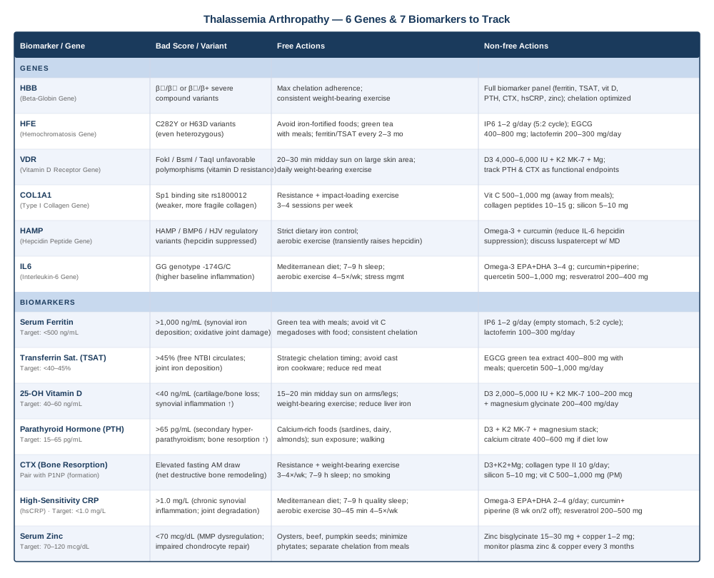
The map provided by biomarkers and genetics only becomes fully useful when it connects to a practical daily strategy — which brings us to a framework that has changed how a growing number of clinicians think about chronic disease management.
What "Outlive" by Peter Attia Reveals About Managing Chronic Disease From the Inside Out
Outlive: The Science and Art of Longevity by Peter Attia (2023) was not written for thalassemia patients. But its central argument — that chronic disease is preceded by measurable biological drift in specific markers years before symptoms become disabling, and that those markers can be individually optimized through lifestyle, nutrition, and targeted interventions — describes the thalassemia arthropathy problem with unusual precision. The book challenges a deeply entrenched medical reflex: the assumption that biomarkers outside the immediately diagnostic range are not worth acting on until they cross clinical thresholds. For thalassemia patients, where the joint damage occurs silently over years before it is visible on imaging, Attia's proactive biomarker framework is directly relevant.
Below are the ten most applicable insights from the book's approach, reframed for the thalassemia arthropathy context.
1. Medicine Two Versus Medicine Three
Attia distinguishes between Medicine 2.0 (reactive care: treat disease once it is diagnosed) and Medicine 3.0 (proactive care: intervene on trajectories before clinical thresholds are crossed). Most thalassemia arthropathy management is 2.0 — joint disease is addressed once it is symptomatic. The 3.0 approach would monitor CTX, PTH, and ferritin trajectory continuously, intervening when the curves are heading the wrong direction rather than waiting for imaging changes.
2. Biomarkers Have Optimal Ranges, Not Just Safe Ranges
Attia emphasizes that conventional lab reference ranges describe the population average, not the optimal biological state. A ferritin of 800 ng/mL might be "within range" for a heavily transfused patient — but it is still meaningfully damaging to joint tissue. A vitamin D of 22 ng/mL is "not deficient" by most lab flags — but it is insufficient for bone protection and inflammation modulation. Reading labs through an optimal lens rather than a normal vs. abnormal binary changes the entire clinical picture.
3. Inflammation Is the Common Currency of Chronic Disease
One of the book's strongest chapters covers how chronic low-grade inflammation — measurable via hsCRP and other cytokine markers — is the shared mechanism driving most of the conditions that eventually disable or kill people. In thalassemia, this inflammation is biologically guaranteed by hemolysis and iron overload; the question is how much amplification your genetics and lifestyle add on top of that baseline. Tracking hsCRP and taking it seriously changes treatment decisions.
4. Exercise Is the Most Powerful Drug Available
Attia's framework, drawing on extensive research, positions structured physical exercise — both aerobic and resistance training — as the intervention with the strongest and broadest evidence base for chronic disease risk reduction of any modality. For thalassemia arthropathy specifically, weight-bearing exercise reduces bone resorption (CTX), lowers hsCRP, stimulates hepcidin, and maintains joint range of motion. The challenge is designing an exercise program that fits within transfusion schedules and energy capacity, not eliminating exercise because it feels difficult.
5. Sleep Is Not Optional; It Is Biological Infrastructure
The book presents sleep as the highest-leverage, zero-cost intervention for inflammation, metabolic health, and tissue repair. Bone remodeling peaks nocturnally. IL-6 and CRP spike reliably with sleep restriction. PTH regulation is affected by circadian disruption. For thalassemia patients already managing significant physiological stress, protecting sleep duration and quality is not a soft recommendation — it is structural.
6. Visceral Fat and Metabolic Dysfunction Drive Joint Degradation
Adipose tissue is metabolically active and proinflammatory — adipokines from visceral fat directly stimulate osteoclast activity and synovial inflammation. Attia's framework measures visceral fat quantitatively (via DEXA or MRI) rather than relying on BMI. For thalassemia patients, who often have altered body composition from anemia and splenomegaly history, visceral adiposity assessment provides a clinically relevant inflammatory signal beyond standard weight measurements.
7. Omega-3 Index Should Be Measured, Not Assumed
Attia consistently argues for measuring the omega-3 index (EPA+DHA as a percentage of red blood cell fatty acids) rather than relying on dietary estimates. A target omega-3 index of 8–12% (versus a typical Western index of 4–5%) requires specific supplementation at doses that most people underestimate. For thalassemia patients seeking to reduce IL-6 and synovial inflammation, knowing your baseline omega-3 index and supplementing to a quantified target is more rigorous than guessing.
8. Bone Density Is a Trajectory, Not a Number
Attia advocates for serial bone density measurements starting earlier than conventional guidelines suggest, because the trajectory matters as much as the absolute value. A DEXA scan showing borderline osteopenia in a 35-year-old thalassemia patient is more informative as a comparison point for a scan two years later than as an isolated number. Combining DEXA with CTX and P1NP as dynamic biomarkers of bone turnover reveals whether the trajectory is improving under current interventions.
9. Supplement Decisions Should Be Evidence-Stratified
The book's approach to supplementation is not dismissive but rigorous — classify each supplement by quality of human evidence, plausibility of mechanism, and relevance to measured biomarker deficits. Vitamin D+K2+magnesium for a patient with low 25-OH-D and elevated PTH is a well-supported decision. IP6 as an adjunct iron chelator in a patient with inadequately controlled ferritin is plausible but early-evidence. This evidence-stratification framework prevents both reflexive supplement dismissal and indiscriminate supplementation.
10. Personalization Over Population-Level Recommendations
Attia's overarching argument is that the most useful medical advice is tailored to the individual's specific biomarker profile, genetics, and lifestyle constraints — not derived exclusively from population averages. A thalassemia patient with HFE co-mutations requires a more aggressive iron management approach than one without; a patient with unfavorable VDR variants requires a different vitamin D strategy than one with normal receptor function. The framework described throughout this article is an application of exactly this principle.
The biomarker and genetic framework presented above provides the analytical foundation. Complementary modalities, discussed next, offer practical tools for the physical and inflammatory dimensions of joint management.
Complementary Approaches Worth Considering
The following four approaches have meaningful human clinical evidence for musculoskeletal, inflammatory, or chronic anemia-related joint conditions. None replaces medical management, but each adds a practical dimension to the overall strategy.
Tai Chi
Tai chi is a low-impact mind-body movement practice originating in Chinese martial arts tradition, characterized by slow, deliberate weight-shifting sequences that simultaneously train balance, joint proprioception, lower extremity strength, and parasympathetic activation. For thalassemia patients with joint involvement, it represents one of the few physical modalities that is gentle enough to be performed on low-energy days near transfusion schedules while still providing measurable benefit to joint function and fall risk — a relevant concern given the bone density deficits common in this population.
A 2016 meta-analysis published in Arthritis Care and Research examining tai chi in osteoarthritis found statistically significant reductions in pain, stiffness, and physical dysfunction. The mechanism is partly mechanical (improved periarticular muscle support reducing joint loading) and partly anti-inflammatory (sustained practice lowers cortisol and IL-6). While direct thalassemia arthropathy trials are absent, the joint protection and inflammation-reducing mechanisms are directly applicable.
The practical protocol for thalassemia patients: begin with 20-minute sessions three times per week using a beginner Yang-style sequence, available through supervised community classes or structured video programs. Focus on sessions scheduled two to three days after transfusion when energy is at its peak. Progress to 30–45 minutes as tolerance improves. Balance-focused sequences specifically (single-leg weight shifts, coordinated arm movements) deliver the most relevant joint proprioception benefit. Avoid sessions on days with significant fatigue or active joint inflammation.
Low-Level Laser Therapy (Photobiomodulation)
Low-level laser therapy (LLLT), also called photobiomodulation, uses specific wavelengths of red (630–700 nm) and near-infrared (780–1100 nm) light at low intensities to stimulate mitochondrial cytochrome c oxidase, reduce oxidative stress, and modulate local inflammatory cytokine production in target tissues. For joint-specific applications, LLLT has demonstrated measurable reductions in synovial inflammation, cartilage degradation markers, and pain scores in clinical trials of various arthropathies. The potential relevance to thalassemia arthropathy is the direct effect on iron-stressed, oxidatively burdened joint tissue — LLLT's mechanism of reducing reactive oxygen species and IL-1β at the cellular level addresses the oxidative and inflammatory environment that iron deposition creates.
A Cochrane-level systematic review published by Brosseau et al. in The Cochrane Database found short-term pain relief and functional improvement in knee osteoarthritis with LLLT using doses in the 4–8 J/cm² range. Though no direct thalassemia arthropathy trials exist, the mechanistic overlap is substantive enough to consider as an adjunct for patients with confirmed synovial inflammation on imaging.
Practically, LLLT devices are available for home use in the 10–200 mW range (handheld panels or probe devices); professional-grade clinical devices deliver treatments in 10–20 minute sessions. For joint applications, targets would include knees, hips, ankles, and wrists — the joints most commonly affected in thalassemia arthropathy. A standard starting protocol is 10–20 minutes per joint area, three times per week for eight weeks, then reassess. Consumer home devices range from $150 to $600; clinical sessions cost $40–150 each. Avoid applying over areas with active infections or areas of known metal implants.
Mindfulness-Based Stress Reduction (MBSR)
MBSR is an eight-week structured program developed by Jon Kabat-Zinn at the University of Massachusetts Medical School, combining formal meditation, body scan practices, and mindful movement to alter the perception and neurobiological processing of chronic pain. For thalassemia arthropathy, MBSR is relevant on two distinct levels: as a chronic pain management tool (addressing the psychological amplification of ongoing joint pain) and as an anti-inflammatory intervention (chronic stress and psychological distress directly elevate IL-6 and CRP through HPA axis activation, compounding the inflammatory drivers already present from iron overload).
A 2016 randomized trial in JAMA Internal Medicine by Cherkin et al. demonstrated that MBSR produced greater reductions in functional limitation from chronic musculoskeletal pain than usual care, with effects persisting at one-year follow-up. The reduction in pain catastrophizing and inflammatory reactivity observed in MBSR are directly applicable to the chronic pain context of thalassemia arthropathy.
The practical approach is engaging with a structured eight-week MBSR program either in-person (offered through many hospital systems and mindfulness centers) or via validated online platforms. Core practices include a 45-minute daily body scan and sitting meditation, plus mindful movement adapted for current joint capacity. For thalassemia patients specifically, the body scan practice is particularly useful for distinguishing baseline joint discomfort from acute inflammatory flares — an important clinical skill when communication with care providers relies heavily on subjective symptom reporting.
Microbiome-Directed Therapies
The gut microbiome is increasingly recognized as a regulator of systemic iron absorption, immune tone, and inflammatory cytokine production — all three of which are central to thalassemia arthropathy pathogenesis. Specific microbiome signatures have been associated with altered iron absorption efficiency (through regulation of hepcidin-modulating gut hormones), systemic inflammation levels (through lipopolysaccharide translocation and short-chain fatty acid production), and bone density (through the gut-bone axis involving serotonin and butyrate signaling). In thalassemia specifically, the chronic use of chelation agents and frequent antibiotic courses disrupts the microbiome in ways that may independently worsen inflammatory and absorptive dysregulation.
Human clinical evidence for microbiome interventions in arthritis and musculoskeletal conditions is growing. A 2018 randomized trial in Rheumatology International found that probiotic supplementation (specifically Lactobacillus casei strains) reduced serum IL-6 and TNF-α levels in patients with rheumatoid arthritis, with accompanying improvements in joint disease activity scores. Direct thalassemia-specific microbiome trials are scarce, but the mechanistic relevance is strong.
The practical starting point is a prebiotic-rich, diverse whole-food diet — the evidence most consistently shows that dietary fiber diversity drives beneficial microbiome shifts more reliably than any single probiotic product. Specifically: aim for 30 or more distinct plant foods per week (vegetables, fruits, legumes, nuts, seeds, whole grains); include fermented foods daily (unsweetened yogurt, kefir, kimchi, sauerkraut); minimize ultra-processed foods and artificial sweeteners, both of which measurably reduce microbiome diversity. If adding a probiotic supplement, strains with the most musculoskeletal and anti-inflammatory evidence in human trials include Lactobacillus rhamnosus GG and Bifidobacterium longum — typically dosed at 5–20 billion CFU daily, taken with a small amount of food. Discuss timing with your hematologist if you are on immunosuppressive therapy.
Conclusion
Thalassemia arthropathy is not an inevitable endpoint, and it is not a single problem with a single solution. It is the downstream result of multiple concurrent biological processes — iron toxicity, impaired bone remodeling, micronutrient depletion, chronic inflammation, and genetic variation — each of which is measurable and, to varying degrees, modifiable. The practical takeaway from everything covered here is straightforward: the more precisely you understand your own biological terrain, the more targeted your interventions can be.
The next smart step is not necessarily adding another supplement. It is measuring what you have not yet measured — starting with serum ferritin, transferrin saturation, 25-OH vitamin D, and hsCRP, if you have not done so recently. Then discuss the results with your hematologist and, if appropriate, a functional medicine physician who is familiar with hemoglobin disorders. Ask specifically about bone turnover markers (CTX, P1NP), zinc status, and PTH if joint health is a primary concern. If genetic panels are accessible, HFE co-mutation status and VDR polymorphisms add meaningful context to your vitamin D and iron management strategy.
Tracking these markers longitudinally — not just as isolated snapshots but as trajectories — gives you and your care team a measurably better foundation for every decision that follows.
Musculoskeletal: Bone Conditions Joint Conditions
Autoimmune: Inflammatory Conditions Connective Tissue Conditions