This article contains AI generated content.
· UpdatedThyroid Acropachy — 4 Genes And 6 Biomarkers To Track
Introduction
Thyroid acropachy sits at an unusual intersection of autoimmune disease, bone biology, and endocrinology. If you've been told you have it—or suspect you might—you already know that most conversations with doctors quickly run out of useful detail. The condition is rare, the research base is thin compared to more common thyroid disorders, and the standard clinical response is often "watch and wait." That answer, while honest, rarely satisfies someone living with unexplained finger clubbing, soft-tissue swelling around the hands and feet, or periosteal bone changes confirmed on X-ray.
What makes acropachy particularly frustrating is that it almost always arrives alongside other manifestations of Graves' disease—pretibial myxedema, exophthalmos, and a history of hyperthyroidism—yet it can persist or even appear after thyroid function has been treated and normalized. This disconnect between thyroid hormone levels and symptom burden confuses patients and, frankly, some clinicians too. The autoimmune process that drives it doesn't stop simply because TSH is back in range.
Generic thyroid management advice—treat the hyperthyroidism, monitor TSH, recheck in six weeks—misses the antibody dynamics, inflammatory signaling, and bone-level changes that drive acropachy specifically. You need a different lens: one focused on which biomarkers actually reflect disease activity, what optimal ranges to aim for, and what interventions have a real evidence base for the underlying autoimmune mechanisms involved.
This article takes that targeted approach. The first section identifies six biomarkers that give a layered picture of the processes driving acropachy, with practical measurement guidance and actionable plans for abnormal results—both with and without supplementation. A genetics section then maps four gene variants with the strongest and most replicated links to Graves' disease susceptibility, with specific compensation strategies. Further sections cover a clinician-authored book directly addressing Graves' disease, and three evidence-informed complementary approaches. The goal is not hope through vague reassurance—it's clarity through better data.
Summary
Thyroid acropachy affects roughly 0.1–1% of people with Graves' disease, yet it remains one of the most poorly understood extrathyroidal manifestations of autoimmune thyroid dysfunction. Most patients never receive guidance beyond monitoring thyroid hormone levels—a strategy that misses the real drivers entirely. This article maps six biomarkers that directly reflect what's actually going on: from TSH receptor antibodies (the single most clinically meaningful test for acropachy activity) to alkaline phosphatase, a bone turnover marker that most thyroid patients have never been asked to check. For each biomarker, you'll find cost estimates, optimal functional ranges, and step-by-step plans both with and without supplementation.
The genetics section covers four gene variants—HLA-DRB1*03:01, CTLA4, PTPN22, and TSHR—that shape how aggressively the immune system targets thyroid tissue and how readily inflammation amplifies the process. These variants don't lock you into a fixed outcome; each one has practical compensation strategies with specific dosing, cycling, and side-effect notes based on published research.
Beyond lab work, this article summarizes the protocol from Dr. Amy Myers' The Thyroid Connection, which directly addresses Graves' disease across 10 high-impact insights, and evaluates three complementary approaches—including Sarah Ballantyne's Autoimmune Protocol—with meaningful mechanistic and clinical rationale for reducing the autoimmune burden underlying acropachy. If you've been told to simply monitor things and come back in six months, there is considerably more you can actually do in the meantime.
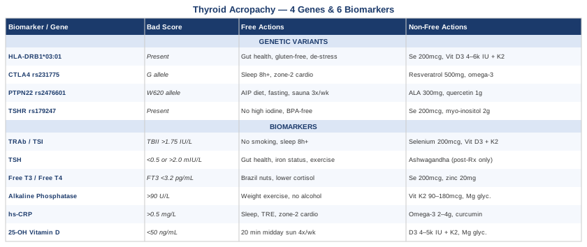
6 Biomarkers to Track for Thyroid Acropachy
No single blood test explains thyroid acropachy. The condition is driven by a convergence of autoimmune activity, inflammatory signaling, and abnormal bone remodeling—each of which has its own measurable fingerprint. The six biomarkers below form a practical monitoring panel that goes significantly beyond the standard thyroid function test. Arranged in order of clinical priority, they give a layered and dynamic picture of where the disease processes stand at any given point.
1. TSH Receptor Antibodies (TRAb)
Why it matters: TRAb—specifically thyroid-stimulating immunoglobulins (TSI)—are the central autoimmune force behind Graves' disease and its extrathyroidal manifestations, including acropachy. In virtually every documented case of acropachy, TRAb levels are elevated, often substantially so. Crucially, acropachy can appear and persist even after thyroid hormone levels have been normalized through antithyroid medication or thyroid ablation, because those treatments address hormone production but not antibody production. Tracking TRAb gives you a direct window into the autoimmune engine that is actually driving the bone and soft-tissue changes.
The correlation between TRAb levels and extrathyroidal manifestations—including acropachy, pretibial myxedema, and Graves' orbitopathy—is well-documented in the clinical literature. A persistently elevated TRAb is the clearest sign that the immune process remains active regardless of what the TSH shows. Conversely, a declining TRAb trend over serial measurements is the most reliable indicator that the condition is coming under control. For this reason alone, TRAb should be the anchor biomarker in any acropachy monitoring panel.
How to measure it: Two assay types are available. The TBII assay (thyrotropin-binding inhibitory immunoglobulin) measures antibody binding to the TSH receptor. The more specific TSI bioassay measures actual receptor-stimulating activity. The TSI bioassay is more clinically informative for acropachy tracking but is slightly less universally available. Cost ranges from approximately $60–$180 depending on which assay is ordered and insurance coverage. Most major reference labs carry both. Test every 3–6 months during active monitoring, extending to every 6–12 months once stable.
Optimal range: TBII below 1.75 IU/L; TSI below 1.3 (index units, varies by lab). Values substantially above these thresholds correlate with higher risk of extrathyroidal manifestation severity and progression.
If TRAb is elevated: plan without supplements
Smoking is the single most potent modifiable risk factor for extrathyroidal Graves' manifestations. Smokers have demonstrably higher TRAb levels and significantly worse outcomes in acropachy, orbitopathy, and pretibial myxedema. Complete cessation is not optional here—it is the highest-yield lifestyle intervention available. Sleep quality is the second lever: T-regulatory cell function, which normally suppresses autoantibody production, is significantly impaired by insufficient or fragmented sleep. Targeting 7.5–9 hours of consolidated sleep per night produces measurable immunological benefits over 4–8 weeks. Chronic low-grade stress shifts immune balance toward Th1/Th17 dominance, promoting autoantibody production—structural stress reduction (not just relaxation techniques) matters meaningfully. Moderate-intensity exercise supports regulatory immune function; excessive intensity (chronic overtraining) can worsen immune dysregulation in autoimmune contexts. A structured 90-day gluten elimination trial has mechanistic rationale through molecular mimicry between gliadin and thyroid antigens.
If TRAb is elevated: plan with supplements or equipment
Selenium (200 mcg/day as L-selenomethionine) is the most evidence-supported supplement for autoimmune thyroid conditions. A randomized controlled trial by Marcocci and colleagues found selenium supplementation significantly reduced TRAb titers in Graves' disease patients compared to placebo. Cycle every 6 months with a 4–6 week break to prevent accumulation; check baseline selenium levels if possible—toxicity risk increases when baseline is already elevated. Vitamin D3 (if deficient, covered in Biomarker 6): 4,000–6,000 IU/day alongside K2 (100–200 mcg MK-7) supports immune regulation and T-regulatory cell production. Myo-inositol combined with selenium has emerging interest from Italian research in autoimmune thyroid conditions—a combination formula at 600 mg myo-inositol plus 83 mcg selenium twice daily has been studied. Avoid high-dose iodine supplementation in any form during active autoimmune phases—it can paradoxically increase receptor antigenicity and worsen antibody production in susceptible individuals.
2. Thyroid Stimulating Hormone (TSH)
Why it matters: TSH is the standard thyroid function marker, but its interpretation in acropachy is more nuanced than usual. During active Graves' disease, TSH is suppressed. After treatment (antithyroid medication, radioiodine, or surgery), it can overcorrect to high-normal or overt hypothyroid values, or land in a normalized range that still isn't functionally optimal. Peter Attia has consistently noted that the standard laboratory reference range for TSH (typically 0.5–4.5 mIU/L in most labs) is derived from population distributions that include many people with subclinical thyroid dysfunction—not a true "optimal" range. Functional targets cluster around 0.5–2.0 mIU/L. In acropachy patients, both ends of the spectrum matter: TSH suppression signals ongoing Graves' activity or overtreatment with thyroid hormone, while TSH elevation signals hypothyroid underperfusion that can worsen fatigue, cognitive function, and immune regulation simultaneously.
How to measure it: Standard blood draw, universally available. Cost: $15–$40. Test every 6–12 weeks when adjusting treatment, every 6 months once stable. Request the result alongside Free T4 and Free T3 (covered in Biomarker 3) for a complete picture.
Optimal range: 0.5–2.0 mIU/L for functional optimization in the acropachy context. Suppression below 0.1 mIU/L or elevation above 3.0 mIU/L each carry implications worth acting on promptly.
If TSH is out of range: plan without supplements
TSH outside the optimal range primarily reflects a medication adjustment need rather than a lifestyle-correctable problem—this is medical territory first. What lifestyle and nutrition can contribute: optimizing gut function matters because gut dysbiosis and intestinal permeability affect thyroid hormone absorption and peripheral conversion. Ensuring adequate iron status (iron deficiency directly impairs thyroid peroxidase function and hormone synthesis). Avoiding prolonged very-low-calorie diets—aggressive caloric restriction suppresses deiodinase activity and can dramatically alter TSH through rT3 elevation. Resistance training improves peripheral thyroid hormone receptor sensitivity over 8–12 weeks of consistent practice.
If TSH is out of range: plan with supplements or equipment
For the post-treatment, stable-hypothyroid state only, adaptogenic herbs with mild thyroid-supportive properties may be considered. Ashwagandha (KSM-66 extract, 300–600 mg/day) has HPA-axis regulation evidence and some data for mild TSH normalization in subclinical hypothyroidism. Cycle: 8 weeks on, 2 weeks off. Critical caveat: ashwagandha has mild thyroid-stimulating properties and is contraindicated in active Graves' disease—it is only appropriate in a stable, adequately treated, hypothyroid post-Graves' state and must be discussed with your prescribing physician before starting.
3. Free T4 and Free T3
Why it matters: TSH alone doesn't capture the full picture of thyroid hormone status at the tissue level. Free T4 (thyroxine, the storage hormone) and Free T3 (triiodothyronine, the active hormone) reflect what is actually circulating and available to cells. The T4-to-T3 conversion step—carried out by deiodinase enzymes in peripheral tissues—is critically sensitive to selenium status, inflammation, caloric intake, and gut health. Many patients treated for Graves' disease feel persistently unwell despite a "normalized" TSH because their Free T3 sits at the low end of the reference range, with insufficient active hormone reaching tissues. Thomas Dayspring and other precision medicine practitioners have emphasized that symptoms track Free T3 levels more closely than TSH in many thyroid patients.
How to measure it: Blood draw, run alongside TSH. Free T4: $20–$50; Free T3: $25–$60. Most physicians can order these together; some may require additional rationale for Free T3 specifically. Optimal Free T4: 1.0–1.5 ng/dL (mid-to-upper portion of the reference range). Optimal Free T3: 3.2–4.2 pg/mL (upper half of the typical reference range). Test every 6–12 weeks when adjusting treatment.
If Free T3 is low or conversion is poor: plan without supplements
When Free T3 is low while Free T4 is adequate, the issue is conversion efficiency rather than thyroid output. Dietary priorities: selenium through Brazil nuts (2–3 per week, not more—toxicity risk at high doses), zinc through oysters and pumpkin seeds, and iron through red meat or organ meats if ferritin is low. Eliminate chronic caloric restriction—dieting below 1,200 kcal/day suppresses deiodinase and sends Free T3 into a floor. Cortisol management is directly relevant: chronically elevated cortisol increases reverse T3 (rT3) production, which competes with and blocks Free T3 receptor binding. Reduce the inputs driving cortisol excess—inadequate sleep, overtraining, high psychological stress load.
If Free T4 or Free T3 is suboptimal: plan with supplements or equipment
Selenium (200 mcg/day selenomethionine): directly supports deiodinase type 1 and type 2 function, which convert T4 to active T3. Zinc picolinate or glycinate (15–25 mg/day): supports thyroid hormone receptor sensitivity; cycle with a copper-containing multivitamin or take copper separately (2 mg/day) to prevent copper depletion from prolonged zinc supplementation. Cycle zinc: 6 weeks on, 1–2 weeks off. Iodine cautiously: 150–250 mcg from food or a minimal supplement is generally safe and supports hormone synthesis, but high-dose iodine (above 500 mcg/day from supplements) can provoke autoimmune flares in individuals with active Graves' antibodies. Pharmaceutical options (levothyroxine, liothyronine, or desiccated thyroid) are the most effective tools when levels remain persistently suboptimal despite lifestyle optimization—these are medical decisions in collaboration with your physician.
4. Alkaline Phosphatase (ALP)
Why it matters: Alkaline phosphatase is a bone turnover marker that most thyroid patients have never been asked to track—yet it is directly relevant to acropachy. ALP is elevated during periods of active periosteal and endosteal bone formation. The radiological hallmark of acropachy is exactly this: abnormal periosteal bone deposition, particularly in the metacarpals and phalanges. Tracking ALP over time, with liver causes excluded, gives a real-time indication of whether the bone remodeling process is active, stable, or resolving. Combined with TRAb trend data, ALP provides a dynamic picture of two converging disease processes simultaneously.
In active and recently treated Graves' disease, ALP can also be elevated due to excess thyroid hormone driving systemic bone turnover—meaning it has informational value both during active hyperthyroid phases and after euthyroid restoration. If ALP remains elevated after thyroid hormone is controlled, bone-specific activity (acropachy-related periosteal formation) becomes the more likely explanation.
How to measure it: Total ALP is included in a standard comprehensive metabolic panel (CMP). Cost: $15–$40 as part of CMP. For higher specificity, request bone-specific alkaline phosphatase (BAP) separately—available through specialized labs at approximately $50–$100. Always check GGT, ALT, and AST alongside total ALP to exclude liver causes. Optimal total ALP: 40–90 U/L (mid-range is preferable to high-normal). Test every 3–6 months during active monitoring phases.
If ALP is elevated: plan without supplements
Rule out liver causes first (elevated GGT alongside ALP points to liver; normal GGT with elevated ALP points to bone). For bone-specific elevation: weight-bearing exercise (30–45 minutes of walking, resistance training, or low-impact activity, 4–5 days per week) promotes organized lamellar bone formation and reduces the disorganized periosteal activity characteristic of acropachy-related remodeling. An anti-inflammatory dietary pattern (Mediterranean structure: olive oil, fatty fish, vegetables, legumes, minimal refined sugar) reduces cytokine-driven osteoclast/osteoblast dysregulation. Eliminate alcohol entirely—alcohol directly elevates ALP, impairs bone matrix quality, and depletes key bone minerals simultaneously. Adequate protein intake (1.2–1.6 g/kg/day) supports collagen synthesis required for normal bone matrix formation.
If ALP is elevated: plan with supplements or equipment
Vitamin K2 (MK-7 form): 90–180 mcg/day activates osteocalcin and matrix Gla protein, which direct mineralization into proper bone structure and inhibit soft-tissue calcification. Continuous use is well-tolerated based on available literature; most human trials have run 12–24 weeks without break requirements. Vitamin D3 (see Biomarker 6 for full protocol): essential cofactor for ALP enzyme function and bone matrix mineralization. Magnesium glycinate: 300–400 mg at night; magnesium is a cofactor for ALP itself and for proper hydroxyapatite crystal formation in bone—deficiency is common and directly impairs normal bone remodeling. Photobiomodulation (low-level laser therapy) has biologically plausible and emerging clinical evidence for periosteal tissue modulation—covered in the complementary approaches section.
5. High-Sensitivity C-Reactive Protein (hs-CRP)
Why it matters: CRP is produced by the liver in response to upstream cytokine signaling—particularly IL-6 and TNF-alpha—that reflects the background inflammatory state of the body. In autoimmune thyroid disease, this background inflammation fuels both antibody production and the connective tissue and fibroblast changes seen in acropachy and pretibial myxedema. Peter Attia consistently places hs-CRP among his core metabolic health biomarkers, with a functional target below 0.5 mg/L—far below the conventional clinical cutoff of under 1.0 mg/L. Even sustained low-grade elevations in the 1–3 mg/L range carry meaningful implications for autoimmune amplification and cardiovascular risk, which is particularly relevant because Graves' disease independently elevates cardiovascular risk.
The distinction between high-sensitivity CRP and standard CRP matters clinically. Standard CRP is calibrated for detecting acute infections and inflammation (ranges in tens of mg/L). hs-CRP is calibrated for detecting low-grade chronic inflammation (ranges in fractions of mg/L)—exactly what's relevant for monitoring chronic autoimmune conditions like Graves'/acropachy.
How to measure it: Standard blood draw; must specifically order high-sensitivity CRP. Cost: $15–$50. Available at any standard reference lab. Optimal: below 0.5 mg/L (Attia's functional target); below 1.0 mg/L is the conventional clinical threshold. Test every 3–6 months alongside the other biomarkers in this panel.
If hs-CRP is elevated: plan without supplements
Sleep is the most powerful zero-cost anti-inflammatory intervention available. Seven or more hours of consolidated sleep reduces IL-6 and TNF-alpha production—the direct upstream drivers of CRP elevation. The impact is measurable within 2–4 weeks of consistent sleep improvement. Dietary elimination of ultra-processed foods, industrial seed oils (soybean, canola, corn, sunflower in large quantities), and refined sugars addresses the primary dietary contributors to low-grade inflammation. Time-restricted eating (10–12 hour eating window, aligned with daylight hours) has consistent evidence for hs-CRP reduction in metabolic studies. Zone 2 cardiovascular exercise (150–180 minutes/week at conversational pace, below lactate threshold) reliably reduces hs-CRP over 8–12 weeks of consistent practice. Treat underlying contributors: periodontal disease, latent infections, and gut dysbiosis all sustain background CRP elevation and are often overlooked.
If hs-CRP is elevated: plan with supplements or equipment
Omega-3 fatty acids (EPA+DHA combined): 2–4 g/day from high-quality triglyceride-form fish oil; multiple meta-analyses confirm dose-dependent hs-CRP reduction with sustained supplementation. Purchase from brands with IFOS certification to avoid rancidity; continuous use is well-tolerated—monitor for anticoagulation interaction at doses above 3 g/day if taking blood thinners. Curcumin as phospholipid complex (BCM-95) or liposomal form: 500–1,000 mg/day; numerous randomized controlled trials confirm hs-CRP reduction; avoid at high doses with anticoagulant or antiplatelet medications. Berberine: 500 mg twice daily with meals; anti-inflammatory and insulin-sensitizing; cycle 8 weeks on, 2 weeks off (gastrointestinal adaptation—bloating, loose stools—is common in the first 1–2 weeks). Sauna use (traditional Finnish or infrared, 3–5 sessions/week, 15–20 minutes at 80–90°C) activates heat shock proteins that suppress inflammatory cytokine signaling—research by Laukkanen and colleagues in Finnish populations provides strong human evidence for cardiovascular and inflammatory benefits.
6. 25-OH Vitamin D
Why it matters: Vitamin D functions less as a nutrient and more as a steroid hormone with profound immunomodulatory properties. Its receptor (VDR) is expressed on virtually every immune cell type, and vitamin D signaling directly promotes the differentiation of T-regulatory (Treg) cells—the exact cell population that normally suppresses autoantibody production. Low vitamin D is disproportionately prevalent in autoimmune thyroid disease populations. A meta-analysis examining vitamin D status in Graves' disease patients found significantly lower 25-OH D levels compared to healthy controls, with lower levels correlating with higher TRAb titers. This isn't a peripheral nutritional detail—it is mechanistically central to the immune regulation that determines how active the antibody-driven processes underlying acropachy remain.
Beyond its direct immune effects, vitamin D participates in bone calcium metabolism, which is separately relevant given acropachy's bone remodeling component. VDR polymorphisms (particularly VDR FokI and BsmI variants) affect how well any given level of circulating vitamin D translates to receptor activity—some individuals need higher serum levels to achieve equivalent functional effects.
How to measure it: Blood draw, measuring 25-hydroxyvitamin D (25-OH D)—the storage form and best indicator of overall vitamin D status. Cost: $30–$70. Available at any reference lab. Optimal range: 50–80 ng/mL. The conventional clinical normal (30–100 ng/mL) is too broad to be useful for autoimmune patients; researchers including Michael Holick and Bruce Hollis argue for the 50–80 ng/mL window for immune optimization. Test every 3–6 months initially, then twice yearly once levels are stable in the optimal range.
If Vitamin D is suboptimal: plan without supplements
Targeted sun exposure: 15–30 minutes of midday sun exposure (UV index 3 or above) on arms, legs, and ideally torso, without sunscreen, 4–5 days per week. This protocol can raise 25-OH D by 10–20 ng/mL over 4–8 weeks depending on skin tone, latitude, and season. Darker skin tones, northern latitudes (above 35°N), and winter months significantly reduce synthesis efficiency—supplementation becomes necessary in these contexts. Fatty fish (wild salmon, mackerel, sardines) consumed 3–4 times per week contributes meaningful dietary D3. Reducing cortisol exposure is indirectly relevant: sustained cortisol elevation impairs VDR sensitivity, meaning the same circulating vitamin D level has reduced functional impact.
If Vitamin D is suboptimal: plan with supplements or equipment
Vitamin D3 + Vitamin K2: Start at 4,000–5,000 IU/day D3 combined with 100–200 mcg K2 (MK-7 form). K2 is critical when supplementing D3 at these doses—it activates matrix Gla protein, which prevents arterial calcification as calcium metabolism is upregulated. Recheck 25-OH D after 8–12 weeks to titrate; some individuals with VDR polymorphisms or significant obesity (vitamin D sequestration in fat tissue) require 8,000–10,000 IU/day to reach the 50–80 ng/mL target. Take with the largest meal of the day for fat-soluble absorption. Magnesium glycinate (300–400 mg/night) is a required cofactor for the enzymatic steps that convert vitamin D to its active form—without adequate magnesium, D3 supplementation may produce lower-than-expected serum level increases. If levels remain low despite adequate supplementation, test for VDR polymorphisms through genetic testing services.
Moving from what blood tests reveal to what your DNA predicts, the genetics of Graves' disease illuminate why some people are dramatically more vulnerable to the autoimmune cascade that leads to acropachy—and what can practically be done to work with those variants rather than against them.
4 Key Genetic Variants in Thyroid Acropachy and Graves' Disease
Thyroid acropachy does not yet have its own dedicated genetic literature—it is too rare for that. But because it occurs almost exclusively within the context of Graves' disease, the genetic architecture of Graves' is directly and entirely relevant. The four variants below have the strongest, most consistently replicated associations across multiple ethnic populations and genome-wide association studies. Collectively, they explain a meaningful portion of individual susceptibility and—crucially—each one points toward specific biological pathways that can be partially compensated for through lifestyle, nutrition, and targeted supplementation.
HLA-DRB1*03:01 (HLA-DR3)
What it does: The human leukocyte antigen system governs antigen presentation—the process by which immune cells recognize and react to both foreign and self-derived proteins. The HLA-DRB1*03:01 allele, commonly called DR3, is the single most strongly and consistently replicated genetic risk factor for Graves' disease across diverse populations. Carriers have approximately 3–5 times the population risk of developing Graves' disease. The mechanism is at the level of antigen presentation: the DR3 molecule is particularly efficient at presenting thyroid-derived peptides—including fragments of the TSH receptor—to CD4+ T helper cells, initiating and perpetuating the autoimmune cascade. Individuals carrying both DR3 and a CTLA4 risk variant (see below) face compounded risk from two complementary immunological pathways.
Understanding this variant helps explain why acropachy patients often have more aggressive autoimmune phenotypes with higher TRAb levels and more pronounced extrathyroidal manifestations—the upstream immunological machinery is running at higher sensitivity from the start.
If HLA-DR3 is present: plan without supplements
This allele cannot be changed, but its expression of risk is significantly modifiable by environmental context. The gut-immune axis is the most powerful lever available. Increased intestinal permeability (leaky gut) dramatically amplifies the antigen load presented to HLA-DR molecules by allowing partially digested food proteins and bacterial fragments to enter systemic circulation and encounter immune cells. A structured elimination diet removing gluten, dairy, soy, and refined sugars for a minimum of 90 days is the highest-impact dietary strategy for reducing the antigenic burden that HLA-DR3 processes most aggressively. Stress is a documented environmental trigger for Graves' onset in genetically susceptible individuals—structural stress reduction (reductions in workload, relationship stressors, and sleep debt) produces immune effects that relaxation techniques alone cannot replicate. Minimize environmental estrogen exposure: plastics containing BPA and phthalates, commercially raised meat, and hormonal contraceptives all add exogenous estrogen, which activates immune pathways that compound HLA-DR3-mediated autoimmune risk.
If HLA-DR3 is present: plan with supplements or equipment
Selenium (200 mcg/day selenomethionine): reduces TRAb in randomized trials—directly relevant to the downstream autoimmune output of this allele. Vitamin D3 at immune-optimizing doses (4,000–6,000 IU/day titrated to 50–80 ng/mL serum): vitamin D promotes T-regulatory cell differentiation and counteracts the Th17/Th1 immune bias that HLA-DR3 facilitates. L-Glutamine (5–10 g/day): supports intestinal barrier integrity by serving as the primary fuel source for enterocytes; cycle 8 weeks on, 4 weeks off. Reducing gut permeability in a DR3 carrier lowers the volume of antigens reaching HLA-DR3 molecules for presentation—a direct upstream intervention on the risk pathway. Zinc carnosine (75 mg twice daily): has specific evidence for mucosal gut barrier repair beyond general zinc supplementation; 12-week cycles are standard in available trials.
CTLA4 (rs231775 / A49G Polymorphism)
What it does: CTLA4 (cytotoxic T-lymphocyte antigen 4) is an immune checkpoint molecule—the biological equivalent of a brake pedal on T cell activation. When CTLA4 functions normally, it prevents T cells from becoming overactivated, including against self-antigens. The rs231775 G allele (the A49G variant) results in reduced CTLA4 expression or impaired function, meaning the brake is less effective. The outcome is a T cell population that is more readily activated against thyroid antigens and less subject to the normal inhibitory controls that prevent autoimmunity. CTLA4 risk variants are present in 60–70% of Graves' disease patients in some European populations and are particularly associated with more severe extrathyroidal manifestations—which includes acropachy. The pharmaceutical drug abatacept (Orencia), used in rheumatoid arthritis, works by mimicking CTLA4 function, which illustrates the clinical significance of this pathway.
If CTLA4 variant (G allele) is present: plan without supplements
Reduced checkpoint function means anything that activates T cells can have disproportionately amplified effects. Concrete practical implications: discuss timing of elective vaccinations and immune-stimulating interventions carefully with your physician during active Graves' phases, as the normal immune modulation from vaccines can be exaggerated in CTLA4-variant carriers. Prioritize sleep rigorously—T cell regulation is significantly and measurably impaired by even a single night of poor sleep, and CTLA4-variant carriers have less regulatory buffering to absorb that impairment. Infections are documented triggers for Graves' exacerbation in genetically susceptible individuals—prompt treatment of any acute infection matters more than in the general population. Shift exercise intensity toward zone 2 cardio and moderate resistance training during active disease phases, as high-intensity exercise transiently activates T cells systemically.
If CTLA4 variant (G allele) is present: plan with supplements or equipment
Resveratrol (trans-resveratrol or pterostilbene form, 500 mg/day): activates SIRT1, which upregulates FOXP3 expression in T-regulatory cells, partially compensating for CTLA4 checkpoint reduction by amplifying an alternative regulatory pathway. Cycle: 12 weeks on, 4 weeks off; pterostilbene has superior bioavailability and a longer half-life than standard resveratrol. EPA+DHA omega-3s (2–4 g/day): promote Th2/T-regulatory immune balance over Th1 activation, offering a relevant compensatory shift for reduced CTLA4 function. Berberine (500 mg twice daily): inhibits the mTOR pathway, reducing T cell hyperactivation; cycle 8 weeks on, 2 weeks off. N-acetylcysteine (NAC) (600 mg twice daily): glutathione precursor reducing oxidative-stress-driven T cell activation; well-tolerated on a continuous basis with no recognized cycling requirement at this dose.
PTPN22 (rs2476601 / R620W Variant)
What it does: PTPN22 encodes the LYP phosphatase enzyme, a critical regulator of T cell receptor signaling. The W620 variant (rs2476601) alters normal calibration of T cell activation thresholds. Despite appearing paradoxically as a gain-of-function variant in some mechanistic studies, it is robustly and repeatedly associated with multiple autoimmune conditions—Graves' disease, rheumatoid arthritis, type 1 diabetes, and lupus among them—across large population studies and GWAS data. The net biological effect in the autoimmune disease context is a T cell population with disrupted self-tolerance. The variant is carried by approximately 8–15% of individuals of European ancestry, making it the most prevalent of the four variants discussed here.
For acropachy specifically, the PTPN22 variant is relevant because it amplifies the autoimmune inflammatory signaling that both generates TRAb and drives the cytokine environment in which connective tissue and periosteal changes develop.
If PTPN22 variant (W620) is present: plan without supplements
Anti-inflammatory dietary patterns are particularly high-priority with this variant, as it amplifies cytokine-driven immune activation. The Mediterranean dietary pattern has meta-analytic evidence for reducing autoimmune flare frequency across several autoimmune conditions and is the most accessible starting point. A structured AIP elimination phase (see Strategy 4) goes further and is worth committing to for 60–90 days as a diagnostic and therapeutic intervention. Intermittent fasting (16:8 daily or periodic 24-hour fasts) activates autophagy, which clears dysfunctional proteins and misfolded peptides that can serve as autoimmune triggers—relevant for PTPN22 carriers because the variant amplifies responses to antigenic stimulation. Sauna (3–4 sessions/week, 15–20 minutes at 80°C) produces post-session immune regulatory shifts and activates heat shock proteins that modulate inflammatory pathways; 3-month consistent use before reassessing impact.
If PTPN22 variant (W620) is present: plan with supplements or equipment
Alpha-lipoic acid (ALA): 300–600 mg/day; antioxidant and immune modulator with evidence across multiple autoimmune contexts; specifically reduces NFkB-mediated inflammatory transcription. Cycle 8 weeks on, 2 weeks off; avoid in thiamine-deficient individuals (rare but worth checking). Quercetin: 500 mg twice daily; inhibits downstream inflammatory signaling pathways relevant to PTPN22-affected T cell activation; take with bromelain (100 mg) to improve absorption. Broad-spectrum probiotics including Lactobacillus rhamnosus GG and Bifidobacterium longum: gut microbiome diversity reduces systemic autoimmune antigen load and supports mucosal immune regulation; 30–90 day course, then maintain diversity through dietary diversity (fermented vegetables, varied plant fiber). Omega-3 fatty acids (EPA+DHA 2–3 g/day): relevant here as in other gene variants for promoting regulatory immune balance.
TSHR Gene Variants (rs179247)
What it does: Genetic variants within and near the TSHR gene (encoding the TSH receptor) influence Graves' disease susceptibility through a mechanistically distinct and particularly relevant pathway: they affect the very tissue structure that becomes the autoimmune target. The rs179247 variant in intron 1 of TSHR is among the most replicated Graves' disease susceptibility loci in genome-wide association studies, appearing across European, Asian, and Chinese ancestry populations. It appears to influence TSHR expression levels in thyroid tissue—and potentially on other cell types expressing the receptor.
This is where the direct connection to acropachy becomes most explicit: the TSH receptor is expressed not only on thyroid follicular cells but also on dermal fibroblasts—the same cells that proliferate abnormally in pretibial myxedema, and that are implicated in the connective tissue changes associated with acropachy. TSHR expression on fibroblasts means that TRAb can directly drive fibroblast activation and glycosaminoglycan deposition in peripheral tissues, which is the underlying mechanism of the soft-tissue manifestations. TSHR variant carriers may have altered receptor expression on these fibroblast populations, potentially explaining some of the variation in extrathyroidal manifestation severity between Graves' patients with otherwise similar antibody levels.
If TSHR variant (rs179247) is present: plan without supplements
Avoid high-dose iodine supplementation unconditionally during active Graves' phases. Excess iodine directly modulates TSHR expression and can increase receptor antigenicity—the Wolff-Chaikoff effect becomes problematic in autoimmune thyroid disease. Dietary iodine through whole foods (seaweed occasionally, seafood 2–3 times/week) is generally acceptable; iodine-containing supplements, high-dose kelp, and Lugol's solution are contraindicated. Environmental thyroid-disrupting chemicals deserve active attention: BPA and phthalates from plastics, organochlorine pesticides, and polychlorinated biphenyls (PCBs) have documented effects on TSHR signaling and thyroid receptor biology. Practical steps: glass food storage, stainless steel water bottles, organic produce prioritization for high-pesticide items (the EWG Dirty Dozen list), and water filtration (reverse osmosis removes fluoride, chlorine, and many endocrine-disrupting compounds).
If TSHR variant (rs179247) is present: plan with supplements or equipment
Selenium (200 mcg/day selenomethionine): protects thyroid follicular cells from oxidative damage during autoimmune attack and reduces TRAb in clinical trials—directly relevant to TSHR-expressing thyroid tissue under immune pressure. Myo-inositol (2 g twice daily): inositol is a secondary messenger in the TSH receptor signaling cascade; myo-inositol supplementation has shown benefit in autoimmune thyroid conditions in a small randomized Italian study by Nordio and Basciani; continuous use is well-tolerated at these doses. N-acetylcysteine (NAC) (600 mg twice daily): reduces oxidative stress specifically in thyroid tissue under autoimmune attack, protecting TSHR-expressing cells from collateral damage. Ashwagandha (KSM-66, 300–600 mg/day): applicable only in post-treatment, stable-hypothyroid state—not in active Graves' disease; may reduce TSH fluctuation and support receptor normalization; cycle 8 weeks on, 2 weeks off.
Both the biomarker monitoring framework and the genetic landscape converge on the same core insight: thyroid acropachy is driven by an autoimmune process with identifiable, partially modifiable mechanisms. The tools to support that modulation extend well beyond standard thyroid medication management.
The Thyroid Connection by Amy Myers, M.D. — 10 Insights That Challenge Standard Thyroid Care
Amy Myers, M.D., a functional medicine physician who developed and reversed her own Graves' disease, wrote The Thyroid Connection (2016) after recognizing that standard endocrinology—antithyroid drugs, radioiodine, thyroidectomy—addresses the organ but not the immune dysfunction driving the organ's destruction. The book synthesizes peer-reviewed research across autoimmune immunology, gastroenterology, environmental medicine, and endocrinology into a clinical protocol directly applicable to Graves' disease, its extrathyroidal manifestations, and the autoimmune processes underlying acropachy. For patients who have been told that controlling thyroid hormone levels is sufficient, it is one of the most useful clinician-authored resources available.
1. Leaky gut is the upstream trigger, not a side effect
Myers argues that increased intestinal permeability precedes and perpetuates autoimmune thyroid disease by allowing partially digested food proteins and microbial fragments to reach systemic immune cells. These antigens drive immune responses that can cross-react with thyroid tissue through molecular mimicry. Restoring gut barrier integrity is therefore not supportive care—it is upstream of the autoimmune process itself, and addressing it first changes the trajectory of what follows.
2. Gluten molecular mimicry is real and specific
Gliadin (a component of gluten) shares structural sequences with thyroid peroxidase and thyroglobulin proteins. In genetically susceptible individuals, immune responses to gliadin can cross-activate against thyroid antigens—a process with documented mechanistic plausibility in the peer-reviewed literature. Myers advocates for complete gluten elimination, not reduction, as partial compliance maintains enough immune activation to sustain the cross-reactive response. A strict 90-day trial produces measurable changes in antibody levels in responsive individuals.
3. Latent infections are underdiagnosed triggers
Epstein-Barr virus (EBV), Helicobacter pylori, and Yersinia enterocolitica have documented associations with Graves' disease onset. Yersinia in particular carries a protein with structural homology to the TSH receptor—meaning immune responses to Yersinia can generate antibodies that cross-react with thyroid receptors. Myers recommends including infectious workup (EBV viral capsid antigen IgG/IgM, H. pylori stool antigen, and Yersinia antibodies) in any comprehensive autoimmune thyroid evaluation. Treating a latent infection in this context can meaningfully reduce antibody burden.
4. Toxin burden disrupts thyroid function at the receptor level
Environmental toxins—mercury from amalgam fillings and high-mercury fish, organochlorine pesticides, PCBs, and halides (fluoride, bromide)—compete with iodine at thyroid receptor sites and disrupt thyroid hormone synthesis and receptor signaling. Myers provides a specific toxin-reduction protocol: water filtration, prioritizing low-mercury seafood (sardines, anchovies, wild salmon), investigating amalgam removal with a biological dentist (carefully and sequentially, not all at once), and supporting hepatic detoxification pathways through cruciferous vegetables and adequate glutathione precursors.
5. The adrenal-thyroid connection shapes hormone conversion
Cortisol and thyroid hormone metabolism are tightly coupled. HPA dysregulation—from chronic stress, sleep debt, prior trauma, or blood sugar volatility—suppresses T4-to-T3 conversion by upregulating reverse T3 (rT3) production. rT3 competes with and blocks Free T3 at receptor sites, producing functional hypothyroid symptoms even when Total T4 appears adequate on labs. This mechanism directly explains why many acropaphy patients who are technically "euthyroid" still feel poorly and continue to have active inflammatory processes—adrenal status must be addressed in parallel with thyroid status.
6. Selenium is the most important thyroid mineral
Myers dedicates substantial discussion to selenium's role across multiple thyroid-critical processes: thyroid peroxidase function (required for hormone synthesis), deiodinase enzymes (required for T4-to-T3 conversion), and glutathione peroxidase activity in thyroid tissue (protecting follicular cells from the hydrogen peroxide generated during hormone synthesis). She cites the randomized controlled trial data supporting 200 mcg/day selenomethionine for TRAb reduction and positions selenium as the foundational supplement for any autoimmune thyroid patient—above and before most other interventions.
7. Standard thyroid panels systematically miss the clinical picture
TSH alone is insufficient. Myers' recommended panel includes TSH, Free T3, Free T4, Reverse T3, TRAb (both TBII and TSI), TPO antibodies, thyroglobulin antibodies, ferritin, selenium, and vitamin D at minimum. Most conventional thyroid workups order only TSH and perhaps Free T4. The gaps—particularly the absence of TRAb, Free T3, and Reverse T3—mean that active autoimmune disease and poor conversion efficiency are routinely missed. This is the most direct and actionable critique of standard thyroid care for acropaphy patients: the monitoring panel is usually too narrow to guide meaningful decisions.
8. The autoimmune spectrum changes how you interpret your labs
Myers frames autoimmune thyroid disease on a spectrum with three stages: early silent dysregulation (abnormal immune markers before antibodies become positive), active autoimmune phase (elevated antibodies with or without frank disease), and burned-out or post-treatment stage. Most clinical protocols treat this as binary—either you have it or you don't. The spectrum framework allows earlier identification of intervention windows and more nuanced interpretation of symptom shifts. For acropaphy patients specifically, understanding which phase they're in changes what to prioritize in terms of intervention focus.
9. Nutrient deficiencies are nearly universal and go unmeasured
Based on her clinical experience with thousands of autoimmune thyroid patients, Myers found vitamin D, selenium, zinc, ferritin, magnesium, and omega-3 fatty acids to be deficient or suboptimal in the vast majority. These deficiencies are not incidental—they are mechanistically relevant at each level of thyroid and immune function. She provides condition-specific dosing ranges and emphasizes that many patients experience meaningful symptom improvement within 60–90 days of correcting these specific deficiencies, independent of any medication adjustment.
10. Radioiodine may worsen extrathyroidal manifestations in high-risk patients
Myers presents evidence and clinical experience suggesting that radioiodine ablation (RAI) may worsen Graves' orbitopathy and potentially other extrathyroidal manifestations—including worsening TRAb levels post-treatment—in patients with high pre-treatment antibody titers. This is a contested area in the endocrinology literature, with ongoing debate between thyroidologists and ophthalmologists. Myers recommends that TRAb levels be measured and evaluated before choosing RAI, that patients with high TRAb and active extrathyroidal manifestations consider antithyroid drug therapy as a first-line option to allow antibody levels to decline before making irreversible treatment decisions, and that the risks and benefits of RAI for extrathyroidal-manifestation patients be discussed explicitly with an endocrinologist familiar with the controversy.
Complementary Approaches with Clinical Relevance for Thyroid Acropachy
No complementary modality has been studied directly in acropaphy patients—the condition is too rare. What follows draws on evidence from Graves' disease, autoimmune thyroid conditions broadly, and the specific physiological mechanisms at play: autoimmune regulation, systemic inflammation, and connective tissue/bone remodeling. Evidence quality is noted honestly for each approach.
The Autoimmune Protocol by Sarah Ballantyne
Sarah Ballantyne, Ph.D., developed the Autoimmune Protocol (AIP) as a comprehensive dietary and lifestyle framework for autoimmune conditions, detailed in The Paleo Approach (2013). The elimination phase removes grains, legumes, dairy, eggs, nuts, seeds, nightshades, processed foods, industrial seed oils, refined sugars, and alcohol—the foods with the highest immune reactivity potential and the greatest evidence for gut permeability disruption. The reintroduction phase systematically reintroduces eliminated foods to identify individual triggers. The dietary foundation emphasizes organ meats, fatty fish, diverse vegetables, bone broth, and fermented foods—prioritizing micronutrient density and gut-supportive substrates. For Graves' disease and acropaphy specifically, the AIP directly addresses the gut-immune axis mechanism that both Myers and the genetic literature implicate as central to autoimmune thyroid disease perpetuation. The autoimmune protocol from Sarah Ballantyne is the most structurally complete available dietary intervention for this condition.
In 2017, a pilot study published in Inflammatory Bowel Diseases evaluated the AIP in Crohn's disease and ulcerative colitis patients, demonstrating significant clinical remission rates alongside measurable reductions in inflammatory markers (CRP, fecal calprotectin). This provides human proof-of-concept that the protocol produces its intended biological effects in autoimmune conditions—not merely in theory. For autoimmune thyroid conditions, evidence remains at the clinical case-report and anecdotal level; Ballantyne is transparent about this limitation while noting that the mechanistic basis is shared across autoimmune conditions.
To apply it realistically: commit to a minimum 60-day full elimination phase with no partial compliance—the protocol works through cumulative reduction of antigenic and inflammatory load, not episodic adherence. Track TRAb, hs-CRP, and symptom severity scores before starting and at 60 and 90 days to evaluate individual response objectively. During the elimination phase, ensure adequate caloric and micronutrient intake through intentional menu planning around the allowed foods—the elimination list is broad enough that nutritional gaps (particularly calcium, zinc, and B vitamins) are possible without planning. Reintroduction should be systematic: one new food every 5–7 days, monitoring for 72 hours post-introduction before adding another.
Mindfulness-Based Stress Reduction (MBSR)
Psychological stress is both a documented trigger for Graves' disease onset and a perpetuating factor in autoimmune exacerbations. The HPA axis response to chronic stress drives cortisol dysregulation, which impairs T-regulatory cell function, promotes Th1/Th17 immune dominance, and suppresses Free T3 through elevated reverse T3 production—all pathways directly relevant to Graves' and acropaphy disease activity. MBSR, the standardized 8-week mindfulness program developed by Jon Kabat-Zinn, is the most rigorously studied mind-body intervention for stress-mediated immune and inflammatory effects, with the largest and best-controlled evidence base among mind-body approaches.
A study by Rosenkranz and colleagues published in Brain, Behavior, and Immunity (2013) demonstrated that MBSR practitioners showed significantly lower post-stress inflammatory cytokine responses compared to participants in a matched health enhancement program. Additionally, preliminary research in Hashimoto's thyroiditis patients found measurable reductions in TPO antibody titers following an 8-week MBSR intervention—evidence that is preliminary but mechanistically credible given the shared autoimmune biology between Hashimoto's and Graves' disease.
To apply it in a way that reaches meaningful dose: join a structured 8-week MBSR program rather than using an informal mindfulness app—the research evidence is for structured, dose-sufficient practice (typically 45 minutes daily). Formal programs are available through hospital integrative medicine centers and validated online platforms. For acropaphy patients, the therapeutic goal is not symptom relief through relaxation in any session but rather the cumulative neurobiological changes—reduced cortisol reactivity, increased T-regulatory cell populations, lower baseline inflammatory cytokine production—that develop over months of consistent practice and that show up in measurable biomarkers over a 3–6 month monitoring period.
Low-Level Laser Therapy / Photobiomodulation
Low-level laser therapy (LLLT), also called photobiomodulation (PBM), applies specific wavelengths of red (630–700 nm) and near-infrared (810–850 nm) light to modulate cellular function at the mitochondrial level—specifically by activating cytochrome c oxidase, increasing ATP production, reducing oxidative stress, and modulating local inflammatory cytokine expression. Its relevance to acropachy is indirect but biologically coherent: the periosteal bone and connective tissue changes underlying acropachy involve osteoblast and fibroblast biology that LLLT has been shown to modulate in other musculoskeletal and inflammatory contexts.
The most directly relevant human evidence comes from a Brazilian randomized controlled trial by Höfling and colleagues published in Lasers in Surgery and Medicine, which applied LLLT to the thyroid area in autoimmune thyroiditis (Hashimoto's) patients and found significant reductions in TPO antibody levels, reductions in thyroid medication requirements, and improved thyroid ultrasound echogenicity at 9-month follow-up compared to sham treatment. This is not an acropaphy-specific study, but it demonstrates that LLLT can produce biologically meaningful effects in autoimmune thyroid tissue, extending the mechanistic case for its potential role in Graves'-associated conditions.
To apply it cautiously and appropriately: seek a practitioner experienced with therapeutic photobiomodulation—clinical-grade devices (typically 200–1,000 mW at appropriate wavelengths) produce different biological effects than consumer LED panels. Discuss a treatment plan targeting both the thyroid area (for systemic autoimmune modulation) and affected extremity soft tissue (for the periosteal and connective tissue component of acropachy). Protocol in available trials typically involves 2–3 sessions per week for 8–12 weeks, followed by reassessment. Evidence specific to acropaphy is absent; treat this as a low-risk, biologically plausible adjunct rather than a primary intervention—and monitor TRAb and ALP to assess whether objective biomarkers shift alongside any symptomatic changes.
Conclusion
Thyroid acropachy doesn't yield to simple approaches—but it does respond to better information. The six biomarkers covered here—TRAb, TSH, Free T4/T3, alkaline phosphatase, hs-CRP, and 25-OH vitamin D—form a monitoring framework that maps the actual processes driving acropaphy rather than just tracking thyroid hormone levels in isolation. The four genetic variants—HLA-DR3, CTLA4, PTPN22, and TSHR—explain why some individuals develop severe extrathyroidal manifestations while others with similar antibody levels do not, and each one points toward specific intervention strategies with realistic, evidence-informed protocols.
The dietary and lifestyle approaches—particularly the Autoimmune Protocol and structured stress reduction—address the same upstream autoimmune mechanisms through complementary routes. None of these approaches replace careful medical management of Graves' disease; they work alongside it to address what standard treatment leaves unaddressed.
The most useful immediate step is concrete: request an expanded panel at your next appointment or through a direct-access lab service—specifically adding TRAb or TSI, Free T3, alkaline phosphatase, hs-CRP, and 25-OH vitamin D to whatever thyroid testing you already receive. Bring the results alongside this framework to a clinician willing to engage with the level of detail that acropaphy demands. Better data is not a replacement for medical partnership—but it is the prerequisite for every intelligent decision that follows.
Eye Skin Endocrine & Metabolic Autoimmune
Musculoskeletal: Bone Conditions
Endocrine & Metabolic: Thyroid Conditions
Autoimmune: Inflammatory Conditions Connective Tissue Conditions