This article contains AI generated content.
· UpdatedTibial Cortical Desmoid — 5 Genes And 7 Biomarkers To Track
Introduction
When a radiologist's report describes a "cortical desmoid" near the tibia, the typical clinical response is measured reassurance: this is most likely a benign periosteal irregularity, probably related to mechanical stress on the bone surface, almost certainly not a tumor. Watch and wait. Return for follow-up imaging if symptoms change. For many people, that is where the conversation ends. And yet the question of why this happened — why the cortex responded this way, whether the underlying environment supports recovery, and whether it is likely to recur — often goes unanswered entirely.
The tibial cortex does not exist in isolation. The periosteum — the thin, densely innervated membrane covering the outer bone surface — responds to a continuous interplay of mechanical loading, hormonal signaling, inflammatory tone, and nutritional input. When one or more of those inputs is chronically suboptimal, the cortex can mount a reactive response: an irregular, thickened, or roughened surface that shows up on MRI or plain film as a desmoid-type irregularity. Two people with identical imaging findings can have radically different metabolic environments driving the picture — and the interventions that matter most are not the same for each person.
Standard bone health advice — take calcium and vitamin D, do weight-bearing exercise — is correct as a starting point but not calibrated to individual biology. A person with a vitamin D receptor gene variant that impairs intracellular signaling may maintain normal serum vitamin D while their periosteal cells receive a fraction of the functional signal. Someone with elevated CTX and low-normal PTH may be experiencing ongoing cortical resorption that standard imaging tracks too slowly to capture. Broad recommendations do not address these differences; targeted testing does.
This article maps the specific data points worth examining. The biomarker section covers the seven blood markers most directly relevant to cortical bone metabolism — with correction protocols for each that are grounded in evidence. The genetics section identifies five gene variants that shape how the periosteum handles stress at a mechanistic level. A third section distills key insights from recent exercise-bone research that challenge the rest-first paradigm. And three complementary modalities with meaningful human clinical evidence round out the picture. More precise information genuinely leads to more useful decisions — and often, to considerably more clarity about what is actually happening.
Summary
This article examines tibial cortical desmoid through four practical lenses. The biomarker section identifies seven measurable blood markers — including bone-specific alkaline phosphatase, CTX, PTH, 25-OH vitamin D, hs-CRP, osteocalcin, and calcium-to-phosphorus ratio — explaining what each reveals about cortical bone health and providing specific protocols for correcting abnormal values through diet, exercise, and supplementation. The genetics section covers five variants — VDR, COL1A1, OPG/RANKL pathway genes, BMP2, and RUNX2 — with gene-specific strategies for compensating when a variant is unfavorable. A third section distills 10 research-backed insights about mechanical loading and bone adaptation that many clinicians do not routinely discuss. Finally, three evidence-supported complementary modalities are reviewed for practical application to this condition. Whether you received this diagnosis recently or have been monitoring a cortical finding for some time, the tools here are meant to make your next conversation with a clinician more precise and your daily habits considerably more targeted.
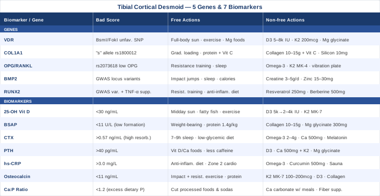
7 Biomarkers That Can Tell You What Is Really Happening in Your Cortical Bone
Most discussions about tibial cortical desmoid focus on what shows up on imaging. But the biology underlying the imaging — bone formation rate, resorption activity, inflammation level, mineralization quality — is visible in the blood, often weeks or months before imaging reflects any change. These seven markers provide the most direct window into the metabolic environment surrounding a cortical irregularity, with enough specificity to guide meaningful intervention.
Biomarker 1: 25-OH Vitamin D — The Non-Negotiable Foundation
Why it matters
Vitamin D is not a minor cofactor in cortical bone health — it is load-bearing infrastructure. The active form (1,25-dihydroxyvitamin D) directly regulates calcium absorption in the gut, modulates osteoblast differentiation, promotes periosteal cell activity, and controls dozens of genes involved in bone matrix synthesis. Insufficient vitamin D impairs the mineralization of bone collagen scaffolding, leaving the cortex less dense, more porous, and more reactive to mechanical stress. In studies of tibial stress reactions and cortical irregularities, low vitamin D is one of the most consistently identified modifiable risk factors. The NIH Office of Dietary Supplements vitamin D reference provides comprehensive evidence for its skeletal roles.
How to measure it
Standard serum 25-OH vitamin D test, available through most labs and physicians. Cost is typically $30–$80 without insurance in the US. Peter Attia targets a minimum of 40 ng/mL for bone health, with 50–70 ng/mL as a stronger target for those with active skeletal concerns. Testing twice per year captures the significant seasonal variation seen at northern latitudes. Always order 25-OH, not 1,25-OH, which is the active form and is not a useful screening marker.
If the score is low — the plan without supplements
Safe midday sun exposure for 15–30 minutes on bare arms and legs is the most natural corrective tool, adjusted for skin tone and latitude. Dietary sources — wild-caught salmon, mackerel, sardines, pastured egg yolks, and fortified dairy — help maintain existing levels but rarely normalize a significant deficiency on their own. Consistent weight-bearing exercise enhances the anabolic signaling that makes vitamin D functionally more effective in bone, even before serum levels fully normalize.
If the score is low — the plan with supplements or equipment
Vitamin D3 (cholecalciferol) is the standard corrective form. For levels under 30 ng/mL, a loading protocol of 5,000 IU per day for 8–12 weeks followed by 2,000–4,000 IU daily maintenance is widely used. Always pair D3 with vitamin K2 as MK-7 (100–200 mcg daily) — K2 activates the proteins that direct calcium into bone matrix rather than arterial tissue. This synergy is well-documented and important. Retest at 3 months. Note that VDR gene variants (covered below) can require higher doses to achieve functional intracellular signaling even with normal serum levels. Side effects at these doses are uncommon; sustained intake above 10,000 IU/day without monitoring carries a small hypercalcemia risk.
Biomarker 2: Bone-Specific Alkaline Phosphatase (BSAP) — The Formation Signal
Why it matters
Bone-specific alkaline phosphatase is produced by active osteoblasts during bone matrix synthesis. It is one of the most direct available readouts of how energetically your bone-building cells are working right now. In adolescents and young adults — who make up the largest share of cortical desmoid presentations — BSAP is naturally elevated due to growth-plate activity. In adults, unexpectedly high BSAP warrants investigation for Paget's disease, hyperparathyroidism, or other metabolic bone conditions. Low BSAP in any age group may indicate impaired bone formation, nutritional deficiency, or suppressed osteoblast activity — meaning the cortical irregularity is not receiving an adequate repair response.
How to measure it
BSAP is measured from a standard blood draw and is more bone-specific than total alkaline phosphatase, which rises with liver conditions as well as bone turnover. Available through physician order or specialty labs at $50–$150. Normal adult reference ranges vary by lab but typically fall between 11–32 U/L. Clinicians focused on advanced bone health assessment, including Thomas Dayspring, routinely include BSAP in comprehensive metabolic panels.
If BSAP is low — the plan without supplements
Progressive mechanical loading is the most potent available stimulus for osteoblast recruitment. Weight-bearing exercise — walking on varied terrain, hiking, resistance training with progressively heavier loads — activates osteoblasts through mechanotransduction signaling cascades in bone. Protein intake is structurally important here: osteoblasts require a constant amino acid supply to produce the collagen matrix that BSAP activity represents. Target at least 1.4–1.6 g of complete protein per kg of body weight daily from varied whole food sources.
If BSAP is low — the plan with supplements or equipment
Hydrolyzed collagen peptides (10–15g daily taken with 50–100 mg vitamin C) supply the proline and glycine most heavily used in bone collagen synthesis. Vitamin C is required for collagen hydroxylation and cannot be omitted. Magnesium glycinate (300–400 mg daily) directly supports alkaline phosphatase enzyme activity, and deficiency in magnesium is among the most common and underdiagnosed factors in low bone formation markers. Whole-body vibration platforms (30–40 Hz, 10–15 minutes daily) have demonstrated osteoblast-stimulating effects in people with limited capacity for high-impact exercise, though quality equipment carries a significant upfront cost. No cycling is required for magnesium; monitor BSAP every 6 months during active correction.
Biomarker 3: CTX — Reading the Bone Resorption Rate
Why it matters
CTX (C-terminal telopeptide of type I collagen) is a collagen breakdown fragment released into blood when osteoclasts are actively resorbing bone matrix. It is the most widely validated bone resorption marker in clinical and research use. For tibial cortical desmoid, persistently elevated CTX indicates the remodeling cycle is running heavily toward breakdown — which can widen or prolong cortical irregularities rather than allowing them to resolve and consolidate. Chronically elevated CTX reflects conditions that upregulate osteoclast activity: low estrogen, elevated cortisol, inadequate dietary calcium, high systemic inflammation, or secondary hyperparathyroidism. Identifying the driver matters because the correction is different in each case.
How to measure it
Fasting morning serum CTX is the gold standard. Timing is critical: CTX follows a strong diurnal rhythm, running 30–50% higher in the morning before eating. Non-fasting or afternoon draws are not interpretable in the same way. Cost ranges from $50–$100 at specialty labs. Normal adult values are typically under 0.573 ng/mL for women under 50 and under 0.584 ng/mL for adult men, though reference ranges vary by lab. CTX should always be interpreted alongside BSAP so the formation-resorption balance can be assessed together.
If CTX is elevated — the plan without supplements
Improving sleep quality and duration is the most underutilized intervention for elevated CTX. Cortisol is the primary systemic activator of osteoclast activity, and disrupted sleep is one of the fastest and most reliable routes to chronically elevated cortisol. Consistently achieving seven to nine hours of quality sleep produces measurable CTX reductions within 4–8 weeks. During periods of elevated CTX, reducing repetitive high-impact mechanical stress on the tibia allows the cortex consolidation time without completely halting exercise. A low-glycemic, whole-food dietary pattern reduces the insulin and cortisol volatility that drives excess resorption through secondary mechanisms.
If CTX is elevated — the plan with supplements or equipment
Omega-3 fatty acids at 2–4g EPA+DHA per day have demonstrated anti-resorptive effects through downregulation of prostaglandin E2 and inflammatory cytokines that activate osteoclasts. Calcium (500 mg elemental, taken with meals and paired with D3 and K2) reduces PTH-driven resorption signaling throughout the day. Melatonin at 1–3 mg at bedtime has direct inhibitory effects on osteoclast differentiation — a mechanism supported by multiple human and controlled studies that is often overlooked. Combining melatonin with sleep optimization addresses CTX from two converging angles simultaneously. Recheck fasting morning CTX after 3 months of consistent intervention before drawing conclusions.
Biomarker 4: Parathyroid Hormone (PTH) — The Hidden Cortical Resorption Driver
Why it matters
PTH is the primary regulator of calcium-phosphorus homeostasis in the body. When serum calcium falls, PTH rises — and its most immediate mechanism for restoring blood calcium is mobilizing it from bone, with the cortex as a primary source. Chronically elevated PTH (secondary hyperparathyroidism, most commonly driven by vitamin D deficiency or persistently low dietary calcium) is an underdiagnosed driver of cortical thinning. What makes this clinically deceptive is that serum calcium itself can remain entirely normal while PTH is quietly resorbing the cortical architecture over months to years. For someone with a tibial cortical irregularity, an elevated PTH finding provides a mechanism that imaging alone cannot reveal. The NIH Calcium fact sheet details the calcium-PTH regulation loop in depth.
How to measure it
Intact PTH via blood draw, ordered alongside serum calcium and vitamin D for full interpretive context. Cost: $40–$100. Most labs report normal range as 15–65 pg/mL, but preventive medicine specialists often target below 40 pg/mL in otherwise healthy adults. PTH rises with age, so values must be interpreted in context of both age-appropriate ranges and concurrent vitamin D and calcium status.
If PTH is elevated — the plan without supplements
Correcting vitamin D deficiency is the first and most important step, since it is the most common root cause of secondary hyperparathyroidism. Increasing dietary calcium through whole food sources — dairy, sardines and salmon with bones, cooked leafy greens, fortified non-dairy alternatives — reduces the ongoing demand on PTH to mobilize skeletal calcium stores. Reducing excess caffeine intake (above 400 mg/day) modestly lowers urinary calcium losses. A basic kidney function panel (creatinine, eGFR) is always worth running to exclude renal causes before attributing elevated PTH to nutritional factors.
If PTH is elevated — the plan with supplements or equipment
Vitamin D3 correction, as described, is primary. Supplemental elemental calcium (500 mg with meals) in combination with D3 and K2 closes the loop by reducing the post-meal calcium dip that triggers repeated PTH spikes. Magnesium is a critical and frequently overlooked co-factor: magnesium deficiency simultaneously impairs PTH secretion dysregulation and blocks the hepatic and renal conversion of vitamin D to its active form — creating a reinforcing cycle. Magnesium glycinate at 300–400 mg before bed produces reliable PTH improvements within 8–12 weeks in people who are deficient. Retest PTH, calcium, and 25-OH vitamin D together every 3 months during active correction.
Biomarker 5: High-Sensitivity CRP (hs-CRP) — The Periosteal Irritability Index
Why it matters
The periosteum — the thin membrane covering the tibial cortex — is among the most densely innervated and vascularized structures in the musculoskeletal system. It is acutely sensitive to systemic inflammatory signals. When hs-CRP is chronically elevated, circulating pro-inflammatory cytokines (primarily IL-1, IL-6, and TNF-alpha) disrupt the osteoblast-osteoclast equilibrium, increase periosteal sensitivity to mechanical loading, and delay the resolution of existing cortical irregularities. Elevated hs-CRP may be the reason a radiologically mild cortical desmoid produces disproportionate clinical symptoms — the periosteum is inflamed in a way that amplifies its sensitivity to normal forces.
How to measure it
Specifically order high-sensitivity CRP (hs-CRP), not standard CRP. The high-sensitivity assay detects the chronic low-level inflammation relevant to metabolic and musculoskeletal conditions, not just the acute-phase responses of infection or injury. Available at most standard labs for $20–$60. Values below 1.0 mg/L are ideal; 1.0–3.0 mg/L is borderline; above 3.0 mg/L warrants investigation into the source.
If hs-CRP is elevated — the plan without supplements
Anti-inflammatory dietary patterns are the most evidence-backed foundation: emphasizing vegetables, legumes, fatty fish (salmon, mackerel, sardines), extra-virgin olive oil, and berries while reducing refined carbohydrates, seed oils, alcohol, and ultra-processed foods. Zone 2 cardio at 150+ minutes per week — a conversational, moderate intensity — is one of the most potent anti-inflammatory interventions available to humans and produces measurable hs-CRP reductions within 8–12 weeks of consistent practice. Reducing chronic psychological stress and improving sleep both reduce baseline inflammatory tone substantially and are often the limiting factor in people who do not respond to dietary changes alone.
If hs-CRP is elevated — the plan with supplements or equipment
Omega-3 fatty acids (2–4g EPA+DHA daily from fish oil or algae-based sources) are among the best-validated supplements for hs-CRP reduction and have direct anti-resorptive effects in bone tissue as well. Bioavailable curcumin complex (500–1000 mg with piperine or phospholipid complex for absorption) reduces inflammatory cytokine production through NF-kB inhibition and has shown CRP-lowering effects in multiple human randomized trials. Regular sauna use — four sessions per week at 80°C for 20 minutes — has shown significant CRP reductions in Finnish population cohort studies. No cycling is needed for omega-3s or curcumin; recheck hs-CRP every 3 months.
Biomarker 6: Osteocalcin — The Bone Formation Quality Marker
Why it matters
Osteocalcin is a protein produced exclusively by osteoblasts, incorporated into bone matrix, with a fraction circulating in blood. Beyond being a bone formation marker, osteocalcin has emerged as a metabolic hormone — it influences insulin sensitivity, muscle function, and energy metabolism. For cortical bone specifically, circulating osteocalcin reflects the quality and activity of bone-forming cells at the sites of active remodeling. Low osteocalcin in the context of a cortical desmoid suggests the repair and remodeling machinery is underperforming — not building new bone effectively at the irregularity site. An important distinction: osteocalcin requires vitamin K2 for carboxylation into its active, functional form; without K2, osteocalcin is produced but cannot properly bind calcium in bone matrix.
How to measure it
Serum osteocalcin is available through physician-ordered metabolic bone panels or specialty labs at $50–$100. Normal adult ranges typically fall between 11–48 ng/mL depending on the lab and assay, with variation by sex and age. It is most useful when run alongside BSAP and CTX as a complete bone turnover assessment panel. Some functional medicine practitioners track it alongside insulin sensitivity markers given its metabolic hormone role.
If osteocalcin is low — the plan without supplements
Exercise is the most reliable osteocalcin stimulator available. Resistance training and impact exercise — particularly walking, running, and jumping — produce acute increases in circulating osteocalcin within hours of a session, and consistent long-term training sustains elevated baseline levels. Adequate dietary protein at 1.4–2g/kg body weight provides the amino acid supply required for osteocalcin synthesis. Reducing excessive caloric restriction is important: significant caloric deficits suppress osteocalcin production as part of a broader anabolic downregulation.
If osteocalcin is low — the plan with supplements or equipment
Vitamin K2 as MK-7 (100–200 mcg daily) is the most direct intervention for osteocalcin function — it activates the carboxylation process that makes osteocalcin functional in bone matrix. Studies have shown that K2 supplementation significantly increases carboxylated (active) osteocalcin over 6–12 weeks. Vitamin D3 is equally required, as it drives osteocalcin gene expression. Hydrolyzed collagen peptides supply the structural substrate for bone matrix into which osteocalcin is incorporated. At the preliminary evidence level, bovine lactoferrin supplements have shown stimulatory effects on osteoblast activity and osteocalcin in small human trials. Recheck osteocalcin every 6 months during active intervention.
Biomarker 7: Calcium-to-Phosphorus Ratio — The Overlooked Mineralization Balance
Why it matters
The ratio of calcium to phosphorus in diet and serum is a chronically under-discussed variable in cortical bone health. Optimal mineralization of cortical bone requires a balanced calcium-to-phosphorus ratio. The modern Western diet — dense in processed foods, soft drinks, and packaged products containing phosphate preservatives — has dramatically shifted this ratio toward excess phosphorus. Excess dietary phosphorus elevates PTH, which then mobilizes calcium from bone to maintain serum balance. This is a slow, ongoing cortical demineralization mechanism that rarely appears on standard lab panels unless calcium and phosphorus are examined together as a ratio. Allan Sniderman and other preventive cardiologists have noted the parallel overlooked importance of similar ratio-based interpretations across metabolic markers.
How to measure it
Serum calcium and phosphorus are both components of a standard comprehensive metabolic panel (CMP) — meaning this is often already being measured without being interpreted together. Cost: included in a CMP at $30–$80. Calculate the ratio by dividing serum calcium by phosphorus, both in mg/dL. A ratio above 1.5 is favorable for bone health; below 1.2 is a concern. Many people with typical Western dietary patterns have ratios below 1.0 without being aware of it.
If the ratio is unfavorable — the plan without supplements
Reducing dietary phosphorus from additive sources is the most impactful single change: eliminating canned processed meats, colas and dark soft drinks, packaged fast food, and preserved snack products containing phosphate additives. Natural food phosphorus from whole sources (legumes, dairy, meat, fish) is bound to organic compounds and absorbed less efficiently than the inorganic phosphate salts used as food additives. Increasing whole food calcium sources — dairy, bone broth, canned fish with bones, cooked kale, bok choy — improves the ratio from the other direction. Tracking dietary intake for two to four weeks with a nutrition app usually reveals the primary phosphorus sources clearly.
If the ratio is unfavorable — the plan with supplements or equipment
Calcium carbonate taken with phosphate-heavy meals acts as a modest gut phosphate binder, reducing absorption. This is a clinical intervention used routinely in kidney disease management but can be adapted cautiously in moderate cases: 500 mg calcium carbonate with the highest-phosphorus meals of the day, not exceeding a total of 1,500 mg elemental calcium daily. Prebiotic fiber (inulin/FOS at 5–10g daily) modestly improves calcium absorption through microbiome-mediated mechanisms. Recheck the CMP and ratio after 8–12 weeks of consistent dietary changes before adding supplemental interventions.
The biomarker picture gives you a real-time metabolic map. The genetic layer adds the structural architecture underneath — the reasons why some people's biology responds to these stressors more readily than others.
The Genetic Layer: 5 Variants That May Shape Your Cortical Bone Response
Genetic testing for bone health has become increasingly accessible through consumer platforms (23andMe raw data, ancestry services) and clinical genetic panels. Interpreting the variants that matter requires a framework — the five genes below represent the strongest current evidence for influencing how the periosteum and cortical bone layer develop, remodel, and respond to mechanical and inflammatory challenge. Evidence levels vary: some, like VDR, have extensive human intervention data; others, like BMP2 and RUNX2, are primarily supported by genome-wide association studies. The distinction is noted throughout.
Gene 1: VDR — The Vitamin D Receptor
What it does and why it matters here
The VDR gene encodes the intracellular receptor that binds active vitamin D and then controls gene expression across dozens of processes including osteoblast differentiation, calcium transport through periosteal cell membranes, and inflammatory modulation. Without a fully functional VDR signal, circulating vitamin D remains largely ineffective at the cellular level — like having abundant fuel but a malfunctioning ignition.
Several VDR single nucleotide polymorphisms (SNPs) are extensively studied in bone health contexts. The most researched are the BsmI (rs1544410), FokI (rs2228570), TaqI (rs731236), and ApaI (rs7975232) variants. Carriers of certain haplotype combinations, particularly around the BsmI locus, have shown consistently lower bone mineral density and altered periosteal stress responses across multiple population studies spanning diverse ethnic groups. A person with an unfavorable VDR genotype can have completely normal serum 25-OH vitamin D while receiving significantly impaired intracellular signaling in bone cells.
If the gene is unfavorable — the plan without supplements
Full-body sun exposure (not just face and arms) maximizes the vitamin D synthesis surface and pushes production as high as the skin can generate naturally. Prioritize midday UV exposure and track winter versus summer levels to understand your personal seasonal swing. Weight-bearing exercise has been shown to upregulate VDR expression in bone tissue through mechanotransduction pathways — providing some compensation for receptor sensitivity impairment. Dietary magnesium (from pumpkin seeds, dark leafy greens, nuts, and legumes) is critical because magnesium activates the enzyme that converts vitamin D to its active form, a bottleneck step that matters more in VDR-variant individuals.
If the gene is unfavorable — the plan with supplements or equipment
For unfavorable VDR variants, the functional target for serum 25-OH vitamin D should be higher than for the general population — targeting 60–80 ng/mL rather than the standard 40–60 ng/mL — because higher circulating levels are needed to drive adequate intracellular receptor occupancy. This typically requires 5,000–8,000 IU D3 daily under medical monitoring. Pair consistently with K2 (MK-7, 200 mcg) and magnesium glycinate (300–400 mg nightly). Retest serum levels every 3 months while titrating to target. No cycling is needed — this is lifelong management for a genetic trait, not a temporary protocol.
Gene 2: COL1A1 — The Collagen Blueprint
What it does and why it matters here
COL1A1 encodes the alpha-1 chain of type I collagen — the principal structural protein of cortical bone. Type I collagen provides the fibrous protein scaffolding onto which hydroxyapatite crystals mineralize, forming the tensile-resistant architecture of the cortical layer. The COL1A1 Sp1 binding site polymorphism (rs1800012) is one of the most studied genetic risk factors for reduced bone mineral density and increased stress fracture susceptibility. Carriers of the "s" allele (heterozygous or homozygous) produce collagen with altered fiber arrangement, resulting in bone matrix that is subtly less resilient under repeated mechanical loading.
For tibial cortical desmoid specifically, the implication is direct: the cortex relies on dense, well-organized collagen scaffolding to distribute tibial load across its surface without focal stress concentration. A COL1A1 variant that disrupts fiber organization may mean the periosteum mounts a more pronounced and persistent reactive response — a desmoid-type irregularity — in response to the same traction stress that would not affect someone with optimal collagen architecture.
If the gene is unfavorable — the plan without supplements
Load management during high mechanical demand periods is important — graduated return to running, jumping, or high-impact sports rather than abrupt increases in volume. Ensuring adequate dietary protein alongside vitamins C and zinc through whole foods provides the raw materials for maximal collagen synthesis despite impaired gene efficiency. Progressive resistance training supports collagen cross-linking improvement over time; longitudinal data shows that consistent training partially compensates for genetically reduced collagen quality through adaptive remodeling.
If the gene is unfavorable — the plan with supplements or equipment
Hydrolyzed collagen peptides at 10–15g per day taken with 50–100 mg vitamin C represent the most direct intervention. Vitamin C is required for two key hydroxylation steps in collagen biosynthesis — without it, collagen fibers form but with reduced tensile strength. Silicon as orthosilicic acid (10–15 mg daily from commercial stabilized preparations) has shown improvements in bone collagen cross-linking quality markers in small human trials and is mechanistically plausible. Cycle silicon at 3 months on, 1 month off, as long-term data in humans is limited. No significant side effects are known at these doses. Recheck BSAP and osteocalcin after 6 months as proxy markers for collagen-supported bone formation quality.
Gene 3: TNFRSF11B and TNFSF11 — The OPG/RANKL Resorption Switch
What these do and why they matter here
The RANKL/OPG system is the molecular master switch for bone resorption. RANKL (encoded by TNFSF11) activates osteoclasts — the cells that dismantle bone. Osteoprotegerin or OPG (encoded by TNFRSF11B) functions as a decoy receptor that binds RANKL before it reaches osteoclast precursors, directly suppressing resorptive activity. The balance between RANKL and OPG determines how aggressively bone is dismantled at any site at any time.
Genetic variants in both genes can shift this balance toward excess resorption. A TNFRSF11B variant (rs2073618) associated with reduced OPG expression has been linked to higher bone resorption rates and lower cortical density in multiple independent studies. A chronically elevated RANKL-to-OPG ratio — whether genetic or acquired through low estrogen, inflammatory cytokines, or physical inactivity — means the cortex is under continuous resorptive pressure that undermines repair.
If the genes are unfavorable — the plan without supplements
Resistance training is the most potent natural regulator of RANKL/OPG balance. Mechanical loading directly increases OPG expression in osteoblasts while locally suppressing RANKL — this effect has been confirmed in human exercise intervention studies. Estrogen (in women) and testosterone both upregulate OPG expression; addressing hormonal optimization through sleep quality, body composition management, and chronic stress reduction supports the RANKL/OPG axis through endocrine pathways without supplementation.
If the genes are unfavorable — the plan with supplements or equipment
Omega-3 fatty acids reduce RANKL-mediated osteoclast activation through anti-inflammatory prostaglandin pathways. Vitamin K2 in the MK-4 form at clinical doses (45 mg three times daily — the dose used in Japanese osteoporosis trials) has demonstrated significant OPG-upregulating effects. This is a pharmaceutical-grade dose far above typical supplemental MK-7 levels; it warrants physician involvement. At lower supplemental doses of MK-7 (200–800 mcg daily), the effect on carboxylated proteins is present but more modest. Whole-body vibration platforms have shown OPG-promoting effects in human studies for populations with activity limitations. No cycling is needed for K2.
Gene 4: BMP2 — The Periosteal Repair Signal
What it does and why it matters here
BMP2 (Bone Morphogenetic Protein 2) is among the most potent known stimulators of bone formation. It is released at sites of skeletal stress and microtrauma, recruiting mesenchymal stem cells to differentiate into osteoblasts and coordinating the local repair response. Multiple SNPs near the BMP2 locus have been identified in genome-wide association studies as influencing variation in bone mass, cortical geometry, and periosteal reactivity. The evidence here is primarily GWAS data rather than mechanistic intervention trial data — the association is established, but the mechanistic picture in humans is still developing.
For tibial cortical desmoid specifically: BMP2 signaling is part of what coordinates the periosteal reaction itself. When the cortex experiences the traction stress underlying a desmoid formation, BMP2 is one of the signaling molecules organizing the repair response. Variants that impair BMP2 function may produce a slower, less well-organized periosteal reaction — explaining why some cortical desmoid presentations are more pronounced or more persistent than the typical clinical course would predict.
If the gene is unfavorable — the plan without supplements
Mechanical loading — particularly impact loading — is the primary driver of endogenous BMP2 release at the periosteal level. Jumping, bounding, and plyometric movements produce stronger BMP2 signaling in cortical bone than non-impact exercise. Even modest jumping protocols (50 countermovement jumps, performed three times weekly) have been studied for their effects on cortical bone adaptation markers. Adequate caloric intake and sleep quality are important since BMP2-mediated periosteal repair occurs primarily during anabolic recovery windows overnight.
If the gene is unfavorable — the plan with supplements or equipment
Creatine monohydrate at 3–5g daily (no loading or cycling required at this dose) has shown support for bone formation markers in some trials, possibly through effects on osteoblast energy metabolism that interact with BMP-adjacent signaling cascades. Adequate zinc (15–30 mg elemental zinc with food) supports BMP pathway signaling and is frequently depleted in people with high training loads. Side effects of creatine at maintenance doses are minimal; zinc above 40 mg daily chronically can impair copper absorption, so stay within recommended ranges. These are promising signals from early research, and framing them as supportive rather than definitive to any clinician you consult is the right approach.
Gene 5: RUNX2 — The Master Osteoblast Switch
What it does and why it matters here
RUNX2 (Runt-related transcription factor 2) is the master regulator of osteoblast differentiation. Without RUNX2 signaling, mesenchymal stem cells cannot commit to the bone-forming lineage. It orchestrates the expression of virtually every major bone formation gene — osteocalcin, osteopontin, type I collagen, bone sialoprotein — making it the apex controller of the entire bone formation cascade. Severe RUNX2 mutations cause cleidocranial dysplasia, a rare skeletal development disorder. Subtler common variants near the RUNX2 locus have been identified in GWAS studies as being associated with differences in bone geometry, cortical thickness, and osteoblast efficiency, though functional intervention data in carriers of these variants remains limited.
RUNX2 activity is also directly suppressed by chronic inflammatory cytokines, particularly TNF-alpha. This creates an important connection: someone with borderline RUNX2 function due to a genetic variant, living with chronically elevated hs-CRP, is experiencing a compounding impairment to osteoblast recruitment — both genetic and acquired simultaneously.
If the gene is unfavorable — the plan without supplements
Progressive resistance training is the best-documented activator of RUNX2 expression in human bone tissue. Mechanical strain from load-bearing exercise directly promotes RUNX2 upregulation in osteoblast precursors. Reducing systemic inflammatory load (targeting hs-CRP below 1.0 mg/L through the dietary and lifestyle strategies described above) removes the TNF-alpha–mediated RUNX2 suppression that compounds genetic limitations. These two interventions — exercise and anti-inflammatory lifestyle — directly converge on RUNX2 pathway optimization.
If the gene is unfavorable — the plan with supplements or equipment
Resveratrol at 250–500 mg daily from high-quality standardized sources has shown preliminary evidence for RUNX2 upregulation in human mesenchymal stem cell studies, though clinical bone outcome data in humans remains limited and this should be treated as early-signal evidence. Berberine at 500 mg twice daily with meals has shown pro-osteogenic effects in in vitro studies through RUNX2-adjacent pathways, and its anti-inflammatory effects address the TNF-alpha pathway simultaneously. Cycle berberine at 8 weeks on, 4 weeks off; mild GI effects (loose stool, bloating) are possible during the first two weeks. Both of these are promising signals from emerging research — useful framing for a clinician conversation rather than standalone self-treatment protocols.
With the biomarker and genetic picture established, it is worth addressing what the most current exercise physiology research says about the single most impactful modifiable factor in cortical bone health — one that challenges the instinctive response to rest and avoid loading.
The Exercise-Bone Connection: 10 Things Current Research Reveals That Most Clinicians Do Not Discuss
The Huberman Lab podcast has dedicated significant coverage to the science of bone adaptation, drawing on mechanobiology, endocrinology, and clinical exercise research to build a framework that is substantially more nuanced than standard musculoskeletal advice. Below are the ten most practically relevant and clinically underappreciated insights from this body of research, with direct application to cortical bone remodeling and periosteal health.
1. Bone Is a Dynamic Organ That Requires Mechanical Challenge to Maintain Itself
Cortical bone does not maintain its density and geometry passively. It requires regular mechanical stress — through ground reaction forces, muscle pull, and impact — to signal that density needs to be preserved. Prolonged rest or unloading triggers rapid cortical thinning through osteoclast-mediated resorption; studies of bed rest and spaceflight consistently show measurable bone loss within days to weeks. For cortical desmoid, the reflex to completely offload the tibia is often counterproductive. Graduated, progressive mechanical loading is not just safe in most cases — it is biologically necessary for recovery.
2. Impact Loading Signals Bone Formation More Powerfully Than Non-Impact Exercise
Not all exercise is equivalent for bone. Cycling, swimming, and low-impact cardio produce cardiovascular benefits but minimal osteogenic signals. High-impact activities — running, jumping, stair climbing with loaded descent — produce the ground reaction forces that most powerfully activate osteoblasts through mechanotransduction pathways. Jumping protocols with 50 countermovement jumps three times weekly have been shown to improve cortical bone geometry markers in controlled studies. The key is gradual dosing, not avoidance.
3. Resistance Training Adds a Second Osteogenic Signal Through Muscle Pull
Muscle contractions generate direct tensile forces on the periosteum through tendon attachments — a separate osteogenic signal from impact. Resistance training that loads muscles crossing the tibia (calf raises, tibialis raises, leg press, lunges) generates these forces at the periosteal surface. Progressive overload over time — not just maintaining current loads — is what sustains the osteogenic signaling. This is underused in cortical desmoid management and typically safe when introduced gradually.
4. Osteocalcin Released During Exercise Is a Systemic Anabolic Signal
Exercise acutely elevates circulating osteocalcin within hours, and osteocalcin acts as a systemic hormone that improves insulin sensitivity, supports muscle function, and provides feedback to bone that active remodeling is occurring. People who are sedentary have chronically suppressed osteocalcin, removing this positive feedback loop entirely. A single resistance training session produces a measurable osteocalcin spike; chronic training sustains elevated baseline levels.
5. Cortisol Timing Matters More Than Most People Realize
Morning cortisol is the primary zeitgeber for bone resorption — CTX peaks in the early morning partly because of cortisol-driven osteoclast activity overnight and in the pre-dawn hours. Chronically elevated cortisol (from sleep disruption, psychological stress, or overtraining) sustains elevated CTX throughout the day. Training periodization matters: high-stress training protocols without adequate recovery windows can paradoxically accelerate cortical resorption rather than stimulate formation.
6. The Protein Threshold for Bone Formation Is Higher Than General Recommendations Suggest
The recommended dietary allowance for protein (0.8g/kg) is a floor, not a target for people actively trying to support bone formation. Evidence from exercise science suggests that 1.6–2.2g/kg is closer to the optimal range for maximizing osteoblast activity and bone matrix synthesis in people doing regular resistance training. Protein quality matters too: leucine-rich complete protein sources (meat, fish, dairy, eggs) most effectively stimulate the mTOR pathway relevant to osteoblast anabolism.
7. Sleep Architecture Directly Governs Bone Repair
Slow-wave (deep) sleep is when the majority of growth hormone pulsatility occurs — and growth hormone is a primary anabolic signal for bone repair at periosteal surfaces. Poor sleep architecture — even without reduced total sleep time — can substantially impair nighttime bone repair processes. Prioritizing sleep hygiene interventions (consistent timing, darkness, cooler temperature, limiting late-night blue light exposure) is not a soft lifestyle recommendation: it is directly relevant to whether bone repair processes complete overnight.
8. Inflammation-to-Anabolism Ratio Determines Net Bone Outcome
Bone adaptation is the result of a competition between anabolic signals (mechanical loading, growth hormone, IGF-1, testosterone, estrogen) and catabolic signals (cortisol, TNF-alpha, IL-6, RANKL). No exercise program outpaces a highly inflammatory baseline biology indefinitely. People with chronically elevated hs-CRP and disrupted sleep can train consistently without improving cortical bone markers because the inflammatory signaling is suppressing anabolism at the cellular level. Fixing the inflammatory environment is a prerequisite for exercise-driven bone improvement.
9. Vitamin D and K2 Work Synergistically and Cannot Be Separated in Bone Protocols
Vitamin D3 drives calcium absorption and osteocalcin gene expression. Vitamin K2 activates the carboxylation of osteocalcin and matrix GLA protein that directs calcium into bone matrix rather than arterial tissue. Running D3 without K2 is a well-intentioned but incomplete protocol — particularly in people with genetic impairment of either the VDR pathway (reducing D3 signaling efficiency) or RUNX2 (reducing osteocalcin production). The combination has substantially more evidence than either alone.
10. Bone Density Changes Lag Behind Biological Changes by 3–6 Months
DEXA scans and MRI findings reflect remodeling that completed 3–6 months before the scan, not current biological activity. This means someone who began an effective bone-building protocol three months ago may show unchanged imaging while their biomarkers (BSAP up, CTX down, osteocalcin improving) confirm the protocol is working. Tracking biomarkers every 3 months gives a far more responsive feedback loop than annual imaging — and prevents premature abandonment of protocols that are actually working.
Additional Approaches with Evidence Worth Considering
Beyond biomarker optimization and genetic compensation strategies, three modalities from the complementary medicine space have meaningful human clinical evidence for musculoskeletal tissue repair, periosteal health, or bone adaptation — and a reasonable evidence base to justify consideration alongside a standard management approach.
Low-Level Laser Therapy (Photobiomodulation) — Accelerating Periosteal Repair
Photobiomodulation (PBM), also called low-level laser therapy (LLLT), uses red and near-infrared light wavelengths (typically 630–1000 nm) to stimulate mitochondrial activity and cellular repair processes in target tissues. In bone, PBM has been shown to accelerate periosteal cell proliferation, stimulate osteoblast activity, reduce local inflammatory markers, and promote cortical healing in several human controlled trials. The biological plausibility for tibial cortical desmoid is grounded in the periosteal specificity of these effects — the periosteum is the first tissue layer reached by transcutaneous light in the tibial region.
A randomized controlled trial published in Photomedicine and Laser Surgery demonstrated that LLLT at 780 nm accelerated tibial stress fracture healing and reduced pain in athletes compared to sham treatment, with measurable differences in imaging at 6 weeks. While this study involved stress fractures rather than cortical desmoid specifically, the periosteal repair mechanisms are overlapping and the evidence is directionally relevant.
A practical PBM protocol for tibial cortical desmoid involves 4–6 sessions per week using a clinical-grade device (10–50 mW/cm² at 810 nm, 60–90 seconds per spot) applied directly over the area of cortical irregularity. Professional LLLT devices used in physiotherapy clinics range from $2,000–$8,000; consumer-grade red-light therapy panels (available from $200–$800) deliver lower irradiance but can provide meaningful effects at longer application times. Contra-indications include active malignancy and direct application over growth plates in skeletally immature individuals — the second point is particularly relevant given that cortical desmoid is most common in adolescents.
Tai Chi — Bone Loading Through Controlled Dynamic Movement
Tai chi is a form of mind-body exercise involving slow, controlled, low-impact movements that create continuous shifting of weight through the lower extremities. Its relevance to bone health stems from the mechanical loading it applies to the tibial cortex through controlled ground reaction forces and the proprioceptive training it delivers, reducing the risk of falls and impact injuries that can exacerbate existing cortical irregularities. Multiple controlled trials and one Cochrane systematic review have examined tai chi's effect on bone mineral density, particularly in postmenopausal women and older adults, finding modest but consistent improvements with regular practice.
A 12-month randomized trial published in the Archives of Physical Medicine and Rehabilitation found that tai chi practice three times weekly (45-minute sessions) was associated with significantly attenuated bone loss at the proximal femur compared to low-activity control, with measurable effects on lower limb bone remodeling markers. The evidence in younger populations and specifically for cortical desmoid is limited, but the loading pattern — gradual, controlled tibial weight-bearing — is mechanically appropriate and carries low injury risk.
A realistic tai chi protocol for someone with tibial cortical desmoid involves three sessions per week of 30–45 minutes each, beginning with the 24-form Yang style which is the most widely studied. Online guided programs are widely accessible and cost-free. Supervision from a trained instructor is beneficial during the first 4–6 weeks to establish correct foot placement mechanics. Tai chi can reasonably complement a progressive mechanical loading program rather than replacing it — particularly in the early phase when high-impact exercise is being introduced gradually.
Massage Therapy — Reducing Periosteal Tension and Local Inflammatory Load
The muscles crossing and attaching near the tibial cortex — particularly the tibialis anterior, posterior tibialis, and gastrocnemius-soleus complex — exert continuous traction on the periosteal surface through their tendinous attachments. Chronic muscle tightness or hypertonicity in these groups increases periosteal tension, potentially perpetuating the mechanical irritation at the desmoid site. Massage therapy, particularly soft tissue mobilization techniques targeting the lower leg musculature, reduces muscle tension, improves local circulation, and modulates the inflammatory environment at periosteal attachment sites.
A randomized controlled trial in the Journal of Athletic Training found that lower-leg soft tissue massage significantly reduced the pain and functional impairment of periosteal stress reactions in athletes compared to rest-only management. The mechanism proposed involved reduced compartment pressure, improved lymphatic drainage, and modulation of local prostaglandin levels. The study used periosteal stress reactions (shin splints) rather than cortical desmoid specifically, but the anatomical and mechanical overlap is substantial.
A practical protocol involves 30–45 minute sessions targeting the tibialis anterior, posterior tibialis, calf complex, and the peroneal group, performed once or twice weekly by a trained massage therapist. Deep tissue and myofascial release techniques are most relevant — lighter Swedish massage is likely insufficient for the mechanical tissue changes needed. Self-massage tools (foam roller and targeted ball massage) can supplement professional sessions at home. The primary caution is avoiding direct pressure over the confirmed cortical irregularity site, particularly if it is symptomatic — work around the perimeter rather than directly on it.
Conclusion
Tibial cortical desmoid is not a diagnosis that demands alarm, but it also does not have to be dismissed with a blanket "don't worry about it." The biology behind the imaging is measurable, and in most cases, improvable. Seven biomarkers — particularly vitamin D, CTX, PTH, and hs-CRP — can tell you what the metabolic environment around the cortical irregularity actually looks like right now. Five gene variants, led by VDR and COL1A1, can explain why some people's periosteum responds more reactively to mechanical stress than others — and point toward specific compensatory strategies. The exercise physiology research confirms that mechanical loading, done intelligently, is not just safe but necessary for cortical repair. And several complementary modalities can support periosteal tissue recovery in ways that fit alongside standard management.
The most useful next step is not a new intervention but a more specific conversation — with a sports medicine physician, an endocrinologist, or a preventive medicine specialist who uses bone turnover markers routinely. Bring your biomarker results. Ask about VDR variants if you have genetic data. Ask whether your current CTX and BSAP values explain the finding. That kind of targeted dialogue is where better data becomes better care.
Musculoskeletal Endocrine & Metabolic
Musculoskeletal: Bone Conditions Sports Injuries
Autoimmune: Inflammatory Conditions