This article contains AI generated content.
· UpdatedTibial Eminence Fracture — 7 Biomarkers and 5 Genes to Track
Introduction
A tibial eminence fracture is a structurally precise injury. The bony intercondylar spine at the center of the tibial plateau — the site where the anterior cruciate ligament inserts into bone — fractures under hyperextension or torsional force. Most commonly seen in children and adolescents between 8 and 14 years old during sports or cycling accidents, it can also occur in active adults under high mechanical demand. If you are reading this with that diagnosis in hand, you likely already know the classification system, the surgical or conservative treatment options, and the general rehabilitation timeline. What you may not have been told is why this happened in your body specifically — and why your recovery trajectory may differ significantly from someone with the same fracture type on paper.
Individual biology shapes both fracture susceptibility and healing capacity in ways that standard protocols cannot account for. Bone mineral quality, collagen architecture at the ligament-bone interface, systemic inflammation, and key nutrient status are not uniform across patients. Two people with a Type II tibial eminence fracture may have completely different biological starting conditions — different bone formation rates, different inflammatory baselines, different genetic variants governing how they build and maintain connective tissue. Generic advice about calcium and rest addresses none of that specificity.
This article takes a more targeted approach. The first section covers seven biomarkers that directly reflect the biological factors most relevant to this injury: bone formation activity, bone resorption balance, systemic inflammation, anabolic capacity, and mineral status. For each, you will find guidance on measurement, optimal ranges, and concrete plans both with and without supplementation for improving suboptimal values. A second section explores the most studied genetic variants that influence connective tissue integrity, bone quality, and injury response — with practical implications for each genotype. Additional sections cover evidence-supported recovery strategies and complementary modalities with meaningful clinical research behind them.
The goal here is not a cure claim. Better biological data leads to sharper decisions — whether that means correcting a vitamin D insufficiency that is silently impairing callus formation, modifying your rehabilitation approach based on a collagen gene variant, or simply arriving at your next appointment with better questions. More information does not replace clinical expertise, but it does make the conversation more precise.
7 Biomarkers That Reflect Your Fracture Risk and Healing Capacity
The following seven biomarkers connect, through well-established mechanisms and clinical research, to the biology that governs bone formation, ligament-bone interface quality, and the inflammatory environment during fracture recovery. They span a wide range of accessibility — from inexpensive tests available at any lab to more specialized panels — and testing even three or four of them will give you a substantially more useful picture than standard blood panels provide.
1. 25-Hydroxyvitamin D (25(OH)D)
Why it matters
Vitamin D is not simply a bone mineral nutrient — it is a steroid hormone with nuclear receptors on osteoblasts, chondrocytes, immune cells, and muscle tissue, all of which are directly involved in fracture healing. Adequate serum 25(OH)D is required for intestinal calcium absorption, osteoblast differentiation, regulation of parathyroid hormone, and resolution of the inflammatory response after tissue injury. At the fracture site itself, vitamin D supports callus formation and mineral deposition in a way that is not replaceable by calcium supplementation alone.
The evidence linking vitamin D deficiency to fracture risk and impaired healing is among the strongest in bone biology. Multiple large meta-analyses confirm that deficiency increases fracture rates across pediatric and adult populations. The critical nuance is that being above the clinical "sufficiency" threshold of 20 ng/mL is not the same as being in the range that supports optimal bone biology — functional bone metabolism and fracture healing benefit from levels between 40 and 60 ng/mL, a target consistently endorsed by researchers including Thomas Dayspring and Peter Attia in their clinical discussions of bone health markers.
How to measure it
Standard serum 25(OH)D test via blood draw. Cost: $30–$80 out of pocket; frequently covered by insurance when fracture or deficiency is documented. Optimal range for bone healing: 40–60 ng/mL — not simply above 20 ng/mL. Test at baseline, then 8–12 weeks after initiating any intervention.
If the score is low — the plan without supplements
Daily midday sun exposure on unprotected arms and legs (15–30 minutes depending on skin tone, latitude, and season) can raise 25(OH)D by 10–20 ng/mL over 4–8 weeks in favorable climates. Dietary sources — fatty fish (salmon, mackerel, sardines), egg yolks, cod liver oil — contribute meaningfully but rarely close a significant gap alone. Equally important is reducing vitamin D depletion: avoiding excessive sunscreen during the primary sun exposure window, managing excess body fat (which traps vitamin D in adipose tissue), and correcting magnesium deficiency (which blocks vitamin D conversion to its active hormonal form).
If the score is low — the plan with supplements
Vitamin D3 at 2,000–5,000 IU/day is the standard correction dose for mild to moderate deficiency. Always co-supplement with vitamin K2 (MK-7 form, 100–200 mcg/day) to direct calcium into bone matrix rather than soft tissue or arterial walls, and with magnesium glycinate (300–400 mg/day) as a required cofactor for D3 activation. Without adequate magnesium, supplemental D3 may have blunted or absent effect. Retest at 8–12 weeks and titrate dose to target. For significant deficiency, some sports medicine physicians use 10,000 IU/day for 4–8 weeks under medical supervision before transitioning to maintenance dosing. Side effects: toxicity is uncommon below 100 ng/mL; hypercalcemia is the primary risk at excessive doses. Monitoring serum calcium alongside 25(OH)D is prudent during aggressive repletion.
2. P1NP (Procollagen Type 1 N-Terminal Propeptide)
Why it matters
P1NP is released into the bloodstream as a direct byproduct of type I collagen synthesis by active osteoblasts. It is considered the most sensitive and specific serum marker of bone formation activity currently available. During tibial eminence fracture recovery — when osteoblasts should be actively laying down new bone matrix at the callus and insertion zone — elevated P1NP indicates healthy formation activity. A persistently low P1NP during the expected healing window signals sluggish bone building due to nutritional deficiencies, hormonal imbalances, or excessive immobilization.
Peter Attia has discussed P1NP as one of two essential bone turnover markers (alongside CTX) that give patients and clinicians not just a static snapshot of bone density, but the direction and speed of remodeling. That dynamic information is precisely what matters during fracture healing, where you need active formation outpacing resorption for weeks to months.
How to measure it
Serum P1NP via blood draw (morning, fasted for reproducibility). May require a specific lab request; not always included in routine panels. Cost: $50–$120 out of pocket. Reference for adults: above 35 µg/L for adequate formation activity, though values during active fracture repair should be higher than baseline. Retest 8–12 weeks after any intervention.
If the score is low — the plan without supplements
Mechanical loading is the most potent stimulus for bone formation marker elevation. Progressive weight-bearing on the healing limb — as cleared by your surgeon — should be advanced as quickly as safely possible rather than defaulted to prolonged non-weight-bearing. Even brief periods of impact (10 moderate jumps per day, where tolerated) have been shown to raise bone formation markers over weeks. Upper-body resistance exercise during lower-extremity recovery provides a systemic anabolic stimulus. Adequate dietary protein (1.2–1.6 g/kg/day from complete sources) provides the amino acid substrate required for type I collagen synthesis.
If the score is low — the plan with supplements
Hydrolyzed collagen peptides (10–15 g/day with 50 mg vitamin C, taken 30–60 minutes before mechanical loading or physical therapy) have demonstrated meaningful increases in collagen synthesis markers in randomized controlled trials. A well-cited study by Shaw et al. (2017) in the American Journal of Clinical Nutrition demonstrated significant increases in collagen synthesis after this timed protocol — available on PubMed. Calcium supplementation (calcium citrate, 500 mg/day with meals) is appropriate if dietary intake falls below 1,000–1,200 mg/day but is insufficient alone without the formation-stimulating context above. Cycling: daily throughout active recovery; reassess necessity at 3–6 months.
3. CTX (C-Terminal Telopeptide of Type I Collagen)
Why it matters
CTX is the complementary counterpart to P1NP: it reflects bone resorption activity by measuring a fragment released during osteoclastic breakdown of existing bone matrix. Together, P1NP and CTX define your bone remodeling balance — the ratio of formation to resorption that determines whether bone mass and quality are net-increasing, stable, or net-declining.
After a tibial eminence fracture, a disproportionately elevated CTX relative to P1NP indicates that breakdown is outpacing repair — a pattern that prolongs healing time and reduces the mechanical quality of the forming callus. High CTX during recovery can signal several correctable problems: vitamin D deficiency, low sex hormone levels, prolonged immobilization, caloric deficit, or chronic psychological stress driving elevated cortisol.
How to measure it
Serum CTX (also called beta-CrossLaps) via blood draw. Critical: must be done fasted in the morning — food intake can suppress CTX by up to 25%, making fed-state results unreliable for comparison. Cost: $50–$100. Optimal adult range: below 0.3 ng/mL. Values above 0.6 ng/mL indicate clinically relevant elevated resorption.
If the score is high — the plan without supplements
Reduce immobilization time to the clinically appropriate minimum — prolonged unloading dramatically increases bone resorption markers through disuse pathways. Even upper-body resistance training and contralateral leg work during recovery from this fracture dampens systemic resorption signals measurably. Ensure caloric sufficiency: a caloric deficit is among the most reliable drivers of CTX elevation through hormonal mechanisms. Improving sleep quality and duration (7–9 hours) reduces both cortisol and downstream resorption markers. Alcohol restriction has a meaningful effect on cortisol-mediated resorption.
If the score is high — the plan with supplements
Vitamin D3 + K2 + magnesium correction (as outlined above) consistently reduces CTX in deficient individuals, typically within 8 weeks. Omega-3 fatty acids (EPA+DHA, 2–4 g/day from high-quality fish oil) modestly reduce bone resorption through anti-inflammatory prostaglandin pathways. Important caution about NSAIDs: while commonly used for post-fracture pain, chronic NSAID use inhibits prostaglandins required for osteoblast recruitment and callus maturation, and has been associated with delayed fracture healing in multiple animal and some human studies. Use NSAIDs for acute pain management only — not as a sustained recovery protocol when CTX is already a concern.
4. High-Sensitivity C-Reactive Protein (hs-CRP)
Why it matters
Acute post-fracture inflammation is biologically necessary: it initiates the healing cascade. But chronic, low-grade systemic inflammation — present before injury or persisting beyond the acute phase — is a different entity with a different biological impact. Elevated hs-CRP has been associated with impaired fracture healing, reduced collagen cross-link quality, and greater re-injury susceptibility in multiple musculoskeletal research contexts.
In the pediatric and adolescent population most commonly affected by tibial eminence fractures, elevated hs-CRP often reflects poor sleep patterns, ultra-processed food consumption, or early metabolic imbalance. In active adults, it more commonly signals visceral adiposity, insulin resistance, chronic psychological stress, or inadequate recovery from high-volume training. All of these are modifiable — often substantially — within the healing timeframe.
How to measure it
Standard hs-CRP via blood draw; included in many routine metabolic panels. Cost: $15–$40. Optimal range for tissue healing context: below 1.0 mg/L (Peter Attia targets below 0.5 mg/L as a general longevity benchmark). Values above 3.0 mg/L indicate meaningful systemic inflammation that warrants direct intervention, not just monitoring.
If the score is high — the plan without supplements
Shifting to a Mediterranean-pattern or whole-food diet reduces hs-CRP by 20–40% within 4–8 weeks in most populations, with consistent evidence across multiple clinical trials. The structural dietary changes driving this reduction: eliminating ultra-processed foods, refined seed oils (high omega-6), and refined sugar. Improving sleep — particularly duration and consistency — reduces circulating IL-6 and downstream CRP reliably. Moderate aerobic exercise (30 minutes, 4–5 days/week), even if temporarily limited to pool-based or upper-body work during tibial fracture recovery, lowers hs-CRP significantly within 8–12 weeks.
If the score is high — the plan with supplements
Omega-3 fatty acids (EPA+DHA, 2–4 g/day from triglyceride-form fish oil) have the strongest and most consistent evidence for hs-CRP reduction across populations. Cycling: 12 weeks minimum; can be continued long-term. Curcumin with enhanced bioavailability formulations (theracurmin 180–360 mg/day, or BCM-95 1,000 mg/day) provides a meaningful anti-inflammatory adjunct. Vitamin D3 correction, if deficient, also reliably reduces hs-CRP through immunomodulatory mechanisms. Side effects: omega-3 at these doses may have mild anticoagulant effects — note for anyone near a surgical procedure.
5. IGF-1 (Insulin-Like Growth Factor 1)
Why it matters
IGF-1 is the primary downstream mediator of growth hormone's anabolic effects on musculoskeletal tissue. It stimulates osteoblast proliferation and differentiation, drives collagen synthesis in tendons and ligaments, supports chondrocyte function, and accelerates fracture callus development. Low IGF-1 is associated with slower fracture healing, reduced bone mineral density, and impaired muscle regeneration — all directly relevant to tibial eminence fracture recovery.
In children and adolescents — who make up the majority of tibial eminence fracture cases — IGF-1 is naturally elevated, which partly explains the generally faster healing observed in this age group. In adults, particularly those over 35, with poor sleep quality, elevated chronic stress, low protein intake, or excess body fat, IGF-1 may be suboptimally low and worth measuring directly rather than assuming adequacy.
How to measure it
Serum IGF-1 via blood draw. Cost: $50–$100. Always use age-adjusted reference ranges — IGF-1 declines substantially with age, and values that look "normal" for a 55-year-old would be low for a 30-year-old. Target the upper half of age-adjusted normal range, not simply above the lower threshold.
If the score is low — the plan without supplements
Sleep is the dominant modifiable lever: the majority of daily growth hormone secretion (which drives IGF-1) occurs during slow-wave sleep stages. Prioritizing 7–9 hours of quality sleep, maintaining consistent bedtimes, and limiting blue light exposure in the 2 hours before sleep can meaningfully elevate IGF-1 over weeks. High-intensity resistance training — compound lifts (squats, deadlifts, rows) with explosive concentric phase — provides a strong growth hormone stimulus; apply this to upper-body and contralateral leg work within recovery constraints. Adequate dietary protein (1.5–1.8 g/kg/day), distributed across meals rather than concentrated in one sitting, supports IGF-1 synthesis and signaling.
If the score is low — the plan with supplements
Zinc (15–25 mg/day with food) and magnesium glycinate (300–400 mg at night) support both growth hormone secretion and downstream IGF-1 signaling — particularly in individuals who are deficient in either mineral, which is common in active people and adolescents. Ashwagandha (KSM-66 extract, 600 mg/day) has shown modest IGF-1 and testosterone elevation in several small but well-designed RCTs. Cycling: 8–12 weeks on, 4-week break, reassess. Side effects: ashwagandha can lower blood pressure and thyroid hormones in a minority of individuals; monitor if clinically relevant. Avoid very high-dose zinc without copper co-supplementation (1–2 mg copper per 15 mg zinc) to prevent copper depletion over time.
6. RBC Magnesium
Why it matters
Magnesium is a cofactor in over 300 enzymatic reactions, including those governing vitamin D activation, collagen cross-linking, ATP production, bone hydroxyapatite crystal formation, and calcium channel regulation. Approximately 60% of total body magnesium is stored in bone, where it influences the structural quality of bone mineral beyond what calcium and vitamin D alone can provide. Magnesium deficiency impairs bone formation, blunts the efficacy of vitamin D supplementation, and increases systemic inflammatory markers — three intersecting problems for fracture healing.
The critical measurement caveat: standard serum magnesium has poor sensitivity for true intracellular deficiency, because the body maintains serum levels at the expense of bone and tissue stores. RBC magnesium, which reflects intracellular status, is the more informative test — and is commonly suboptimal even when serum magnesium reads as "normal."
How to measure it
Serum magnesium: standard, $15–$30, widely available. RBC magnesium: $50–$80, requires a specialty lab request (available through Spectracell, Boston Heart Diagnostics, or functional medicine-oriented panels). Optimal serum level: 2.0–2.5 mg/dL (not simply above 1.7 mg/dL). Optimal RBC magnesium: above 5.5 mg/dL. Retest after 10–12 weeks of targeted intervention.
If the score is low — the plan without supplements
Increase dietary magnesium through dark leafy greens (spinach, Swiss chard), pumpkin seeds, dark chocolate (85%+), avocado, black beans, and quinoa. Reduce factors that chronically deplete magnesium: excess caffeine increases urinary magnesium excretion; alcohol amplifies this effect; high sugar intake activates pathways that draw on intracellular magnesium. Soaking and sprouting legumes and grains before cooking reduces phytate content and meaningfully improves magnesium bioavailability from these sources.
If the score is low — the plan with supplements
Magnesium glycinate or magnesium malate (providing 300–400 mg elemental magnesium/day) offer the best combination of absorption and gastrointestinal tolerability. Take at night for an additional benefit to sleep quality via GABA receptor modulation. Avoid magnesium oxide — less than 4% is absorbed systemically and it functions primarily as a laxative. Topical magnesium oil (applied to the skin) and Epsom salt baths (20–30 minutes, 2–3x/week) are useful adjuncts for localized muscle tension during rehabilitation. Cycling: daily use throughout recovery is appropriate; retest RBC magnesium at 10–12 weeks. Side effects: loose stools at higher doses — splitting the dose or reducing it resolves this immediately.
7. Parathyroid Hormone (PTH)
Why it matters
Parathyroid hormone is the master regulator of calcium homeostasis and exerts powerful direct effects on bone remodeling. When serum calcium falls — most commonly because vitamin D deficiency impairs intestinal calcium absorption — the parathyroid glands respond by secreting PTH to mobilize calcium from the skeleton. This secondary hyperparathyroidism creates a chronic resorptive state that degrades bone quality throughout the skeleton, including the tibial spine, making avulsion fracture more likely under loading stress and callus formation less robust after fracture occurs.
PTH elevation is also the most sensitive functional indicator that vitamin D supplementation is not working as intended — whether because the dose is insufficient, magnesium is depleted (blocking D3 activation), or a vitamin D receptor gene variant requires higher serum D levels for equivalent receptor activation. Measuring PTH alongside 25(OH)D is therefore far more informative than measuring either alone.
How to measure it
Serum intact PTH via blood draw. Cost: $30–$70. Optimal range: 15–40 pg/mL. Values above 65 pg/mL warrant clinical evaluation to determine whether secondary or primary hyperparathyroidism is driving the elevation. Always test simultaneously with 25(OH)D for maximum interpretive value.
If the score is high — the plan without supplements
Increasing dietary calcium through dairy, sardines with bones, collard greens, and calcium-fortified plant milks provides direct negative feedback on PTH secretion within days to weeks — a rapid and meaningful effect. Increasing sunlight exposure to raise vitamin D addresses the upstream driver. Weight-bearing exercise directly influences calcium-sensing mechanisms in bone and has a modest PTH-lowering effect independent of serum calcium changes.
If the score is high — the plan with supplements
Correcting vitamin D deficiency with D3 + K2 + magnesium (as described) typically normalizes PTH within 8–12 weeks when deficiency is the driver — and retest at that interval should confirm the response. Calcium citrate (500 mg/day with meals) provides direct PTH suppression and is preferred over calcium carbonate for individuals with variable stomach acid. Critical note: if PTH remains elevated despite thorough vitamin D correction and adequate calcium intake, clinical evaluation for primary hyperparathyroidism (a parathyroid adenoma) is warranted. This is a different pathology requiring different management, and continued mineral supplementation without investigation is not the appropriate response.
With a clearer picture of your current biological state from these biomarkers, the next layer of understanding comes from the genetic architecture underlying them — the variants that shape your baseline collagen quality, bone metabolism efficiency, and injury susceptibility at a structural level.
Genetics and Tibial Eminence Fracture — 5 Key Variants That Shape Your Risk and Recovery
Biomarkers show you where your biology stands today. Genetics reveals the underlying architecture — the baseline conditions your body is working from, and why certain interventions may work better for you than for another person with the same injury. You cannot change your genotype, but knowing which variants you carry enables more targeted intervention selection and more realistic expectation-setting for recovery timelines.
The genetic research specific to tibial eminence fracture as an isolated entity is limited. The variants discussed below draw primarily from human genetic studies on ACL injuries, connective tissue integrity, and bone fragility — conditions sharing directly relevant biological substrate with tibial eminence fracture at the ligament-bone interface. Evidence quality is noted for each gene.
How to Access Your Genetic Data
Consumer genetic tests (23andMe, AncestryDNA) provide raw SNP data that can be analyzed through third-party platforms such as Promethease, SelfDecode, or Genetic Genie. Sports genetics panels (DNAfit, Athletigen, Genomics for Life) specifically include many of the variants below and present them in a performance/injury context. Whole-genome sequencing now costs $200–$500 through services such as Nebula Genomics. Ali Torkamani of Scripps Research, whose work has shaped clinical genomics approaches to personalized health, has emphasized that the value of these variants lies not in determinism but in targeted prevention — knowing a risk variant means you can act on it before injury occurs, or optimize recovery after it does.
COL5A1 — The Ligament Architecture Gene
What it does
COL5A1 encodes the alpha-1 chain of type V collagen, a regulatory collagen that controls fibril diameter in type I collagen-rich connective tissues — including the ACL, patellar tendon, and the fibrocartilaginous attachment zone at the tibial eminence. Type V collagen acts as a nucleating template for fibril assembly; variants reducing its function produce larger, less uniform fibrils with altered mechanical stiffness and reduced tensile strength under repetitive or acute loading.
The most studied polymorphism is rs12722 (C/T SNP in the 3' UTR of COL5A1). Multiple studies by Posthumus, Collins, and colleagues demonstrated across South African, European, and Australian athletic cohorts that individuals with the TT genotype are significantly overrepresented in ACL ruptures and ligament-bone avulsion injuries compared to uninjured athletic controls. This is among the most consistently replicated findings in sports injury genetics, with effect sizes meaningful at both the individual and population level.
If the gene is suboptimal — the plan without supplements
When connective tissue structural genetics favor greater laxity and reduced passive stiffness at the ligament-bone interface, muscular dynamic stabilization becomes more critical — the burden shifts from passive structure to active neuromuscular protection. Priority training targets: VMO (vastus medialis oblique) strengthening, eccentric hamstring loading (Nordic curls), hip abductor power, and single-leg reactive stability. Neuromuscular training programs — FIFA 11+ and similar evidence-based protocols — have demonstrated 30–50% reduction in ACL and knee avulsion injury rates in multiple RCTs and are particularly high-value for TT carriers. Return-to-sport timelines should be extended beyond standard guidelines, and reactive hop and agility tests should be used as functional clearance criteria rather than relying solely on strength thresholds.
If the gene is suboptimal — the plan with supplements
Hydrolyzed collagen peptides (10–15 g/day with 50 mg vitamin C, taken 30–60 minutes before loading) provide direct substrate for type I and V collagen synthesis and have demonstrated increased collagen synthesis markers in RCT settings. Vitamin C (500–1,000 mg/day as ascorbic acid) is essential for proline and lysine hydroxylation during collagen cross-linking — without it, collagen peptides provide limited structural benefit. Glycine (5–10 g/day as a supplement or through bone broth) provides an additional collagen building-block substrate at low cost and excellent tolerability. Frequency/cycling: daily during active recovery; reassess at 6 months. Side effects at these doses are minimal.
COL1A1 — The Primary Bone and Tendon Structural Gene
What it does
COL1A1 encodes the alpha-1 chain of type I collagen — the most abundant structural protein in bone, tendon, and ligament. The Sp1 polymorphism (rs1800012) produces an "s" allele where a G-to-T substitution alters the binding efficiency of transcription factor Sp1, reducing COL1A1 expression and thereby altering collagen stoichiometry in bone matrix. The "ss" genotype (homozygous for the s allele) is associated with reduced bone mineral density, lower biomechanical quality of bone matrix, and elevated fracture risk across multiple skeletal sites.
Multiple meta-analyses covering thousands of subjects confirm the COL1A1 Sp1 polymorphism association with fracture susceptibility, particularly at cortical bone sites. For the tibial eminence specifically, reduced bone matrix quality at the ligament insertion zone — a point of concentrated mechanical stress during hyperextension — increases the probability of avulsion fracture under loading that might not fracture higher-quality bone.
If the gene is suboptimal — the plan without supplements
Progressive weight-bearing and impact loading remain the most evidence-supported interventions for improving bone matrix quality regardless of genetic starting point. Resistance training with axial loading (squats, deadlifts, lunges — progressed as surgically cleared), plyometric loading, and high-impact sport all activate mechanotransduction pathways that drive osteoblast activity through mechanisms at least partially independent of COL1A1 expression. The critical implication for ss carriers is that recovery timelines should not be compressed: reduced COL1A1 expression may mean slower callus mineralization than expected from fracture type alone, and premature return to loading increases re-fracture risk. DEXA scan at 6- to 12-month intervals is warranted for ongoing bone density monitoring in ss individuals engaged in high-impact sports.
If the gene is suboptimal — the plan with supplements
The core stack — collagen peptides + vitamin C + D3 + K2 + magnesium — applies here as for COL5A1, and provides the substrate and cofactor environment for maximizing what COL1A1 expression remains. Beyond this: orthosilicic acid (stabilized bioavailable forms such as BioSil, 6 mg/day) has small but replicated RCT evidence for stimulating type I collagen synthesis and improving bone formation markers in individuals with lower bone density. Cycling: 6 months on, 2-month break; generally well-tolerated with no significant known interactions. Boron (3–6 mg/day from food or supplementation) modestly supports sex hormone levels and has demonstrated bone density effects in postmenopausal studies. Side effects: both silicon and boron are well-tolerated at these doses.
VDR (Vitamin D Receptor) — How Efficiently You Use the Vitamin D You Have
What it does
The VDR gene encodes the nuclear receptor through which vitamin D exerts most of its effects on bone metabolism, calcium absorption, immune modulation, and inflammation. Multiple VDR polymorphisms have been characterized: FokI (rs2228570), BsmI (rs1544410), ApaI (rs7975232), and TaqI (rs731236) are the most studied. The FokI "ff" genotype produces a receptor isoform with reduced transcriptional activity, meaning individuals with this variant may need substantially higher serum 25(OH)D levels to achieve equivalent biological effects on bone formation, calcium absorption, and immune regulation.
Gary Brecka, who has extensively discussed genetic-informed nutrient protocols in clinical and public health contexts, specifically highlights VDR variants as a key reason why standard vitamin D recommendations may be functionally inadequate for a significant subset of the population. His framework emphasizes calibrating vitamin D dosing not to a fixed blood number but to biomarker response — PTH normalization, bone turnover marker improvement, and inflammatory marker reduction — a position increasingly reflected in functional medicine practice.
If the gene is suboptimal — the plan without supplements
Target the upper range of optimal 25(OH)D through consistent sun exposure and dietary sources, while simultaneously optimizing cofactors. Magnesium is required for VDR activation — without adequate intracellular magnesium, even normal serum vitamin D may not translate to sufficient receptor activation. Zinc influences VDR gene expression and should be adequate. Regular weight-bearing exercise independently upregulates VDR expression in osteoblasts through mechanical loading pathways, providing partial compensation for reduced baseline receptor activity.
If the gene is suboptimal — the plan with supplements
Target serum 25(OH)D at 55–70 ng/mL rather than the minimum sufficiency threshold, using D3 at 4,000–6,000 IU/day + K2 (200 mcg MK-7) + magnesium glycinate (400 mg). Do not treat the target as a fixed blood level — use functional markers (PTH, P1NP, CTX) as the endpoint indicators that vitamin D is working at the receptor level. If PTH remains elevated and bone turnover markers remain suboptimal despite 25(OH)D above 50 ng/mL, further VDR genotyping and clinical evaluation is warranted. Side effects: at these doses with K2 and magnesium co-supplementation, toxicity risk is low; monitor serum calcium at the 12-week retest.
MMP3 — The Matrix Remodeling Gene
What it does
Matrix metalloproteinase 3 (MMP3, also called stromelysin-1) degrades extracellular matrix components — collagen, fibronectin, proteoglycans — and plays a central role in tissue remodeling after injury. The critical balance is between adequate MMP3 activity (needed to clear damaged matrix and remodel the injury site) and excessive activity (which degrades the provisional repair matrix before it can be properly replaced and mineralized).
The 5A/6A promoter polymorphism (rs3025058) directly controls transcriptional activity: the 5A allele drives significantly higher MMP3 expression than the 6A allele. The 5A/5A genotype is associated with more aggressive matrix turnover, greater articular laxity following ligament injury, and elevated re-injury risk in multiple sports injury cohort studies. In the tibial eminence fracture context, chronically elevated MMP3 activity — particularly in an inflammatory environment — can compromise callus quality by degrading the collagen scaffold before adequate mineralization.
If the gene is suboptimal — the plan without supplements
The primary non-supplement approach is reducing the inflammatory and stress inputs that drive MMP3 upregulation through NF-κB and glucocorticoid response pathways. Chronic psychological stress, poor sleep, and dietary inflammatory load are the three most important targets — each independently upregulates MMP3 expression. Structured stress management (consistent sleep-wake timing, reducing chronic workload stressors), anti-inflammatory diet, and active recovery approaches that promote healthy matrix turnover without excessive acute inflammation (contrast temperature therapy, light mobilization) are more appropriate for 5A/5A individuals than complete rest, which allows inflammatory stagnation.
If the gene is suboptimal — the plan with supplements
Curcumin (theracurmin 180–360 mg/day or BCM-95 1,000 mg/day) inhibits NF-κB, a primary transcription factor for MMP3 induction, and has demonstrated MMP3 suppression in human inflammatory joint studies. Omega-3 fatty acids (EPA+DHA, 3–4 g/day) reduce MMP expression through anti-inflammatory prostaglandin and resolvin pathways. Boswellia serrata (600 mg of standardized extract, 30%+ AKBA, 2–3x/day) has been shown to inhibit MMP activity in articular tissue in several human clinical trials with joint inflammatory conditions. Cycling: 12 weeks on for curcumin and boswellia, 4-week break, reassess; omega-3 can be maintained long-term. Side effects: curcumin at high doses may interact with anticoagulant medications; boswellia is generally well-tolerated; mild gastrointestinal effects are possible with high-dose fish oil.
ACTN3 (R577X) — Fast-Twitch Muscle Protection of the Knee
What it does
ACTN3 encodes alpha-actinin-3, a structural protein expressed exclusively in type IIx (fast-twitch glycolytic) muscle fibers — the fibers responsible for rapid, explosive force production and joint stabilization during sudden, unexpected loading. The R577X polymorphism (rs1815739) creates a premature stop codon in the X allele; the XX genotype, present in approximately 16–18% of the global population, results in complete absence of alpha-actinin-3.
Alpha-actinin-3 absence does not cause disease, but it significantly reduces high-power explosive force capacity in the fast-twitch fibers responsible for reflex joint protection in the milliseconds of a hyperextension event — precisely the mechanism of tibial eminence fracture. MacArthur and colleagues demonstrated in a landmark 2007 Nature Genetics study that XX individuals show lower sprint performance and reduced peak power, with compensatory metabolic shifts toward oxidative fiber characteristics — see PubMed PMID 17767153. Subsequent work has linked the XX genotype to modified injury risk profiles in collision and reactive sports.
If the gene is suboptimal — the plan without supplements
XX individuals benefit disproportionately from reactive neuromuscular training: plyometrics with emphasis on landing mechanics and deceleration, sprint intervals, jump squats, and reactive agility drills. These stimulate the nervous system and remaining type IIa fibers to compensate for reduced type IIx capacity — a trainable deficit, not a fixed ceiling. Return-to-sport clearance should include specific reactive testing (single-leg hop series, reactive agility assessments) rather than relying on strength ratios alone. Injury prevention programs with a neuromuscular emphasis (FIFA 11+, PEP Protocol) have demonstrated 30–50% injury rate reductions in multiple RCTs and carry especially high utility for XX carriers due to the compensatory neuromuscular development they generate.
If the gene is suboptimal — the plan with supplements
Creatine monohydrate (3–5 g/day, no loading phase required) is the most directly relevant and evidence-supported supplement for fast-twitch fiber performance enhancement. Several analyses examining genotype-specific creatine responses suggest XX individuals may experience greater relative power performance improvements, potentially through enhanced phosphocreatine availability in type IIa fibers compensating for type IIx absence. Frequency: daily and continuously; creatine is among the most extensively safety-tested sports supplements available. Beta-alanine (3.2–4.8 g/day in sustained-release form, or divided doses with food) supports high-intensity repeat performance through carnosine buffering. Side effects: paresthesia (harmless tingling) with immediate-release forms; sustained-release or divided dosing eliminates this in most users.
The genetic and biomarker picture provides a solid foundation for decision-making. The following section draws on broader research insights — many emerging from sports science and physiology research communicated through Andrew Huberman's work — that challenge several default assumptions in orthopedic recovery.
10 Research Insights About Bone and Connective Tissue Recovery That Challenge Default Thinking
Andrew Huberman, a neuroscientist and professor at Stanford, has built a substantial audience by translating primary research literature into actionable protocols — particularly in the domains of sleep, recovery, inflammation, and musculoskeletal health. His podcast discussions on bone biology and connective tissue recovery draw from dozens of primary studies and offer several perspectives that diverge from what most patients hear at discharge from orthopedic care. The following ten insights synthesize key research themes from that body of work.
1. Timing of Vitamin C Intake Relative to Loading Changes Collagen Synthesis Outcomes
Research discussed by Huberman and directly tested in the Shaw et al. (2017) RCT demonstrates that vitamin C taken 30–60 minutes before mechanical loading dramatically amplifies collagen synthesis at the stressed tissue site. The mechanism involves vitamin C's role as a cofactor for prolyl hydroxylase, an enzyme required for collagen stabilization. Simply taking vitamin C at any time of day does not produce the same response as strategic pre-load timing. This applies to physical therapy sessions, rehabilitation exercises, and progressive weight-bearing progression during fracture recovery.
2. NSAIDs During Bone Healing Are a Trade-off, Not a Standard of Care
Multiple studies reviewed in sports medicine literature — and discussed extensively in Huberman's coverage of injury recovery — demonstrate that prostaglandins (specifically COX-2-derived prostaglandins) are required for osteoblast recruitment and proper callus formation. NSAIDs block these pathways. Short-term acute use for pain management is a defensible trade-off; chronic NSAID use during the active healing phase is not benign, and the clinical habit of recommending ibuprofen throughout fracture recovery deserves more scrutiny than it typically receives in standard patient discharge instructions.
3. The Inflammatory Phase Should Not Be Suppressed Immediately
Acute inflammation after fracture is not a problem to be eliminated — it is the initiation signal for the healing cascade. Aggressive early icing, high-dose anti-inflammatories, and premature return to loading can each interrupt this signaling phase before it has established the cellular scaffolding for repair. Huberman has discussed research supporting the idea that allowing the acute inflammatory response to proceed unimpeded for the first 24–48 hours, while managing pain through positioning, compression, and acetaminophen rather than aggressive anti-inflammatory measures, may improve downstream healing quality. This remains an area of active research debate, but the evidence is directionally clear.
4. Blood Flow Restriction Training Maintains Muscle Mass During Immobilization
One of the most significant losses during tibial fracture recovery is muscle atrophy of the quadriceps and posterior chain — atrophy that dramatically increases re-injury risk upon return to sport. Blood flow restriction (BFR) training, applied to the uninvolved leg and upper body during immobilization of the injured limb, has demonstrated significant contralateral limb atrophy-prevention effects through cross-education mechanisms. BFR allows low-load exercise to produce high-force muscle adaptation signals, making it suitable during phases when direct loading of the injured limb is restricted. Huberman has highlighted BFR as among the most underutilized tools in musculoskeletal rehabilitation.
5. Adequate Protein Intake Is Almost Always Underestimated in Recovery Contexts
The research position on protein requirements during musculoskeletal healing has shifted substantially. What was long taught as "adequate" (0.8 g/kg/day) is now understood by most sports medicine and longevity researchers — including Peter Attia and researchers whose work Huberman covers — to be a minimum to prevent deficiency, not a target for recovery. During active fracture healing, 1.6–2.2 g/kg/day of high-quality complete protein supports osteoblast function, collagen synthesis, and muscle preservation simultaneously. Most patients recovering from fractures are not close to this intake without intentional effort.
6. Zone 2 Aerobic Fitness Predicts Systemic Healing Capacity
Aerobic base fitness — measured by the capacity to sustain moderate-intensity exercise (Zone 2: roughly 60–70% of maximum heart rate, or the highest intensity at which a full conversation remains possible) — directly influences the systemic healing environment through its effects on mitochondrial function, capillary density, anti-inflammatory cytokine balance, and insulin sensitivity. Huberman has discussed research suggesting that individuals with poor Zone 2 fitness have significantly higher systemic inflammatory markers and worse tissue healing outcomes across multiple contexts. Pool running, cycling with the injured leg protected, or upper-body ergometer work during recovery maintains aerobic base and directly supports the healing environment.
7. Sleep Architecture Is as Important as Sleep Duration for Tissue Repair
Growth hormone — the upstream driver of IGF-1 and the most potent systemic tissue repair signal — is secreted in pulses primarily during deep slow-wave sleep. A person sleeping 8 hours with fragmented or light sleep may receive far less growth hormone stimulus than a person sleeping 7 hours with intact sleep architecture. Huberman has discussed research on optimizing sleep architecture: consistent sleep-wake timing (the most powerful regulator), cool sleeping environment (67–70°F), avoiding alcohol within 4 hours of sleep (which dramatically suppresses slow-wave sleep), and limiting heavy meals within 3 hours of bedtime. These behavioral targets are free and, in the context of fracture healing, directly relevant.
8. Deliberate Cold Exposure Timing Relative to Exercise Affects Adaptation
Huberman has covered research demonstrating that deliberate cold water immersion (ice baths, cold showers) immediately after resistance training suppresses some of the hypertrophic and strength adaptation signals that make training effective — specifically by blunting the inflammatory phase that triggers myofibrillar synthesis. For fracture recovery, this research suggests that cold exposure should be timed away from, rather than immediately after, rehabilitation sessions that aim to drive bone and muscle adaptation. Cold may still be useful for pain management on non-training days or for general systemic inflammatory load reduction when timed appropriately.
9. Sunlight Exposure Has Benefits Beyond Vitamin D That Affect Healing
Huberman has particularly emphasized research on non-visual photoreceptors in the skin and their sensitivity to UVB and near-infrared light, producing effects on nitric oxide release, serotonin regulation, and circadian signaling that are independent of vitamin D synthesis. For fracture healing specifically, near-infrared light (the same spectrum as photobiomodulation devices) penetrates several centimeters into tissue and has demonstrated accelerated bone healing in multiple clinical trials. Natural sunlight exposure, particularly in the late morning UVA-dominant range, supports many of these mechanisms simultaneously and at no cost — a point often overlooked in purely pharmaceutical approaches to recovery.
10. Psychological Stress Has Direct and Measurable Effects on Fracture Healing Speed
Elevated cortisol from chronic psychological stress directly drives bone resorption (through MMP3 upregulation and osteoclast activation), suppresses collagen synthesis, elevates hs-CRP, and reduces slow-wave sleep — hitting virtually every biomarker discussed in this article simultaneously. Research reviewed across multiple Huberman Lab episodes demonstrates that brief daily practices reducing perceived stress (physiological sighing, 5–10 minutes of non-sleep deep rest, brief walks in natural settings) produce measurable cortisol reductions that are biologically significant even if their duration is short. For someone recovering from a tibial eminence fracture, stress management is not a soft recommendation — it is a physiologically direct intervention.
These research-based insights combine with the hands-on clinical approaches outlined in the next section, which offer additional evidence-supported modalities for pain management, rehabilitation quality, and tissue healing during fracture recovery.
Complementary Approaches With Meaningful Clinical Evidence
The following modalities were selected for having genuine clinical evidence relevant to tibial eminence fracture recovery — not for general wellness claims but for bone healing, rehabilitation quality, and pain management during orthopedic recovery. Only modalities with at least some meaningful human clinical data for musculoskeletal or bone contexts are included.
Low-Level Laser Therapy (Photobiomodulation)
Low-level laser therapy (LLLT), also called photobiomodulation (PBM), delivers red and near-infrared light (typically 600–1,000 nm wavelength) to tissue at intensities that stimulate cellular activity without generating significant heat. In bone healing contexts, LLLT activates cytochrome c oxidase in mitochondria, increasing ATP production in osteoblasts, chondrocytes, and fibroblasts — directly stimulating the cellular machinery of fracture repair. It also reduces local inflammatory cytokines (IL-1β, TNF-α) while supporting the early macrophage signaling that initiates the healing cascade.
A 2014 systematic review and meta-analysis by Renno et al. published in the Journal of Photochemistry and Photobiology B: Biology found consistent evidence across multiple RCTs that LLLT accelerates bone repair and callus formation in fracture healing models, with results in both animal models and human pilot trials supporting the mechanism. A 2020 clinical trial published in the Journal of Orthopaedic Surgery and Research examined LLLT applied to tibial fractures and found significantly accelerated healing markers in the treated group compared to control. The evidence for LLLT specifically in pediatric tibial fractures remains preliminary but directionally positive.
Practically, LLLT for tibial eminence fracture recovery involves applying a clinical-grade device (typically 810–980 nm, 100–200 mW output) directly over the anterior knee for 5–10 minutes, 3–5 times per week, for 4–8 weeks following fracture reduction and stabilization. Sessions can be performed in physical therapy settings or using consumer-grade near-infrared panels (devices in the $200–$600 range from reputable manufacturers). Caution: do not use over active growth plates in young children without specialist guidance; do not apply directly through a cast. No significant adverse effects have been reported at standard dosing parameters, but consult your orthopedic surgeon before beginning.
Biofeedback
Biofeedback involves providing real-time physiological information — most commonly from surface electromyography (sEMG) or force plates — to patients so they can consciously modify muscle activation patterns during rehabilitation exercises. In knee rehabilitation contexts, sEMG biofeedback specifically targeting the VMO (vastus medialis oblique) has demonstrated consistent improvements in achieving adequate quadriceps activation during post-surgical and post-fracture recovery, addressing the inhibitory neuromuscular suppression that occurs after knee injury and that significantly impairs return to function.
A Cochrane-influenced systematic review on biofeedback-assisted physical therapy for knee rehabilitation found that sEMG biofeedback significantly improved VMO activation ratios and earlier return to functional weight-bearing compared to standard physical therapy alone. While studies specific to tibial eminence fracture rehabilitation are limited, the neuromuscular suppression mechanisms involved are identical to those documented extensively in ACL and tibial plateau injury rehabilitation — which share the same quadriceps inhibition pathology.
Clinically, biofeedback is incorporated by trained physical therapists using surface electrode placement over the VMO and lateral quadriceps to provide real-time activation feedback during terminal knee extension exercises, step training, and progressive closed-chain loading. Home-based sEMG devices (Compex or similar consumer-grade options) allow patients to continue biofeedback-guided VMO activation exercises between clinical sessions. The approach is most useful during weeks 4–12 of recovery, when the goal shifts from fracture immobilization to active rehabilitation of neuromuscular control. No adverse effects; the primary requirement is working with a therapist trained in EMG-guided neuromuscular rehabilitation.
Massage Therapy
Massage therapy is directly relevant to tibial eminence fracture recovery not because it treats the fracture itself, but because it addresses the surrounding soft tissue environment that governs the quality of rehabilitation. After several weeks of immobilization or limited weight-bearing, the musculature surrounding the knee — quadriceps, hamstrings, calf complex, and iliotibial band — develops adaptive shortening, trigger points, and circulatory compromise that impair range of motion recovery, proprioceptive retraining, and pain tolerance during progressive loading. Addressing these tissues accelerates the rehabilitation process.
A systematic review published in the Journal of Athletic Training (2011) examining massage therapy in musculoskeletal rehabilitation found consistent evidence for reduced delayed-onset muscle soreness, improved range of motion, and decreased perceived pain during post-injury rehabilitation. Research on massage specifically after lower extremity fractures shows improved lymphatic drainage from the surgical or immobilized region and faster resolution of peri-fracture soft tissue edema. Evidence specific to tibial eminence fracture is limited, but the mechanism and patient population overlap are strong.
Practical application: Swedish or deep tissue massage of the quadriceps, iliotibial band, calf, and hip flexors, delivered by a licensed massage therapist in sessions of 30–60 minutes, 1–2 times per week during weeks 4–12 of recovery. Particularly valuable in the 2 weeks following cast or brace removal, when soft tissue restriction is typically most pronounced. Avoid: deep tissue work directly over the fracture site during active healing; direct anterior tibial and patellar tendon work should wait until cleared by the orthopedic surgeon. Self-massage with a foam roller or massage gun can be used independently for quadriceps and iliotibial band tissue work on non-clinical days.
Mindfulness Meditation / MBSR
Mindfulness-Based Stress Reduction (MBSR) is an 8-week structured program developed by Jon Kabat-Zinn that combines mindfulness meditation, body scanning, and gentle yoga. Its relevance to tibial eminence fracture recovery lies primarily in two documented mechanisms: reduction in cortisol and inflammatory cytokines (directly relevant to the CTX, hs-CRP, and MMP3 pathways discussed above), and improvement in pain tolerance, sleep quality, and psychological resilience during prolonged rehabilitation.
A meta-analysis by Hilton et al. published in JAMA Internal Medicine (2016), covering 38 RCTs and over 3,500 participants, found that mindfulness meditation programs produced significant improvements in pain intensity, depression, and anxiety in chronic pain populations. More relevant to fracture recovery specifically, a 2019 RCT in Pain Medicine found that an 8-week MBSR program in patients recovering from orthopedic surgery reduced reported pain scores by 30% and improved sleep quality significantly compared to standard care. While tibial eminence fracture-specific RCTs do not exist, the pain and stress mechanisms addressed are directly applicable.
Practically, formal MBSR programs are available online (Palouse Mindfulness offers a free, complete 8-week MBSR course following Kabat-Zinn's curriculum) and in clinical settings. The minimum effective dose for measurable cortisol reduction and improved pain tolerance appears to be 10–20 minutes of daily mindfulness practice, including body scan meditation and breath-focused awareness — readily achievable during periods of physical restriction during fracture recovery. Caution: MBSR is a complement to, not a replacement for, medical pain management; do not deprioritize appropriate analgesic treatment in favor of meditation practice for poorly controlled post-fracture pain.
Conclusion
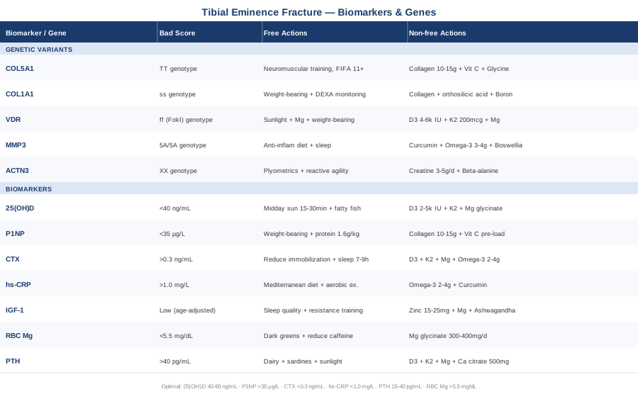
Tibial eminence fracture recovery is rarely as straightforward as the classification system implies. The seven biomarkers covered here — vitamin D, P1NP, CTX, hs-CRP, IGF-1, magnesium, and PTH — give you a functional window into the biological conditions that will either support or impede your healing trajectory. The five genetic variants — COL5A1, COL1A1, VDR, MMP3, and ACTN3 — help explain why your baseline starting point differs from someone with the same fracture type, and where targeted intervention delivers the most return.
The most useful next step is not implementing everything at once, but choosing your highest-leverage starting point. For most people, that means testing 25(OH)D, hs-CRP, and either P1NP or CTX — three relatively accessible and affordable tests that often reveal the most actionable gaps. Discuss the results with a sports medicine physician, orthopedic specialist, or functional medicine practitioner who can contextualize them relative to your fracture stage, age, and rehabilitation timeline. Precise data, discussed with qualified professionals, is where the gap between generic recovery and genuinely optimized healing begins to close.
Musculoskeletal: Bone Conditions Joint Conditions Tendon & Ligament Conditions Sports Injuries
Autoimmune: Connective Tissue Conditions