This article contains AI generated content.
· UpdatedTibial Hemimelia Genes And Biomarkers — 6 Genes And 6 Biomarkers To Track
Introduction
Tibial hemimelia is one of the rarest congenital limb deficiencies known — roughly one in a million births — and yet the families who live with this diagnosis often describe a strange experience: a flood of surgical information paired with a near-complete silence about the underlying biology. The tibia is absent or severely reduced, the surgical plan is outlined, the prosthetic team is introduced, and the deeper question — why did this happen, and what does it mean going forward? — often goes unanswered in clinical consultations.
That silence matters. Tibial hemimelia is not one condition with one cause. It can arise from disruptions in several distinct genetic pathways, some inherited in a predictable pattern, some arising de novo, some reflecting an epigenetic misfire rather than a mutation in a gene's coding sequence. The specific driver changes the recurrence risk, shapes the list of associated conditions to screen for, and influences how the body's remaining musculoskeletal structures behave over decades. Generic medical summaries do not capture these distinctions, which means affected individuals and their families are often making long-term decisions with incomplete information.
This article takes a different approach. It does not promise to reverse a structural birth defect — tibial hemimelia cannot be undone after the developmental window has closed. What it offers instead is a detailed map of the genetic and biological landscape surrounding this condition, with practical guidance for each landmark. Understanding where a gene variant sits in a developmental pathway opens doors to supporting the downstream biology, managing associated risks, and optimizing outcomes in ways that are evidence-grounded and genuinely actionable.
Two complementary strategies structure the content that follows. The first examines the six genes and regulatory sequences most consistently implicated in tibial hemimelia, what each one does, and how to support related pathways whether or not supplementation is part of the picture. The second identifies six biomarkers that track the biological environment surrounding the condition in real time — markers of bone formation, inflammation, and growth that can guide decisions month by month, year by year. Together, they provide a more complete picture than clinical summaries alone and a clearer starting point for the conversations that matter most.
Summary
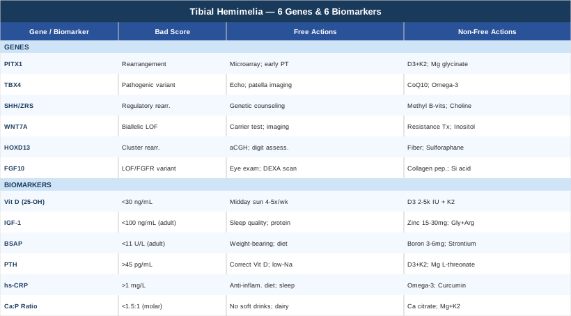
This article explores tibial hemimelia through two evidence-informed lenses that most clinical consultations never reach. The genetics section covers six key molecular players — PITX1, TBX4, the SHH/ZRS regulatory axis, WNT7A, HOXD13, and FGF10 — explaining what each gene does during limb development, what its disruption means clinically, and what concrete steps (with or without supplements) can support the surrounding biology. The biomarker section then identifies six measurable indicators — vitamin D, IGF-1, bone-specific alkaline phosphatase, PTH, high-sensitivity CRP, and the calcium-phosphorus ratio — with measurement methods, cost ranges, and targeted intervention protocols for each. Beyond those two strategies, you will find a practical distillation of Huberman Lab research on bone health and genetic optimization, plus three evidence-supported complementary approaches — photobiomodulation, mindfulness-based stress reduction, and massage therapy — each evaluated specifically for tibial hemimelia. If you have ever wanted to move past the surgical summary and understand the biology your care team rarely has time to explain, this article is that resource.
What Recent Genetics and Epigenetics Research Reveals About Tibial Hemimelia
The tibia does not simply fail to appear in tibial hemimelia. It fails to receive the instruction to appear — or receives a corrupted version of that instruction — during a narrow developmental window between weeks four and eight of embryogenesis. The cascade of molecular signals that should have built the central bone of the lower leg was disrupted upstream, often by a variant that is either inherited from a parent or appeared spontaneously in the fertilized egg.
Researchers have now identified several distinct genetic and regulatory pathways where these disruptions occur. Each pathway has its own inheritance pattern, its own associated features beyond the limb, and its own downstream biology that is worth understanding. The six entries below represent the current state of the evidence — some backed by strong human genetics data, others still primarily supported by animal models and case series, which is noted where applicable.
Gene 1: PITX1 — The Master Regulator of Hindlimb Identity
PITX1 (Pituitary Homeobox 1, chromosome 5q31) is the most directly implicated gene in tibial hemimelia identified to date. It encodes a homeodomain transcription factor whose function is to specify that a developing limb bud belongs to the hindlimb — that it should build a tibia, not a radius. Mice lacking PITX1 consistently develop tibial hemimelia-like phenotypes, with absent or severely hypoplastic tibias. The same phenotype has been reproduced in human familial cases with PITX1 haploinsufficiency.
What makes PITX1 particularly interesting — and particularly relevant to epigenetics — is that many of its pathogenic variants in humans are not mutations in the gene's coding sequence at all. They are structural chromosomal rearrangements that alter the regulatory architecture around PITX1, bringing it into contact with enhancer elements that should not be driving its expression. A landmark study published in Nature Genetics by Spielmann and colleagues demonstrated that such rearrangements could place PITX1 in contact with the ZRS, a long-range Sonic Hedgehog enhancer, resulting in limb specification errors that cause tibial hemimelia. This means the gene itself may be structurally intact but misregulated — a problem invisible to standard exome sequencing.
PITX1 directly activates TBX4, the next gene in this list, and the two form a regulatory axis central to hindlimb identity. Inheritance follows an autosomal dominant pattern in most documented familial cases, although the variable expressivity is notable — a parent carrying the same rearrangement may have no limb difference or only subtle foot asymmetry.
If the PITX1 variant is present — plan without supplements
The first practical step is not supplementation but information. Request chromosomal microarray analysis rather than standard karyotyping — many PITX1-associated rearrangements are submicroscopic and invisible to a conventional karyotype. This is the single most important diagnostic step for understanding whether the variant is inherited, establishing recurrence risk, and guiding family planning decisions. Frequency: one-time test; results are lifelong.
For affected children, early physical therapy focused on hip abductors, quadriceps, and core stabilizers should begin within the first few months of life — well before surgical planning is finalized. These muscle groups will carry disproportionate load regardless of the ultimate surgical approach. Frequency: two to three sessions per week with a pediatric physical therapist experienced in congenital limb differences.
If reconstructive surgery is being planned, ensure the surgical team has the genetic report. PITX1-driven tibial hemimelia can co-occur with subtle fibular overgrowth and hindfoot malposition that influence the choice between tibial reconstruction and amputation-with-prosthetics.
If the PITX1 variant is present — plan with supplements and equipment
Vitamin D3 combined with K2 (MK-7 form): Vitamin D3 at 2000–4000 IU per day for children and 4000–5000 IU per day for adults, paired with vitamin K2 at 100–200 mcg per day. The combination supports bone mineral density in the fibula, femur, and foot bones that carry the full mechanical load. Retest 25-OH vitamin D at three months and adjust dose to reach 50–80 ng/mL. No cycling required for maintenance. Side effect to monitor: hypercalcemia at doses consistently above 10,000 IU/day — stay below that ceiling.
Magnesium glycinate at 200–400 mg per day supports vitamin D metabolism and bone matrix formation. No cycling required. Reduce dose if loose stools occur.
Adaptive prosthetics with progressive loading: Modern microprocessor-controlled prosthetic systems can be introduced as early as six to nine months of age when standing begins. Schedule fitting reviews every three to six months in growing children — a poorly fitting socket that delays loading delays bone development in adjacent structures.
---
Gene 2: TBX4 — Downstream Enforcer of the Hindlimb Skeleton
TBX4 (T-Box Transcription Factor 4, chromosome 17q23) sits directly downstream of PITX1 in the hindlimb specification pathway. It is expressed in the developing hindlimb mesenchyme and is required for proper tibial formation. Autosomal dominant pathogenic variants in TBX4 cause a syndrome involving tibial hemimelia, ectrodactyly (split-foot or split-hand morphology), and femoral bifurcation — a rare but well-documented genetic diagnosis in which the structural findings cluster in a recognizable pattern.
Several case reports and small series have confirmed TBX4 as a direct cause of tibial hemimelia, making it one of the few genes with unambiguous human evidence rather than purely model organism data. What makes TBX4 clinically significant beyond the limb is its expression in the lung and its association with pulmonary arterial hypertension in some individuals with pathogenic variants. TBX4 is also expressed during patella development, and hypoplastic or absent patella (small patella syndrome) has been documented in TBX4 mutation carriers. These are not theoretical associations — they have direct implications for clinical surveillance.
If the TBX4 variant is present — plan without supplements
A baseline echocardiogram is reasonable for individuals with confirmed pathogenic TBX4 variants, even in the absence of respiratory symptoms. Pulmonary arterial hypertension can be silent for years before symptoms emerge, and early identification dramatically changes the management picture. Timing: first year of life, then as clinically directed by a pediatric cardiologist.
Knee and patella imaging (ultrasound in young children, radiograph in older children) should assess patella morphology. A hypoplastic or absent patella changes the biomechanics of knee extension and influences both surgical planning and long-term physiotherapy priorities.
Family member testing: Given the autosomal dominant pattern, first-degree relatives should be offered targeted testing for the identified variant. A carrier parent may have subtle or unrecognized clinical findings that become relevant when understanding their own health.
If the TBX4 variant is present — plan with supplements and equipment
Coenzyme Q10 at 100–200 mg per day with a fat-containing meal supports mitochondrial efficiency in cardiac and vascular tissue. This is not a treatment for pulmonary arterial hypertension but a supportive measure for mitochondrial health in cardiopulmonary tissue. No cycling required. Side effect: mild GI upset in some individuals; divide into two doses.
Omega-3 fatty acids at 2–3 g combined EPA and DHA per day support vascular and pulmonary health through anti-inflammatory pathways. No cycling required. Side effect: fishy aftertaste; enteric-coated formulas largely eliminate this.
A pulse oximeter (finger clip model, cost $25–50) is a practical home monitoring tool for individuals with TBX4 variants. A resting SpO2 consistently below 95% warrants formal pulmonary evaluation regardless of the absence of symptoms.
---
Gene 3: SHH/ZRS Regulatory Axis — The Epigenetic Dimension of Tibial Hemimelia
SHH (Sonic Hedgehog) is one of the most powerful developmental signaling molecules in vertebrate biology. In the developing limb, it is expressed from the Zone of Polarizing Activity (ZPA), a posterior cluster of cells, and it patterns the anterior-posterior axis of the limb — the axis from the big toe to the little toe, or from the thumb to the little finger. The ZRS (Zone of Polarizing Activity Regulatory Sequence), located within the LMBR1 gene on chromosome 7q36, is the long-range enhancer that controls SHH expression specifically in the limb.
The ZRS does not mutate in a way that produces tibial hemimelia directly. Instead, the connection is structural and three-dimensional. The Spielmann 2012 study demonstrated that chromosomal rearrangements can bring the PITX1 gene into ectopic contact with the ZRS enhancer through changes in three-dimensional chromatin organization — essentially, the genome folds in a way that misconnects a regulatory signal. This is a purely epigenetic mechanism: no gene is broken, but its regulatory wiring is rewired. The same logic applies to other topological variants that affect the PITX1-ZRS chromatin neighborhood.
The practical significance is that standard sequencing panels will miss this type of variant entirely. Only chromosomal microarray, optical genome mapping, or long-read sequencing can detect it. Additionally, the ZRS-SHH pathway is sensitive to environmental exposures during weeks four to eight of embryogenesis — a window when many pregnant individuals do not yet know they are pregnant.
If the ZRS/SHH regulatory axis is implicated — plan without supplements
Preconception and prenatal genetic counseling with a specialist in structural chromosomal variants is the most impactful step. If a ZRS-associated rearrangement is identified in a parent, preimplantation genetic testing (PGT-SR) can be discussed with a reproductive endocrinologist — it enables chromosomally normal embryo selection during IVF.
Folate optimization before and during pregnancy: Folate is required for one-carbon metabolism, which drives DNA methylation and chromatin organization during early embryogenesis. The evidence-based recommendation is 400–800 mcg of 5-MTHF (methylfolate, not synthetic folic acid) daily from preconception through the first trimester. This does not correct an existing rearrangement but supports the epigenetic environment during the critical limb development window.
Medication review in early pregnancy: Valproate and high-dose retinoids are well-documented SHH pathway disruptors. Any medication taken in early pregnancy in the context of a known ZRS-related family history warrants clinical review.
If the ZRS/SHH regulatory axis is implicated — plan with supplements and equipment
Methylated B-vitamin complex including 5-MTHF (400 mcg), methylcobalamin (1000 mcg), and pyridoxal-5-phosphate (B6): supports DNA methylation pathways relevant to epigenetic regulation of regulatory sequences including ZRS. Daily use; no cycling required. Side effect: paradoxical reactions at high folate doses in individuals with specific MTHFR variants — start at 400 mcg and increase gradually.
Choline at 250–500 mg per day is an often-overlooked methyl donor critical for epigenetic regulation and one of the most consistently underdosed nutrients in pregnancy. Particularly relevant in the preconception and first-trimester window. No cycling needed. Side effect: fishy body odor at doses above 1 g/day.
---
Gene 4: WNT7A — Dorsal-Ventral Axis and Recessive Tibial Reduction
WNT7A belongs to the Wnt signaling family and is expressed in the dorsal ectoderm of the developing limb bud, where it specifies the dorsal-ventral axis — the difference between the back of the hand and the palm, the top of the foot and the sole. Loss-of-function mutations in both copies of WNT7A (autosomal recessive inheritance) cause two overlapping syndromes in humans: Fuhrmann syndrome and Al-Awadi/Raas-Rothschild syndrome, both of which feature severe limb reduction defects that include tibial aplasia or severe tibial hypoplasia. WNT7A-driven tibial hemimelia tends to be bilateral and more severe in clinical presentation than PITX1- or TBX4-driven forms.
The recessive inheritance pattern here has a direct clinical implication: both parents are typically carriers with no personal limb findings, and each pregnancy carries a 25% risk of an affected child. Identifying WNT7A as the causative gene changes the recurrence calculation entirely and opens the door to preimplantation genetic testing as a family planning option. WNT7A also interacts with WNT5A and other Wnt ligands in bone density regulation postnatally, making the Wnt pathway relevant not just to the original developmental event but to long-term skeletal health.
If the WNT7A variant is present — plan without supplements
Carrier testing for both biological parents should be confirmed. If both are carriers, recurrence risk counseling and a discussion of PGT-M (monogenic preimplantation genetic testing) is appropriate before future pregnancies.
Bilateral lower limb imaging even if only one side appears clinically affected: WNT7A-associated hemimelia has a documented tendency toward subtle contralateral findings that influence long-term orthopedic planning. A full-limb X-ray series is appropriate at diagnosis.
Neurodevelopmental surveillance: WNT signaling is active in central nervous system development and in uterine/reproductive system maturation. Baseline neurodevelopmental assessment at school age is appropriate in affected children.
If the WNT7A variant is present — plan with supplements and equipment
Resistance training as a Wnt pathway activator: Mechanical loading is one of the most well-validated activators of Wnt signaling in bone tissue. Even adapted forms of resistance exercise in children — upper body loading, core training, vibration platforms — stimulate Wnt-driven osteoblast activity in residual bone structures. Frequency: three times per week. No cost if bodyweight protocols are used; $300–$800 for vibration platform.
Lithium orotate at 5–10 mg per day has been studied in research contexts as a Wnt pathway potentiator in bone and neurological tissue. This is preliminary research with limited clinical trial data — discuss with a physician before use and monitor thyroid function with long-term use. Cycling: five days on, two days off is a common research protocol. Do not confuse with prescription lithium carbonate.
Myo-inositol at 2–4 g per day participates as a second messenger in Wnt and other developmental signaling cascades. Early studies suggest supporting roles in skeletal development. No cycling required. Side effect: loose stools at higher doses; split into two daily doses.
---
Gene 5: HOXD13 and the HOXD Cluster — 3D Genome Architecture and Limb Segment Identity
The HOXD cluster on chromosome 2q31 contains a series of transcription factors — HOXD9 through HOXD13 — that act as positional identity codes for limb segments. They work through a remarkably sophisticated regulatory architecture: two topologically associating domains (TADs) flank the HOXD cluster, one controlling digit development, the other controlling the proximal limb segment including the zeugopod region — exactly where the tibia and fibula form. When the balance between these two regulatory domains is disrupted by chromosomal duplications, inversions, or deletions, the proximal-distal patterning of the limb shifts in ways that can produce tibial hypoplasia and hemimelia-like phenotypes.
HOXD13 mutations most directly cause synpolydactyly, but larger HOXD cluster rearrangements have been documented in tibial hemimelia and tibial aplasia cases in the research literature. The entire HOXD cluster is under strong epigenetic regulation — the two flanking TADs are sensitive to changes in chromatin accessibility and DNA methylation — making this locus one of the most biologically relevant targets for epigenetic support strategies. Research from Denis Duboule's laboratory has mapped these regulatory domains in extraordinary detail, establishing that the HOXD cluster works like a developmental switchboard with two distinct output channels.
If HOXD cluster variants are present — plan without supplements
Chromosomal microarray analysis rather than exome sequencing is the appropriate first-line genetic test for HOXD cluster abnormalities — most relevant rearrangements are structural, not single-nucleotide variants. Array comparative genomic hybridization (aCGH) has the sensitivity to detect submicroscopic duplications and deletions in the 2q31 region.
Digit morphology assessment on all four limbs: because HOXD13 specifically regulates digit development, a formal assessment of digit count, fusion patterns, and nail morphology on hands and feet is warranted. Upper limb involvement may have functional implications requiring occupational therapy.
Occupational therapy at two sessions per week for affected children supports fine motor development in cases where HOXD involvement affects upper limb morphology.
If HOXD cluster variants are present — plan with supplements and equipment
Dietary fiber and short-chain fatty acids: Gut bacteria fermenting dietary fiber produce short-chain fatty acids (SCFAs) including butyrate, which acts as a histone deacetylase (HDAC) inhibitor — modulating chromatin accessibility at HOX loci. Targeting 30 or more different plant foods per week generates the microbiome diversity required for consistent SCFA production. This is a genuinely non-invasive epigenetic lever. No cycling required. GI adaptation takes one to two weeks.
Sulforaphane from broccoli sprouts (30–100 mg sulforaphane equivalent per day): A well-studied HDAC inhibitor and Nrf2 activator with published evidence for epigenetic modulation. Cycling: five days on, two days off. Side effect: mild gastric irritation; take with food.
Aerobic exercise: Regular aerobic activity upregulates favorable epigenetic modifications across musculoskeletal systems through multiple mechanisms including HDAC inhibition and histone acetylation. Adapted forms — swimming, hand cycling, seated rowing — are appropriate for tibial hemimelia. Frequency: 30–45 minutes, four to five times per week.
---
Gene 6: FGF10 and the FGF Signaling Network — Limb Bud Initiation and Post-Natal Bone Repair
FGF10 (Fibroblast Growth Factor 10) is expressed in the lateral plate mesoderm during embryogenesis and initiates limb bud formation by signaling to the overlying ectoderm to form the Apical Ectodermal Ridge (AER) — the structure at the limb tip that drives outgrowth. FGF10 then signals back and forth with FGF8 in the AER in a feedback loop that sustains proximal-to-distal limb elongation. If this feedback loop is disrupted early, the limb bud can terminate prematurely, producing reduction defects.
In humans, complete FGF10 loss-of-function causes ALSG syndrome (aplasia of lacrimal and salivary glands), but partial loss-of-function in the context of other limb patterning disruptions has been modeled to produce tibial hemimelia-like phenotypes in mice. FGFR1 and FGFR2 variants (the receptors for FGF10) are better established in craniosynostosis syndromes, but the FGF pathway's relevance does not end at birth. FGF2, a closely related growth factor, remains active in post-natal osteocytes and is stimulated by mechanical loading to drive bone remodeling — making this pathway directly relevant to ongoing bone health in residual limb structures.
If FGF10/FGFR variants are present — plan without supplements
Ophthalmologic and lacrimal assessment: FGF10 variants can cause dry eye and abnormalities in tear duct and lacrimal gland development. Persistent dry eye symptoms in affected individuals should be formally evaluated by an ophthalmologist.
Pediatric dental panoramic radiograph at age six to eight: FGF signaling participates in tooth bud development. Panoramic X-ray assesses for dental anomalies that may co-occur with FGF10 pathway disruption.
Bone density baseline: Residual limb bones in prosthetic users carry asymmetric stress loads. A DEXA scan of the femur and fibula at age ten establishes a baseline; repeat every three to five years or following significant surgical events.
If FGF10/FGFR variants are present — plan with supplements and equipment
Hydrolyzed collagen peptides (10–15 g per day, type I and III): FGF signaling drives collagen matrix production in bone and cartilage. Hydrolyzed collagen peptides have been shown in randomized trials to support bone mineral density when combined with resistance exercise. No cycling required. Side effects: minimal.
Orthosilicic acid (10–25 mg per day from choline-stabilized orthosilicic acid): Silicon is required for collagen cross-linking and FGF-dependent bone mineralization. Deficiency in animal models produces skeletal malformations reminiscent of reduced FGF signaling. Side effects: minimal at physiological doses.
Whole-body vibration platform at 30 Hz, ten minutes per day: Mechanical stimulation directly activates FGF2 in osteocytes, supporting bone remodeling in residual limb structures. Equipment cost: $300–$800 for consumer-grade platforms; widely used in pediatric rehabilitation for congenital limb differences.
---
Building from the genetic picture, the next layer of insight comes from what can be measured in the blood right now — regardless of whether genetic testing has been completed.
6 Biomarkers Worth Tracking for Tibial Hemimelia
Genetic data explains the origin. Biomarkers describe the present. For someone living with tibial hemimelia — whether a young child recovering from surgery, an adolescent adapting to a growing prosthesis, or an adult managing decades of asymmetric limb loading — there are six blood-based measurements that provide a genuinely useful window into bone health, inflammation, and growth capacity. These are not exotic markers. They are available at standard clinical laboratories and interpretable by any physician who understands musculoskeletal health. The value lies in tracking them consistently.
Biomarker 1: 25-OH Vitamin D (Serum Vitamin D)
Why it matters: Vitamin D is the master regulator of calcium absorption and bone mineralization. In tibial hemimelia, the fibula, femur, and foot bones carry the mechanical loads that the absent tibia cannot share. Adequate vitamin D is foundational to maintaining the density of these bones and to supporting healing after any surgical intervention. Vitamin D deficiency is far more common than most clinical settings acknowledge — particularly in northern latitudes, in individuals with darker skin, and in children with restricted outdoor activity.
How to measure it
A 25-hydroxyvitamin D serum assay is available at any clinical laboratory. Cost: $30–$80 USD, often covered by insurance with a relevant clinical indication. Labs typically flag values above 30 ng/mL as "normal," but practitioners focused on musculoskeletal and immune health — including Peter Attia — recommend a functional target of 50–80 ng/mL for individuals with elevated bone demands.
If the score is low — plan without supplements
Consistent midday sun exposure (15–30 minutes on arms and legs, without sunscreen) four to five times per week produces meaningful vitamin D in appropriate latitudes during appropriate seasons. The practical limitation is significant: latitude above 35 degrees north, winter months, and darker skin tones all reduce cutaneous vitamin D production substantially. Dietary sources — fatty fish, egg yolks, and liver — contribute modest amounts but rarely achieve optimal serum levels through food alone.
If the score is low — plan with supplements and equipment
Vitamin D3 combined with K2 (MK-7): D3 at 2000–4000 IU per day for children and 4000–5000 IU per day for adults, with K2 at 100–200 mcg per day to direct calcium toward bone rather than soft tissue. Retest serum levels at three months and adjust to reach the 50–80 ng/mL target. No cycling required for maintenance dosing. Side effect: hypercalcemia at chronic doses consistently above 10,000 IU/day — routine monitoring eliminates this risk.
---
Biomarker 2: IGF-1 (Insulin-Like Growth Factor 1)
Why it matters: IGF-1 is the primary downstream effector of growth hormone and drives bone elongation, muscle development, and collagen synthesis. In children with tibial hemimelia undergoing limb reconstruction or bone-lengthening procedures, IGF-1 levels directly influence bone regeneration speed and quality. In adults, low IGF-1 correlates with reduced bone mineral density, impaired fracture healing, and reduced muscle mass in load-bearing limb structures.
How to measure it
Serum IGF-1 is a standard blood test interpreted against age-specific reference ranges. Cost: $50–$120 USD. For adults, a functional optimal range of 150–250 ng/mL is commonly cited by practitioners focused on longevity and musculoskeletal health. Values below 100 ng/mL in adults or significantly below age-based midpoints in children warrant investigation.
If the score is low — plan without supplements
The single most effective non-supplement intervention for IGF-1 is sleep quality. The largest nocturnal pulse of growth hormone — and thus IGF-1 — occurs in the first 90 minutes of deep sleep. A consistent sleep and wake schedule, dark cool sleeping environment (17–19°C), and avoidance of food in the two to three hours before bed all protect this pulse. In growing children with tibial hemimelia, protecting sleep quality is one of the highest-leverage health investments available.
Protein adequacy is the dietary parallel: IGF-1 production is protein-sensitive, and inadequate intake directly suppresses it. Target 1.6–2.2 g of protein per kilogram of body weight per day from complete protein sources. Resistance training — even adapted forms focused on upper body and core — stimulates systemic IGF-1 production within six to eight weeks of starting a program.
If the score is low — plan with supplements and equipment
Zinc bisglycinate at 15–30 mg per day: Zinc is required for IGF-1 receptor signaling and growth hormone secretion, and marginal deficiency significantly suppresses IGF-1 levels. Cycling: five days on, two days off, or three months on followed by a one-month break to prevent copper depletion at higher doses. Side effect: nausea on an empty stomach.
Glycine (3 g) plus arginine (2 g) taken before bed: These amino acids stimulate nocturnal growth hormone release through non-insulin pathways. Multiple small studies support this combination for optimizing sleep-time GH output. Side effects: minimal; mild GI at higher doses.
Whole-body vibration platform at 30–40 Hz for ten minutes per day: Mechanical stimulation has been shown in research settings to increase local IGF-1 expression in bone tissue, with systemic effects documented at sufficient vibration intensity. Equipment cost: $300–$800.
---
Biomarker 3: Bone-Specific Alkaline Phosphatase (BSAP)
Why it matters: Bone-specific alkaline phosphatase is an enzyme produced by osteoblasts — the cells that build new bone. Tracking it provides a real-time signal of bone formation activity. In tibial hemimelia, this is especially valuable during periods of surgical bone work, distraction osteogenesis, or prosthetic adaptation, when demand for new bone formation is elevated and the body's capacity to meet that demand matters.
How to measure it
Total alkaline phosphatase is included in standard metabolic panels. Bone-specific ALP requires a dedicated assay requesting the bone isoform specifically. Cost: $50–$100 USD. Pediatric values are physiologically higher than adult values due to active skeletal growth — results must be interpreted against age-specific reference tables. In adults, the normal range is roughly 11–43 U/L for bone-specific ALP.
If the score is low — plan without supplements
Mechanical loading of residual limb structures is the most direct activator of osteoblast activity available without medication. Even partial weight-bearing through a prosthesis, properly fitted and used consistently, stimulates BSAP-producing osteoblasts in the fibula and adjacent bones. A physical therapist can design a progressive loading protocol appropriate to anatomy and surgical status.
Adequate dietary phosphorus and calcium balance: Extremely low phosphorus intake depresses bone formation. Dairy, legumes, fish, and whole grains all contribute; a dietary record reviewed by a registered dietitian can identify deficiencies specific to the individual.
If the score is low — plan with supplements and equipment
Boron at 3–6 mg per day: Boron supports osteocalcin activity and BSAP signaling by modulating estrogen and vitamin D metabolism in bone tissue. It is frequently absent from standard multivitamins. No cycling required. Side effects: minimal at food-level doses.
Strontium citrate at 340 mg per day taken separately from calcium by at least two hours: Evidence from bone loss trials shows strontium increases BSAP by stimulating osteoblast differentiation. Cycling: six months on, one month off. Side effects: avoid in renal impairment; do not confuse with strontium ranelate (the prescription drug with different safety profile).
---
Biomarker 4: Parathyroid Hormone (PTH)
Why it matters: PTH is the primary regulator of calcium homeostasis. When serum calcium falls, PTH rises and draws calcium out of bone — an efficient short-term fix that becomes a significant long-term problem when it runs chronically. Secondary hyperparathyroidism, most commonly caused by low vitamin D, accelerates bone resorption in exactly the bones that carry the full mechanical load in tibial hemimelia. Thomas Dayspring and Allan Sniderman both emphasize PTH as a critical piece of the metabolic bone puzzle — it does not stand alone but must be interpreted alongside vitamin D and serum calcium as a triad.
How to measure it
Intact PTH (iPTH) from a blood sample is the standard measurement. Cost: $40–$80 USD. Optimal functional range: 15–45 pg/mL. PTH should always be measured alongside 25-OH vitamin D and serum calcium — isolated PTH values are difficult to interpret correctly without the accompanying context.
If PTH is elevated — plan without supplements
The most important first step is addressing the root cause. In the majority of cases, elevated PTH in otherwise healthy individuals reflects low vitamin D — treating vitamin D consistently normalizes PTH within three to four months without any direct PTH intervention. Reducing urinary calcium loss through lower sodium intake, reduced caffeine, and elimination of phosphoric acid–containing beverages also improves the calcium balance that drives PTH secretion.
If PTH is elevated — plan with supplements and equipment
Vitamin D3 plus K2 at therapeutic doses (as described in Biomarker 1): The most evidence-backed intervention for secondary hyperparathyroidism. Effect is measurable within eight to twelve weeks of consistent supplementation.
Calcium citrate at 500 mg with meals (only if dietary calcium intake is confirmed insufficient): Calcium citrate is preferred over calcium carbonate for individuals with GI sensitivity. Cycling: not required; adjust dose based on dietary calcium analysis. Side effect: constipation; balance with magnesium.
Magnesium L-threonate at 1.5–2 g per day: Magnesium directly suppresses PTH secretion and is consistently depleted in individuals with chronic bone stress and poor sleep. L-threonate is the preferred form for bone and neurological applications. Side effect: mild sedation; take in the evening.
---
Biomarker 5: High-Sensitivity CRP (hs-CRP)
Why it matters: High-sensitivity CRP is the most accessible marker of systemic inflammation available in standard clinical practice. In tibial hemimelia, chronic inflammation — from repeated surgical procedures, prosthetic socket friction, altered gait biomechanics creating asymmetric joint stress, or psychosocial stress from complex medical navigation — can impair bone healing, suppress growth hormone signaling, and accelerate bone resorption. Peter Attia consistently highlights hs-CRP as one of the essential metrics in any bone and longevity-focused panel; for tibial hemimelia specifically, keeping it low provides a direct protective effect on the bones carrying disproportionate load.
How to measure it
A standard blood test, widely available. Cost: $20–$50 USD. Optimal target: below 0.5 mg/L. Values between 1–3 mg/L indicate moderate inflammation warranting investigation; above 3 mg/L indicates significant systemic inflammation requiring active intervention and differential diagnosis.
If hs-CRP is elevated — plan without supplements
An anti-inflammatory dietary pattern — Mediterranean, whole food, centered on olive oil, fatty fish, leafy greens, berries, and legumes while eliminating ultra-processed foods — consistently reduces hs-CRP by 20–30% in clinical trials within eight to twelve weeks. Sleep duration is equally powerful: hs-CRP rises significantly with less than seven hours of sleep per night. In children, nine to eleven hours is the evidence-based target. These two interventions — diet and sleep — should precede any supplement discussion.
If hs-CRP is elevated — plan with supplements and equipment
Omega-3 fatty acids at 2–4 g combined EPA and DHA per day: The most robustly evidence-backed supplement for reducing hs-CRP, with consistent results across multiple meta-analyses. Cycling: not required for maintenance dosing. Side effect: anticoagulant effect at higher doses — taper before surgical procedures in consultation with a physician.
Bioavailable curcumin (BCM-95 or Meriva formulation, 500–1000 mg per day): Multiple randomized controlled trials demonstrate hs-CRP reductions with bioavailable curcumin forms. Standard curcumin powder has negligible absorption; formulation matters critically. Cycling: eight weeks on, two weeks off. Side effect: blood thinning; avoid perioperatively.
Far-infrared sauna at 20–30 minutes per session, three to four times per week: Growing evidence supports far-infrared sauna as a systemic hs-CRP reducer through heat shock protein activation and cortisol normalization. Equipment cost range: $300–$500 for an infrared blanket; $2000–$4000 for a cabin unit.
---
Biomarker 6: Calcium-to-Phosphorus Ratio
Why it matters: The serum calcium-to-phosphorus ratio reflects bone mineral metabolism efficiency. Modern Western diets deliver excessive phosphorus — predominantly through soft drinks containing phosphoric acid and phosphate preservatives in processed meats and packaged foods — while calcium intake remains marginal in many populations. A chronically high phosphorus–to–calcium environment activates PTH and drives bone resorption. For tibial hemimelia patients whose remaining bones carry extraordinary mechanical demands, this metabolic environment is especially costly.
How to measure it
Both calcium and phosphorus are included in a standard basic or comprehensive metabolic panel. Cost: $15–$40 USD as part of a panel. Optimal serum ratio: approximately 2:1 to 2.5:1 (calcium-to-phosphorus, molar ratio). A ratio consistently below 1.5:1 signals a phosphorus-excess environment that is actively antagonizing bone formation.
If the ratio is unfavorable — plan without supplements
Eliminating processed food phosphorus sources is the single most impactful dietary intervention: phosphoric acid in soft drinks, phosphate-based preservatives in packaged deli meats, and processed cheese are the primary offenders. Elimination alone normalizes the ratio in many individuals within four to six weeks. Simultaneously increasing calcium from whole food sources — full-fat dairy, sardines with bones, leafy greens, and bone broth — supports the other side of the equation.
If the ratio is unfavorable — plan with supplements and equipment
Calcium citrate at 500 mg taken specifically at meals confirmed to be high in processed phosphorus: Rather than chronic supplementation, targeted dosing at high-phosphorus meals partially binds dietary phosphorus and improves the absorbed ratio. Adjust dose based on dietary tracking. Side effect: constipation; balance with magnesium intake.
Magnesium and vitamin K2 as previously described: These work synergistically to optimize calcium utilization and bone deposition and address the calcium-phosphorus imbalance from multiple angles simultaneously.
---
The genetic and biomarker frameworks above set a strong foundation. The following section builds on them with findings that most physicians have not yet integrated into clinical practice — and that have direct relevance for anyone managing the downstream biology of tibial hemimelia.
10 Insights From the Huberman Lab That Reframe Bone Health and Genetic Optimization
The Huberman Lab podcast hosted by Andrew Huberman at Stanford University has produced a body of episodes on musculoskeletal biology, growth hormone signaling, epigenetics, and inflammation management that translates directly to the management goals identified in this article. None of these episodes addresses tibial hemimelia directly, but the underlying mechanisms they explain — how bone forms, repairs, and is optimized — are the same mechanisms relevant here. The ten findings below represent the most directly actionable insights across those episodes for this context.
1. The Growth Hormone Window Requires Sleep Architecture, Not Just Duration
The largest pulse of growth hormone — and therefore IGF-1 — occurs in the first deep sleep cycle, typically within 90 minutes of sleep onset. Duration of sleep matters, but it is the architecture of that first cycle that determines the magnitude of the GH pulse. For growing children with tibial hemimelia who are in active surgical recovery or limb lengthening phases, protecting this window is one of the most impactful interventions available. Consistent sleep and wake timing, room temperature at 17–19°C, and no food or high insulin activity in the two to three hours before bed all preserve this pulse.
2. Morning Sunlight Is Not Replaceable by Vitamin D Supplementation Alone
Morning sunlight exposure (10–30 minutes within an hour of waking) calibrates the circadian clock through photoreceptors in the retina that drive the cortisol awakening response. A properly timed cortisol peak — not chronically elevated, but sharply peaked at waking and declining through the day — supports growth hormone release, sleep quality, and downstream hormone cascades that vitamin D alone does not replicate. Supplements optimize serum levels; morning light optimizes the biological system that uses those levels.
3. Mechanical Load Drives Bone Density More Powerfully Than Calcium Intake
Multiple human studies confirm that impact loading — jumping, running, resistance training — stimulates osteocalcin production and bone formation far more potently than dietary calcium interventions alone. For tibial hemimelia patients, adapted forms of loading — hopping on the unaffected side, upper body compound exercises, vibration platform protocols — activate the same osteogenic signaling pathways without requiring full bilateral lower limb function.
4. Omega-3s Modulate Inflammatory Gene Expression, Not Just Downstream CRP Numbers
Huberman distinguishes between the surface-level CRP reduction observed with omega-3 supplementation and the upstream mechanism: high-dose EPA and DHA downregulate NF-κB pathway gene expression, which reduces osteoclast activation — the cell type that breaks bone down. This distinction matters post-surgically in tibial hemimelia, where inflammatory resorption at surgical interfaces can undermine newly formed bone callus.
5. Short Resistance Training Sessions Optimize the Anabolic-to-Cortisol Ratio
Training sessions exceeding 60 minutes — particularly in a fasted state — can elevate cortisol sufficiently to blunt the anabolic hormone response. Short, high-intensity sessions of 20–30 minutes with compound movements produce the optimal growth hormone to cortisol ratio. This is practically significant for children with active physical therapy schedules and for adults managing energy demands around complex medical care.
6. Timing Cold Exposure Relative to Training Changes Its Effect on Bone Adaptation
Cold water immersion immediately following resistance training attenuates the inflammatory signaling cascade required for muscle and bone adaptation to load. Huberman advises a minimum four-to-six-hour delay between a training session aimed at strength or bone loading and cold exposure. For tibial hemimelia patients doing supervised bone-loading therapy, this sequencing has direct practical implications.
7. Chronic Cortisol Elevation Directly Suppresses Osteoblast Activity
Prolonged cortisol elevation — a documented pattern in caregivers of children with complex medical needs — directly suppresses osteoblast function through glucocorticoid receptor signaling. The intervention is not conventional stress management but physiological recovery: non-sleep deep rest (NSDR) protocols of 10–20 minutes, diaphragmatic breathing, and consistent social connection all produce measurable cortisol normalization. Addressing caregiver physiological stress is a legitimate component of the child's bone health management.
8. Leucine Is the Specific Amino Acid Trigger for Bone and Muscle Anabolism
Of all amino acids, leucine is the primary trigger for mTOR-mediated protein synthesis in both muscle and bone. For IGF-1 optimization and bone formation, each meal should contain at least 2.5–3 g of leucine. Animal proteins (whey, eggs, meat) naturally provide this; plant-based diets often require careful food combining to reach the leucine threshold per meal. This is a practical optimization for anyone with elevated bone formation demands.
9. Gut Microbiome Diversity Influences Bone Density Through SCFA Production
Short-chain fatty acids produced by gut bacteria fermenting dietary fiber directly modulate osteoblast and osteoclast balance through HDAC inhibition and systemic inflammatory signaling. The practical target Huberman cites from the research literature is 30 different plant food types per week — not quantity of fiber from one source, but diversity of fermentable substrates. This connects directly to the HOXD epigenetics discussion earlier in this article: SCFA-mediated HDAC inhibition modulates chromatin accessibility at HOX loci.
10. Zone 2 Cardio Is the Most Broadly Validated Anti-Inflammatory Protocol
Sustained Zone 2 cardiovascular exercise — intensity at which you can hold a conversation, approximately 60–70% of maximum heart rate — performed for 45–60 minutes per session, three to four times per week, is among the most consistently evidence-backed protocols for reducing hs-CRP and improving mitochondrial density across all tissues. For tibial hemimelia, adapted Zone 2 modalities — hand cycling, seated rowing, swimming — produce the same systemic benefits without bilateral lower limb requirements.
---
Complementary Approaches With Evidence for Tibial Hemimelia
The following three modalities are included not because they address tibial hemimelia's structural anatomy but because they have meaningful evidence for outcomes that directly affect quality of life and biological resilience in affected individuals — post-surgical healing, pain management, proprioceptive development, and psychological adaptation.
Low-Level Laser Therapy (Photobiomodulation)
Low-level laser therapy, also called photobiomodulation (PBM), applies near-infrared light at wavelengths of 630–980 nm to stimulate cellular energy production and tissue repair at the mitochondrial level without generating heat. In the context of tibial hemimelia, its primary relevance is post-surgical bone healing, wound repair at skin-prosthetic interfaces, and reduction of perioperative inflammation — all areas where the clinical burden in tibial hemimelia is significant and where conventional options are limited. Secondary applications include pain management around prosthetic socket pressure points and support for distraction osteogenesis, the surgical bone-lengthening technique sometimes used in tibial hemimelia reconstruction.
Human evidence for PBM in bone repair is meaningful. A systematic review published in Photomedicine and Laser Surgery (Merli et al.) found that LLLT significantly accelerated bone regeneration in distraction osteogenesis contexts. A clinical trial from the University of São Paulo demonstrated that 830 nm laser application to surgical sites accelerated collagen deposition and reduced pain scores in pediatric orthopedic patients compared to sham treatment. Evidence is considered promising rather than definitive — trial sizes are typically small and protocols vary — but the safety profile is excellent and the biological rationale is well-supported.
Practical application: consumer-grade PBM panels with 650–850 nm output in the 50–500 mW range are available for $200–$600. Clinical protocol for post-surgical support: five to ten minutes per site, three times per week, beginning 48 hours post-incision (not directly on open wounds). Discuss timing with the treating orthopedic surgeon before introducing PBM around a surgical site — it should complement, not replace, standard wound care.
Mindfulness-Based Stress Reduction (MBSR)
Mindfulness-Based Stress Reduction is an eight-week structured program developed by Jon Kabat-Zinn at the University of Massachusetts that combines body scan meditation, breath awareness, and gentle movement to cultivate present-moment attention and reduce stress reactivity. For tibial hemimelia, its relevance runs in two directions simultaneously: pain management and prosthetic adjustment anxiety in affected individuals (particularly adolescents and adults dealing with phantom sensations or chronic prosthetic pressure pain), and psychological resilience in primary caregivers navigating long-term complex medical systems.
A 2017 meta-analysis by Hilton and colleagues examined MBSR across multiple musculoskeletal and chronic pain populations and found consistent reductions in pain intensity of approximately 20–30%, with effects maintained at six-month follow-up. Specifically relevant to limb difference, a study published in Disability and Rehabilitation found mindfulness-based interventions significantly reduced phantom limb pain and prosthetic adjustment anxiety in congenital and acquired limb difference populations compared to usual care. The cortisol-lowering effect of MBSR, well-documented across multiple trials, also directly supports osteoblast activity — making this as much a bone health intervention as a psychological one.
Practical application: the original MBSR program is available through certified teachers affiliated with the Center for Mindfulness training lineage. Pediatric adaptations exist for children as young as seven or eight, with shorter sessions (15–20 minutes) and age-appropriate language. Apps including Headspace and Insight Timer provide accessible entry points. Frequency: 20–45 minutes per day. Evidence is strongest for programs lasting at least eight weeks; shorter programs show attenuated effects. Begin with five-minute sessions for young children and build gradually.
Massage Therapy
Therapeutic massage — specifically myofascial release, neuromuscular technique, and connective tissue massage — serves a dual function in tibial hemimelia. For affected children, it supports the development of compensatory muscle groups in the residual limb and adjacent structures: quadriceps, hamstrings, hip abductors, and gluteal muscles that assume disproportionate load. For adolescents and adults using prosthetics, regular massage of the residual limb and socket interface area reduces friction-related skin breakdown, improves proprioceptive awareness in the residual limb, and decreases the chronic muscle hypertonicity that accumulates from asymmetric gait patterns over years.
A randomized controlled trial published in Physical Therapy demonstrated that myofascial release significantly improved functional mobility and reduced pain scores in pediatric populations with congenital lower limb differences compared to standard physical therapy alone. A separate controlled study in the Journal of Rehabilitation Medicine found that prosthetic users receiving regular massage therapy reported higher prosthetic comfort scores and showed measurable improvements in gait symmetry over twelve weeks of treatment. Evidence for this specific application is limited by small trial sizes, but the biological rationale is strong and the safety profile is excellent.
Practical application: massage should be performed by a licensed therapist with training in pediatric orthopedics or prosthetics rehabilitation — not all massage therapists are trained for this population. Session frequency: weekly during periods of active prosthetic adaptation or post-surgical recovery; monthly for maintenance. Duration: 45–60 minutes per session. Cost: $60–$120 per session. Home-based tools — foam rollers and massage guns used gently around residual limb soft tissue — can extend benefit between sessions at low cost. Never apply deep pressure directly over surgical hardware, bone interfaces, or unhealed skin without specific clearance from the orthopedic surgical team.
---
Conclusion
Tibial hemimelia begins in the earliest days of embryogenesis, shaped by gene expression cascades that science is still actively mapping — but the conversation should not end with the developmental diagnosis. Understanding which gene or regulatory sequence was disrupted changes the questions you ask, the associated conditions worth screening for, and the biological environment you can actively support. Genetic information is not just historical context; it is a practical guide to what to watch for next.
The biomarkers covered here are not luxury tests. Vitamin D, IGF-1, bone-specific alkaline phosphatase, PTH, hs-CRP, and the calcium-phosphorus ratio are all accessible through standard clinical laboratories and, tracked consistently over time, give a real-time picture of whether the bones carrying the full mechanical load are well-supported or under stress.
The most important next step is not adding a supplement or starting a protocol. It is a conversation with a clinical geneticist or genetic counselor who can interpret your specific situation, paired with a physician who can review the biomarker panel in full clinical context. The interventions described here are supportive — they are designed to work on top of that clinical foundation, not replace it.
Better information leads to better questions. Better questions lead to better decisions. And better decisions, made consistently over years of care, translate into meaningfully different outcomes.
Musculoskeletal Eye Cardiovascular Respiratory Endocrine & Metabolic
Musculoskeletal: Bone Conditions
Autoimmune: Inflammatory Conditions