This article contains AI generated content.
· UpdatedTibial Muscular Dystrophy - 4 Genes And 6 Biomarkers To Track
Introduction
Living with tibial muscular dystrophy means navigating a condition that arrives quietly, progresses slowly, and is poorly understood even by many clinicians. For most people, the diagnosis comes after years of unexplained foot weakness — appointments where the working theory was a compressed nerve, an aging tendon, or simply bad luck. By the time the right word, titinopathy, finally enters the picture, the window for early intervention has often narrowed considerably.
What follows diagnosis is frequently a second disappointment: there is very little practical guidance. Genetic counseling covers inheritance patterns. Neurology appointments measure decline. But the question most people are actually asking — what can I do, starting today, to stay as functional as possible for as long as possible — tends to go unanswered. Broad advice like "stay active" or "eat well" is not wrong, but it treats this condition the same way it treats everything else, which misses the point entirely.
Tibial muscular dystrophy is a monogenic disease with well-characterized genetic drivers. That specificity is actually an advantage: the molecular pathways involved are known well enough to reason about compensation strategies, not just symptom tracking. Understanding which genes are disrupted, how they fail, and which biological markers reflect current muscle health gives you a more honest and more useful map than general wellness advice ever could.
This article covers four key genes relevant to TMD — what they do in healthy muscle, how their dysfunction causes harm, and what plans (with and without supplementation) may help support affected pathways. It also covers six biomarkers worth tracking over time to get an ongoing picture of muscle function and systemic health. Beyond that, you will find a synthesis of what leading longevity medicine thinking suggests about muscle preservation, along with a few evidence-informed complementary modalities that are worth knowing about. None of this is a cure. But better information consistently leads to better decisions — and that remains true even for a rare disease with no approved pharmacological treatment.
Summary
This article examines tibial muscular dystrophy through two complementary lenses: genetics and biomarker tracking. The four genes covered — TTN, MYOT, ANO5, and FLNC — represent the primary cause of TMD and its closest clinical mimics. For each, you will find a concrete plan with and without supplements, including dosing, cycling, and side effects. The six biomarkers — CK, Vitamin D, Aldolase, hsCRP, CoQ10, and Ferritin — form a practical monitoring panel that any physician can order, with clear targets and actionable steps if results fall outside optimal ranges. The article also synthesizes what Peter Attia's work on muscle longevity reveals that most neurologists will never mention, and it closes with three complementary approaches — photobiomodulation, yoga, and breathing therapy — that have meaningful (if limited) human evidence specifically for neuromuscular conditions. Whether you are newly diagnosed or years into managing TMD, what follows is more specific and more useful than what most people receive.
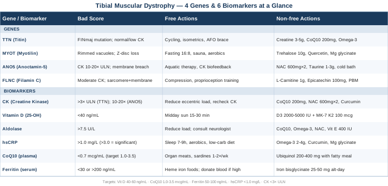
What the Genetics Behind Tibial Muscular Dystrophy Reveal — And What You Can Do About It
Tibial muscular dystrophy is one of the few neuromuscular conditions where genetics gives a precise entry point rather than a vague probabilistic risk. The molecular architecture of the sarcomere — the contractile unit of every skeletal muscle fiber — is now well mapped, and TMD and its closest clinical relatives all converge on the same structural question: what happens when the proteins that build and maintain that architecture begin to fail? The four genes below represent the primary driver of TMD and three overlapping conditions that are frequently confused with it. Understanding the mechanism behind each one makes it possible to target the right pathways — not generically, but specifically, based on the proteins involved.
Gene 1: TTN (Titin) — The Sarcomere's Molecular Spring
What this gene does in healthy muscle
Titin is the largest protein in the human body, a 3.6-megadalton molecular spring that spans the entire length of the sarcomere from the Z-disc to the M-band. Its mechanical function is foundational: during muscle contraction, titin stores elastic energy and returns it during relaxation, maintaining structural tension and preventing sarcomere overstretching. Beyond its passive elastic role, titin's C-terminal M-band domain serves as a critical signaling hub, detecting mechanical stress and transmitting those signals to the nucleus to regulate protein synthesis and degradation.
In tibial muscular dystrophy, the primary pathogenic mechanism is a specific mutation in the last coding exon of TTN. The most common variant — the FINmaj mutation, an 11-base-pair insertion in exon 363 — disrupts the M-band domain precisely, compromising titin's anchoring function within the sarcomere. This mutation was confirmed as the genetic basis of TMD in a landmark study published in The American Journal of Human Genetics by Hackman and colleagues in 2002, establishing TMD as a titinopathy with autosomal dominant inheritance.
Why the tibialis anterior is the first victim
The tibialis anterior is unique among lower limb muscles: it works primarily under eccentric load — meaning it contracts while lengthening, braking the foot during each step. Eccentric loading places vastly greater stress on the sarcomere than concentric (shortening) contractions, and titin bears the majority of that eccentric force. When the M-band is disrupted by the FINmaj mutation, this high-eccentric-load environment accelerates sarcomere failure specifically in the tibialis anterior, which is why foot weakness and difficulty walking on heels are the defining early symptoms rather than proximal (hip or thigh) weakness.
If the TTN Gene Is Affected — The Plan Without Supplements
The single most impactful non-pharmacological strategy for TTN mutation carriers is exercise modality selection. Since titin is the primary bearer of eccentric muscle force, any training that emphasizes eccentric loading — downhill running, heavy slow negatives, depth jumps — directly accelerates sarcomere damage in muscle groups already under structural stress. The principle is not to avoid exercise but to shift toward concentric-dominant and isometric movement patterns.
Recommended modalities: - Cycling: The pedaling motion is predominantly concentric (quadriceps and tibialis load during the downstroke rather than during the return phase). It builds lower-limb endurance and cardiovascular reserve without significant eccentric sarcomere stress. Aim for 30–45 minutes, 4–5 sessions per week, at moderate intensity. - Swimming: Water resistance allows meaningful muscle engagement with dramatically reduced gravity-related eccentric load. Freestyle and breaststroke engage the tibialis indirectly; backstroke is often better tolerated. - Isometric resistance training: Wall sits, planks, and isometric ankle dorsiflexion exercises maintain motor unit recruitment and muscle mass without the stretch-under-tension that damages compromised sarcomeres. - Ankle-foot orthoses (AFOs): A custom-fitted AFO is the most evidence-supported functional intervention in TMD. It compensates for tibialis anterior weakness mechanically, reduces fall risk, and extends years of independent ambulation. Assessment should begin as soon as any foot drop is detectable on clinical examination. - Physical therapy: Quarterly gait analysis to identify compensatory muscle use (hip flexors, peroneals) and adjust exercises accordingly. Peroneals and hip flexors often hypertrophy compensatorily and deserve specific strengthening attention.
What to systematically avoid: Downhill walking without orthotic support, heavy eccentric leg curls and tibialis exercises, plyometric movements, prolonged standing on uneven surfaces without bracing.
If the TTN Gene Is Affected — The Plan With Supplements and Equipment
Creatine monohydrate is the starting point for almost any discussion of supplementation in muscular dystrophy. A systematic review and meta-analysis of creatine use in muscular dystrophies (Tarnopolsky and Mahoney, published in the Annals of Neurology) found consistent improvements in muscle strength and function in multiple dystrophy subtypes with good tolerability. Mechanism: creatine phosphate buffers rapid ATP turnover in muscle fibers, reducing metabolic stress during contraction. Dose: 3–5 g/day, taken continuously. No cycling required. Minor initial water retention is common and harmless.
CoQ10 (Ubiquinol form): Titin dysfunction elevates mitochondrial reactive oxygen species in affected muscle. CoQ10 is an electron carrier within the mitochondrial respiratory chain and a potent membrane antioxidant. Ubiquinol — the reduced, active form — is substantially better absorbed than standard ubiquinone, especially in adults over 40. Dose: 200–400 mg/day of ubiquinol, taken with a fatty meal. No formal cycling needed; consistent use over months is more relevant than short bursts.
Omega-3 fatty acids (EPA + DHA): EPA and DHA reduce prostaglandin-mediated inflammation downstream of muscle fiber membrane damage and also modulate autophagy — particularly relevant given that titin dysfunction impairs the clearance of damaged sarcomeric proteins. Target: 2–4 g/day combined EPA+DHA from high-quality fish oil or algal oil. Can be taken continuously. If you take blood thinners, discuss dose with your physician.
Vitamin D3 + K2: Vitamin D receptor signaling is directly involved in muscle fiber type composition, neuromuscular junction function, and myosin heavy chain expression. Deficiency accelerates sarcopenic processes that compound the underlying genetic deficit. Target serum 25-OH-D: 40–60 ng/mL (per Peter Attia's framework). Typical supplementation dose: 2,000–5,000 IU D3 daily alongside 100–200 mcg MK-7 K2 for calcium routing. Monitor serum levels every 6 months.
Equipment — Neuromuscular Electrical Stimulation (NMES): NMES devices applied transcutaneously over the tibialis anterior and surrounding muscles can maintain motor unit recruitment and reduce disuse atrophy in muscles affected by foot drop. Clinical protocols typically involve 20–30 minutes per session, 3 times per week, at parameters that produce a visible muscle twitch. Evidence in TMD specifically is anecdotal, but NMES is low-risk and is used in clinical neuromuscular programs for atrophic distal muscles.
Gene 2: MYOT (Myotilin) — When the Z-Disc Collapses
What this gene does
Myotilin is a Z-disc component that crosslinks actin thin filaments and stabilizes the entire Z-disc lattice during muscle contraction. It acts as a structural anchor for alpha-actinin and is involved in the proper spacing and registration of sarcomere components. Mutations in MYOT cause myofibrillar myopathy and a specific distal variant (Markesbery-Griggs myopathy) that presents with late-onset weakness in the anterior leg compartment — an almost identical clinical picture to TTN-related TMD.
The distinguishing pathology: While titin mutations disrupt the M-band, myotilin mutations cause the Z-disc to deteriorate. Mutant myotilin forms abnormal aggregates within the Z-disc, progressively disorganizing the sarcomere from the opposite end. On muscle biopsy, this appears as rimmed vacuoles, cytoplasmic protein inclusions, and Z-disc streaming — hallmarks that are visible under electron microscopy but often missed on standard light microscopy.
The challenge of protein aggregation: Unlike structural deficits that might respond to mechanical compensation alone, aggregate-based pathology also impairs the muscle cell's protein homeostasis machinery. When myotilin aggregates accumulate, they physically block proteasomal and autophagic pathways, creating a compounding toxic effect on top of the structural dysfunction.
If the MYOT Gene Is Affected — The Plan Without Supplements
Because MYOT dysfunction involves progressive intracellular protein aggregate accumulation, the most mechanistically relevant non-pharmacological approaches target the cell's clearance systems.
Intermittent fasting (16:8 or alternate-day 5:2): Sustained fasting periods potently upregulate autophagy through the AMPK pathway and reduce mTORC1 activity — both shifts promote clearance of aggregated proteins. A 16-hour overnight fast (eating within an 8-hour window) is the most sustainable version. Evidence for autophagy induction in humans within fasting windows of 14–18 hours is well-established; evidence specific to MYOT aggregate clearance is mechanistic and animal-model-based, not yet clinical.
Sauna (heat shock protein induction): Regular sauna exposure induces HSP70 and HSP90 — molecular chaperones that recognize misfolded and aggregated proteins and assist in either refolding or directing them to degradation pathways. Finnish-protocol sauna (80°C, 15–20 minutes per session, 3–4 sessions per week) has epidemiological evidence for cardiovascular and neuromuscular benefits. For aggregate-prone myopathies, the heat shock response is mechanistically compelling, though direct clinical evidence in MYOT myopathy remains limited.
Exercise modification: Apply the same concentric-dominant approach as for TTN, with additional emphasis on avoiding exhausting sessions that overwhelm the protein homeostasis machinery. Moderate aerobic exercise appears to be beneficial for autophagy (it activates AMPK); high-volume eccentric resistance training is counterproductive.
If the MYOT Gene Is Affected — The Plan With Supplements and Equipment
Trehalose: A disaccharide naturally found in some plants and fungi, trehalose induces autophagy through a pathway independent of mTOR — making it particularly relevant in cells where mTOR-dependent autophagy may already be impaired by aggregate toxicity. Animal studies of myofibrillar and neurodegenerative conditions with protein aggregates have shown aggregate reduction with trehalose supplementation. Human data is early-stage; preliminary studies exist in neurological conditions. Dose used in experimental contexts: 5–15 g/day, taken with meals. Generally recognized as safe; no established cycling protocol for this indication.
Quercetin: A plant flavonoid with well-documented autophagic and anti-inflammatory effects. Quercetin activates AMPK and SIRT1, both of which promote protein quality control. It also has mild mitochondria-protective properties relevant to the secondary mitochondrial stress that protein aggregate pathology induces. Dose: 500–1,000 mg/day with a fatty meal for absorption. Cycling recommendation: 8 weeks on, 2 weeks off is reasonable to prevent receptor downregulation.
Magnesium glycinate: Myotilin mutations affect calcium handling in the sarcomere indirectly. Adequate magnesium supports calcium channel regulation and reduces cramp frequency, which is common in myofibrillar myopathies. Dose: 300–400 mg/day, taken in the evening. Long-term use at this dose is safe and well-tolerated. The glycinate chelate avoids the laxative effect of magnesium oxide.
Gene 3: ANO5 (Anoctamin-5) — Membrane Repair Gone Wrong
What this gene does
Anoctamin-5 is a calcium-activated chloride channel embedded in the muscle cell membrane. Its principal role in skeletal muscle is membrane repair: when a muscle fiber's plasma membrane sustains micro-trauma during intense contraction (a normal and constant event in active muscle), ANO5 coordinates the resealing process by facilitating vesicle fusion and calcium regulation at the injury site. Without functional ANO5, membrane repair is impaired, calcium floods into the fiber through the breach, and the resulting calcium overload triggers protease activation and fiber death.
Mutations in ANO5 cause Miyoshi-type muscular dystrophy 3 (MMD3) and LGMD2L, both of which can present with lower limb distal weakness mimicking TMD. Genetic characterization of ANO5-related disease was detailed in a landmark paper by Bolduc and colleagues (2010). A critical distinguishing biomarker: unlike TTN-related TMD, ANO5 mutations typically cause significantly elevated CK — often 10–20 times the upper limit of normal — because membrane fragility leads to ongoing fiber leakage.
Why this distinction matters for your approach: If your CK is markedly elevated and your TTN sequencing is negative, ANO5 deserves priority attention. The management strategy for membrane-fragile myopathies is meaningfully different from that for sarcomere-structural myopathies.
If the ANO5 Gene Is Affected — The Plan Without Supplements
The core priority with ANO5 dysfunction is minimizing membrane trauma per unit of exercise while maintaining as much muscle mass as possible. This requires more conservative exercise programming than for TTN or MYOT mutations.
Aquatic therapy (hydrotherapy): Water buoyancy reduces gravitational load on the muscle during contraction, allowing meaningful muscle activation while dramatically reducing the shear forces that cause membrane microtrauma. Sessions of 30–45 minutes, 3 times per week, are appropriate. Pool temperature matters: 30–32°C is generally well-tolerated for neuromuscular conditions.
CK monitoring as a biofeedback tool: Unlike TMD where CK may be near normal, ANO5 patients can use CK as a direct indicator of membrane damage load. Baseline CK should be established and monitored every 3–4 months. If a new exercise causes CK to rise significantly above personal baseline, that activity is causing excessive membrane damage and should be modified or discontinued.
Avoiding eccentric overload situations: Heavy downhill walking, deep barbell squats with rapid descent, plyometric movements, and intense eccentric loading of the calf and tibialis are particularly damaging for ANO5 patients. Cold weather (which increases muscle membrane stiffness) and dehydration (which concentrates intracellular ions) should also be managed.
If the ANO5 Gene Is Affected — The Plan With Supplements and Equipment
NAC (N-Acetylcysteine): A glutathione precursor that reduces oxidative stress from calcium influx through membrane breaches. Increased intracellular calcium drives downstream reactive oxygen species production; NAC helps buffer this oxidative insult. Dose: 600 mg twice daily, taken with food. Cycling: 6–8 weeks on, 2–4 weeks off is a reasonable precaution to avoid glutathione feedback suppression. Well-tolerated; mild GI symptoms at higher doses.
Taurine: An amino acid with documented membrane-stabilizing properties, taurine modulates calcium handling in muscle cells. It has been studied specifically in the context of membrane-fragile muscular dystrophies (notably Duchenne), where it showed some protective effects on membrane integrity. Dose: 1–3 g/day, taken in divided doses. Long-term use is safe. Start at 1 g and increase gradually to assess tolerance.
Cold water immersion (post-exercise): Brief cold immersion (12–15°C, 10–15 minutes) applied after exercise sessions has been shown to reduce post-exercise inflammatory signaling and may transiently reduce membrane permeability increases that follow intense muscle contraction. This is used empirically in neuromuscular conditions; direct ANO5-specific evidence is absent, but the mechanism is sound and the risk is minimal. Contraindicated if cardiovascular disease is present without physician clearance.
Gene 4: FLNC (Filamin C) — The Sarcomere-Membrane Bridge
What this gene does
Filamin C is a large actin-binding protein expressed almost exclusively in skeletal and cardiac muscle. It crosslinks actin filaments within the Z-disc and simultaneously connects the internal sarcomere to the sarcolemma (muscle cell membrane), forming a mechanical bridge that allows the contractile machinery to exert force on the cell as a whole. It is also involved in signal transduction pathways that regulate muscle repair and hypertrophic responses to mechanical load.
Mutations in FLNC cause filaminopathy, a form of myofibrillar myopathy that typically presents in the third to fifth decade with distal or proximal limb weakness. Missense mutations tend to cause aggregate-forming pathology (similar to MYOT), while haploinsufficiency mutations cause a milder, more slowly progressive phenotype. In either case, the sarcomere-to-membrane mechanical coupling is compromised, making the muscle fiber structurally vulnerable during force generation, particularly under eccentric conditions — the same category of vulnerability as TTN mutations.
A subtle but important distinction: Because filamin C bridges both the Z-disc and the membrane, filaminopathy combines elements of sarcomere structural failure (like TTN) and membrane vulnerability (like ANO5). Patients may show moderately elevated CK (not as high as ANO5, not as near-normal as TTN) and histology with both Z-disc changes and membrane abnormalities.
If the FLNC Gene Is Affected — The Plan Without Supplements
The combined sarcomere-and-membrane vulnerability of FLNC mutations calls for a similarly combined management approach. Load management remains central: slow, controlled concentric movement throughout the full range of motion, with deliberate avoidance of sudden deceleration during weight-bearing activities (since deceleration applies maximal eccentric force to the sarcomere-membrane bridge).
Compression sleeves and bracing: External mechanical support provided by compression garments distributes load across the limb during exercise, reducing peak stress at any single sarcomere-membrane junction. This is particularly relevant for calf muscles and the anterior tibial compartment.
Proprioception and balance training: When the sarcolemmal integrity is compromised, the mechanoreceptors embedded in the muscle membrane (responsible for proprioception and postural feedback) are also affected. Specific balance and proprioception exercises — single-leg stance on unstable surfaces, BOSU ball work, Tai chi — maintain neuromuscular coordination and reduce fall risk as weakness progresses.
Physical therapy emphasis: 3–4 sessions per year with a physiotherapist specializing in neuromuscular disease to reassess compensatory patterns, adjust AFO or bracing needs, and update the exercise program to current functional capacity.
If the FLNC Gene Is Affected — The Plan With Supplements and Equipment
L-Carnitine: Filamin C dysfunction reduces the mechanical efficiency of contraction, increasing the metabolic cost per movement and making muscle more dependent on fat oxidation as a fuel source during sustained activity. L-Carnitine supports the transport of long-chain fatty acids into mitochondria for oxidation. Dose: 1,000–2,000 mg/day as L-carnitine tartrate, taken with meals. No cycling required; well-tolerated. Mild GI symptoms possible at higher doses. Note: trimethylamine N-oxide (TMAO) concerns exist with very high long-term doses; 1–2 g/day is considered a reasonable and safe range.
Epicatechin: A flavanol naturally found in dark chocolate and green tea. Epicatechin has been shown to increase follistatin, a myostatin inhibitor. Myostatin is the primary negative regulator of muscle growth — by reducing myostatin activity, epicatechin may help offset the muscle wasting driven by filamin C dysfunction. Small human trials have shown increases in follistatin/myostatin ratios and improvements in hand grip strength. Dose: 50–200 mg/day of purified epicatechin (or 30–40 g/day of 85% dark chocolate as a practical alternative). Cycle: 8 weeks on, 2 weeks off. Side effects: minimal.
Photobiomodulation (PBM) devices: Red light (630–680 nm) and near-infrared (808–850 nm) delivered transcutaneously to affected muscle groups has been shown in multiple human studies to reduce muscle damage markers after exercise and improve mitochondrial function. For a condition where membrane and sarcomere integrity are both compromised, PBM's anti-inflammatory and mitochondrial-stimulating effects are mechanistically relevant. Protocols of 10–20 minutes over the anterior tibial compartment and calf, 3 times per week, are commonly studied. Full discussion in the complementary approaches section below.
With the four major gene pathways covered, the next step is understanding what your blood work can actually reveal about the current state of your muscle health — independent of which genetic variant you carry.
Six Biomarkers Worth Tracking Closely
Genetic diagnosis confirms the underlying mechanism of TMD. But it tells you nothing about how aggressively the disease is progressing in your body right now, how much compensatory stress your other muscle groups are under, or which systemic factors might be accelerating the process. That is where biomarkers come in. The six below were selected for their practical value: each can be measured with a standard blood test, each has meaningful interpretive guidance for TMD and related muscle diseases, and each has actionable steps associated with abnormal results.
Biomarker 1: Creatine Kinase (CK)
Why it matters
Creatine kinase is released from damaged or leaking muscle fibers into the bloodstream. It is the standard first-line biomarker for muscle disease activity and has important diagnostic and monitoring value in TMD. The level and its pattern over time provides meaningful clinical information: in classic TTN-related TMD, CK is typically normal or mildly elevated (1–3× the upper limit of normal), which distinguishes it from DYSF or ANO5 myopathies where CK is often dramatically elevated. Understanding where your CK sits gives both diagnostic clarity and a longitudinal benchmark.
How to measure it
Standard serum CK through any clinical laboratory. Cost range: $20–50 as part of a muscle enzyme panel. Important note: CK is physiologically elevated for 24–72 hours after intense exercise. Blood should be drawn after at least 48 hours of rest from any strenuous physical activity to get a meaningful baseline.
What it may reveal: Serial CK measurements every 3–4 months can detect acceleration of disease activity, reveal an exercise program causing excessive muscle damage, or flag an intercurrent illness that is stressing muscle.
If the CK Score Is Elevated — The Plan Without Supplements
The first step when CK is above expected range for TMD (i.e., above 3× ULN) is identifying the source. Is a recent exercise bout responsible? Has the exercise program changed? Is a new medication (especially a statin) being taken? Once iatrogenic or lifestyle causes are excluded, persistently elevated CK warrants re-examination of exercise modality.
Reduce eccentric exercise intensity immediately. Shift fully to aquatic or cycling-based sessions for 4–6 weeks and recheck CK. If it normalizes with exercise reduction, the previous exercise plan was causing excessive membrane damage and needs permanent modification.
If the CK Score Is Elevated — The Plan With Supplements and Equipment
CoQ10 (ubiquinol, 200–400 mg/day): Reduces mitochondrial ROS, which is elevated downstream of muscle fiber membrane damage. NAC (600 mg twice daily): Supports glutathione production to buffer oxidative stress from calcium influx. Curcumin with piperine (500–1,000 mg/day): Anti-inflammatory effect that reduces downstream inflammatory signaling from elevated CK without blunting the repair response. Cycle curcumin 8 weeks on, 2 weeks off.
Biomarker 2: 25-OH Vitamin D (Serum Vitamin D)
Why it matters
Vitamin D is not just a bone mineral. Vitamin D receptors are expressed throughout skeletal muscle, and adequate vitamin D signaling is essential for maintaining muscle fiber type composition (particularly fast-twitch Type IIa fibers), neuromuscular junction function, and IGF-1-mediated muscle protein synthesis. In a disease where muscle mass is already under genetic threat, vitamin D deficiency functions as an additional and entirely preventable accelerant.
Peter Attia consistently cites 40–60 ng/mL (100–150 nmol/L) as the optimal range for muscle and longevity outcomes, substantially above the 20 ng/mL threshold many physicians consider "sufficient." Thomas Dayspring's lipidology framework similarly treats vitamin D as a metabolic modulator worthy of consistent monitoring.
How to measure it
Serum 25-OH-D (the stable storage form). Cost range: $30–80. Measure twice yearly — once at the end of winter (when levels are at their nadir) and once at end of summer (at peak). This captures the seasonal swing and allows meaningful dosage adjustment.
If the Vitamin D Score Is Low — The Plan Without Supplements
Midday sun exposure: 15–30 minutes of skin exposure (arms and legs exposed) at solar noon produces 10,000–20,000 IU of vitamin D in lighter skin tones and somewhat less in darker skin tones. This is the most bioavailable form and comes with additional benefits (nitric oxide release, circadian regulation). Regular midday sun is the most sustainable vitamin D optimization strategy where climate allows.
If the Vitamin D Score Is Low — The Plan With Supplements and Equipment
Vitamin D3 + K2: D3 is the biologically active cholecalciferol form. Start at 2,000–4,000 IU/day and recheck in 12 weeks. If still below 40 ng/mL, increase to 5,000 IU/day. Pair with 100–200 mcg of MK-7 K2 to direct calcium appropriately and avoid arterial calcification at higher vitamin D doses. Take D3 with the largest fat-containing meal of the day for optimal absorption. Magnesium (300–400 mg/day) also supports vitamin D activation — do not supplement D without ensuring adequate magnesium.
Biomarker 3: Aldolase
Why it matters
Aldolase is a glycolytic enzyme expressed in muscle that is released into the bloodstream when muscle fibers are damaged or their membranes become permeable. It is less commonly ordered than CK but has important supplementary diagnostic value: aldolase can be elevated in some muscle diseases even when CK is within normal range, and it is more sensitive than CK for certain forms of myopathy — including some titinopathies where CK elevation is modest.
In clinical practice, ordering aldolase alongside CK gives a more complete picture of muscle enzyme leakage. Some neurologists use the aldolase-to-CK ratio as a tool to differentiate inflammatory from degenerative myopathies.
How to measure it
Serum aldolase through standard clinical laboratory. Cost range: $30–60. Normal range: approximately 1.0–7.5 units/L (varies by laboratory). Same pre-test precautions apply as for CK: no vigorous exercise for 48 hours before blood draw.
If the Aldolase Score Is Elevated — The Plan Without Supplements
Elevated aldolase with normal or near-normal CK warrants the same first-line investigation: rule out exercise-induced elevation, then assess exercise load and modality. Sustained aldolase elevation in the context of known TMD may indicate more active disease and should prompt consultation with the treating neurologist to discuss whether the current disease management plan needs revision.
If the Aldolase Score Is Elevated — The Plan With Supplements and Equipment
Address the underlying drivers of membrane permeability: optimize CoQ10, omega-3s, and antioxidant status (NAC, vitamin E as mixed tocopherols at 400 IU/day). Reduce eccentric exercise load and reassess after 8 weeks with a repeat panel. No specific supplement directly lowers aldolase; the target is reducing muscle damage load to bring aldolase back to the lower portion of its range.
Biomarker 4: hsCRP (High-Sensitivity C-Reactive Protein)
Why it matters
High-sensitivity CRP is the most accessible blood marker of systemic low-grade inflammation. In the context of a degenerative myopathy, chronic inflammation is a compounding problem: inflammatory cytokines (particularly IL-6, TNF-alpha) directly activate the ubiquitin-proteasome degradation pathway in muscle, accelerating the breakdown of sarcomeric proteins. In a muscle where titin or filamin C is already structurally compromised, superimposed inflammatory protein degradation becomes clinically meaningful — it narrows the already-limited buffer between functional reserve and functional loss.
Peter Attia and many longevity medicine practitioners target hsCRP below 1.0 mg/L, with values below 0.5 mg/L representing an optimal low-inflammation state. Values above 3.0 mg/L indicate significant systemic inflammation warranting root-cause investigation.
How to measure it
Serum hsCRP. Cost range: $20–40. Measure every 4–6 months as part of a routine panel. Draw in the morning in a fasting state for best reproducibility. A single elevated hsCRP result should be repeated 2–4 weeks later, as any acute illness or injury temporarily elevates it.
If the hsCRP Score Is Elevated — The Plan Without Supplements
The most impactful lifestyle levers for hsCRP reduction: sleep optimization (7–9 hours of quality sleep reduces IL-6 and CRP more than almost any single supplement), reduction of visceral adiposity (adipose tissue is a major source of inflammatory cytokines), and moderate aerobic exercise (consistently shown to reduce hsCRP over 6–12 weeks of regular practice). Eliminating ultra-processed foods and reducing refined carbohydrate load also produces measurable CRP reductions.
If the hsCRP Score Is Elevated — The Plan With Supplements and Equipment
Omega-3 fatty acids (2–4 g EPA+DHA/day): The most consistently replicated anti-inflammatory supplement in human clinical trials. Curcumin with piperine (500–1,000 mg/day): Blocks NF-kB inflammatory signaling; cycle 8 weeks on, 2 off. Magnesium glycinate (300–400 mg/day): Magnesium deficiency is independently associated with elevated CRP; repletion is often sufficient to produce measurable reductions in 8–12 weeks.
Biomarker 5: CoQ10 (Plasma Coenzyme Q10)
Why it matters
CoQ10 is synthesized in every cell of the body and functions as a critical electron carrier within the mitochondrial respiratory chain (Complex I to Complex III). Muscle tissue — even at rest — has among the highest mitochondrial density of any tissue type, making adequate CoQ10 particularly important for myocyte energy production. In a degenerating muscle, mitochondrial dysfunction is an early and consistent finding: damaged sarcomeres generate increased reactive oxygen species, which deplete the mitochondrial CoQ10 pool, creating a vicious cycle of energy failure and oxidative stress.
Plasma CoQ10 is especially relevant for anyone in the TMD age group (typically 35+) who is also taking a statin. Statins deplete endogenous CoQ10 synthesis by inhibiting the same mevalonate pathway used to produce CoQ10 — this depletion can exacerbate muscle symptoms and contribute to statin-associated myopathy on top of the underlying TMD.
How to measure it
Plasma or serum CoQ10 (total CoQ10 or ubiquinol fraction). Cost range: $50–150 (often not covered by standard insurance panels; available through functional medicine laboratories and some specialty labs). Optimal plasma CoQ10 level: 1.0–3.5 mcg/mL. Values below 0.7 mcg/mL are considered deficient.
If the CoQ10 Score Is Low — The Plan Without Supplements
Dietary CoQ10 is found in organ meats (heart, liver), sardines, mackerel, and beef. Organ meat consumption of 1–2 servings per week provides meaningful dietary CoQ10 (70–100 mg per serving of heart), though this is rarely sufficient to correct significant deficiency. Prioritizing these foods is meaningful but insufficient as a standalone strategy.
If the CoQ10 Score Is Low — The Plan With Supplements and Equipment
Ubiquinol (reduced form), 200–400 mg/day with a fatty meal: Substantially better absorbed than standard ubiquinone, especially in adults over 40 where the conversion of ubiquinone to active ubiquinol becomes less efficient. Plasma levels should be re-checked after 8–12 weeks of supplementation to confirm dose adequacy. No cycling required. Well-tolerated; no meaningful adverse effects reported at doses up to 600 mg/day in clinical studies.
For statin users with confirmed low CoQ10: discuss the statin-CoQ10 interaction with your prescribing physician. Some clinicians recommend dose reduction or switching to a lower-myotoxic-risk statin (pravastatin, fluvastatin) in patients with pre-existing muscle disease.
Biomarker 6: Serum Ferritin
Why it matters
Ferritin is the primary intracellular iron storage protein, and its serum level reflects both total body iron stores and inflammatory status (as an acute-phase reactant). For muscle health, iron is foundational in multiple ways: iron-containing proteins (myoglobin, cytochromes) are essential for muscle oxygen delivery and mitochondrial electron transport. Iron deficiency — even without frank anemia — impairs aerobic capacity, accelerates fatigue, and reduces mitochondrial function. In a disease already characterized by progressive mitochondrial dysfunction, iron deficiency is an entirely correctable compounding factor.
The challenge is interpreting ferritin in both directions. Very low ferritin (below 30 ng/mL) signals iron deficiency. Very high ferritin (above 200–300 ng/mL in men, above 150 ng/mL in women) signals chronic inflammation, potential iron overload, or metabolic dysfunction — and high ferritin is independently associated with worse muscle and cardiovascular outcomes. The therapeutic window is meaningful: optimal ferritin is approximately 50–100 ng/mL for most adults.
How to measure it
Serum ferritin as part of a standard iron panel (also includes serum iron, TIBC, and transferrin saturation). Cost range: $30–60. Measure every 6–12 months.
If the Ferritin Score Is Low — The Plan Without Supplements
Dietary iron prioritization: heme iron from red meat and organ meats is absorbed 2–3× more efficiently than non-heme iron from plant sources. Pairing non-heme iron foods with vitamin C sources enhances absorption. Reducing coffee and tea consumption with meals (which chelate iron) may have a meaningful impact on iron status over months.
If the Ferritin Score Is Low — The Plan With Supplements and Equipment
Iron bisglycinate (25–50 mg elemental iron every other day): Alternate-day dosing has been shown in clinical trials to achieve better absorption than daily dosing by preventing the hepcidin spike that daily iron triggers. Take on an empty stomach for best absorption; if GI intolerance, take with a small amount of food. Recheck ferritin and hemoglobin after 8–12 weeks.
If ferritin is high (above 200 ng/mL): Do not supplement iron. Investigate the source of elevation: rule out hereditary hemochromatosis (HFE gene testing), chronic infection, or metabolic syndrome. Regular blood donation (every 2–3 months) reduces iron stores effectively in confirmed iron overload and has additional cardiovascular benefits.
With genetics and biomarker tracking covered, there is one more framework worth understanding — not from a laboratory, but from the practice of proactive longevity medicine.
What Peter Attia's Work on Muscle Longevity Reveals That Most Neurologists Will Never Mention
Peter Attia's Outlive: The Science and Art of Longevity (2023) is not a book about muscular dystrophy. But its framework for understanding muscle as the central organ of longevity — and its prescription for protecting muscle function across a lifetime — is strikingly applicable to TMD. Attia's approach synthesizes decades of research on exercise physiology, metabolic medicine, and aging into a practical system. Several of his most important insights have direct relevance for someone managing a progressive neuromuscular condition.
1. Muscle Is the Organ of Longevity — Not Just a Side Concern
Attia argues that skeletal muscle mass and strength are the most powerful predictors of all-cause mortality, more predictive than cholesterol, blood pressure, or BMI. For TMD patients, this reframes the entire management project: preserving muscle is not cosmetic or quality-of-life enhancement, it is the primary determinant of long-term survival outcomes. This makes every muscle preservation strategy a longevity intervention, not a palliative measure.
2. VO2 Max Is the Most Important Trainable Biomarker
Cardiorespiratory fitness, measured as VO2 max, predicts survival outcomes more robustly than any disease-specific marker in almost every population studied. In TMD, VO2 max tends to be preserved longer than lower limb strength because the condition is initially distal and lower limb-specific. This creates an important window for high-intensity cardiorespiratory training (cycling, swimming) before the disease progresses to proximal muscles. Attia recommends targeting the 75th percentile or above for age-matched VO2 max — an achievable goal for many TMD patients in the early to middle stages of the disease.
3. Zone 2 Training Is the Foundation, Not a Warm-Up
Zone 2 training — low-intensity aerobic exercise at 60–70% of maximum heart rate, sustained for 45–90 minutes — is Attia's primary recommendation for mitochondrial health and metabolic flexibility. For TMD, Zone 2 exercise (particularly cycling or swimming) serves a dual purpose: it builds mitochondrial density in unaffected muscle groups while remaining compatible with the low-eccentric-load requirements of affected muscles. Attia recommends 3–4 sessions per week of Zone 2 as a non-negotiable baseline.
4. Strength Training Must Be Preserved Until the Last Possible Moment
Attia emphasizes that strength training — particularly in the 40–70 age range — is the primary defense against the sarcopenic trajectory that converts functional independence to dependence. For TMD patients, this means maintaining structured resistance training for unaffected muscle groups (hip abductors, quadriceps [concentric-dominant], upper limbs) even as tibialis anterior function declines. Not doing any strength training because "the disease will take the muscle anyway" is a profoundly counterproductive framework.
5. Protein Intake Is Chronically Underestimated
Attia consistently cites 1.6–2.2 g of protein per kilogram of body weight per day as the target for anyone trying to preserve muscle mass — far above the 0.8 g/kg recommended daily allowance. In a condition where muscle protein synthesis is already compromised by structural dysfunction, ensuring adequate substrate for the protein synthesis that is occurring is a foundational priority. Leucine-rich protein sources (animal proteins, whey) are particularly important because leucine directly activates mTOR-mediated protein synthesis.
6. Sleep Is a Non-Negotiable Anabolic Driver
Growth hormone — the primary driver of overnight muscle repair and protein synthesis — is released almost entirely during slow-wave sleep. Sleep deprivation directly reduces growth hormone secretion, accelerates muscle protein catabolism, and elevates cortisol, which activates muscle-degrading pathways. Attia treats 7–9 hours of quality sleep as non-negotiable for muscle health — and for a TMD patient, this is one of the highest-leverage interventions available.
7. Insulin Sensitivity Protects Muscle from Inflammatory Degradation
Insulin resistance — the basis of metabolic syndrome — elevates inflammatory cytokines that directly activate muscle protein degradation pathways (the ubiquitin-proteasome system). This creates a situation where metabolic dysfunction amplifies the genetic-driven muscle loss of TMD. Maintaining insulin sensitivity through Zone 2 exercise, adequate sleep, and a low-refined-carbohydrate diet is mechanistically protective for anyone with a degenerative myopathy.
8. Grip Strength and Gait Speed Are Practical Proxies for Systemic Muscle Status
Attia uses grip strength and gait speed as accessible, instrument-free measures of functional muscle status. In TMD, gait speed is directly compromised, but grip strength and handgrip dynamometry remain excellent biomarkers of whole-body muscle reserve and nervous system integrity. Serial grip strength measurements every 3–6 months (inexpensive dynamometers are available for home use) provide meaningful longitudinal data on compensatory muscle health.
9. The First 10 Years of Decline Are the Most Consequential Window
Attia frames the decade immediately following the onset of functional decline as the most leverage-rich period for intervention — not because the disease can be reversed, but because the difference between doing nothing and doing everything in this window compounds dramatically over the following decades. For TMD patients typically diagnosed in their 40s–50s, this is the decade to build maximum cardiovascular and muscular reserve in unaffected systems.
10. Longevity Medicine Thinks in Decades, Not Appointments
The central insight of Outlive is that medicine typically intervenes reactively — after function is lost — while the optimal strategy is proactive investment in biological reserves before they are needed. For a disease with a predictable trajectory like TMD, this reframing is particularly actionable: every month of structured cardiovascular and muscular investment in unaffected systems is a deposit against future functional withdrawals.
Complementary Approaches with Meaningful Evidence
The approaches below are not replacements for neurological care, physical therapy, or genetic management. They are adjuncts — selected specifically because human clinical evidence, even if limited, exists for neuromuscular conditions. Each has a plausible mechanism, a practical protocol, and an honest assessment of where the evidence is strong and where it is preliminary.
Low-Level Laser Therapy and Photobiomodulation
Photobiomodulation (PBM) is the application of red and near-infrared light (typically 630–850 nm) to tissue, where it is absorbed by cytochrome c oxidase in mitochondria, increasing ATP production, reducing oxidative stress, and modulating inflammatory signaling. The mechanism is directly relevant to TMD: damaged muscle mitochondria that are producing excess reactive oxygen species and insufficient ATP are the precise targets of PBM's primary mechanism of action.
Multiple human randomized controlled trials have evaluated PBM before and after exercise in healthy subjects and in patients with neuromuscular conditions. A systematic review of PBM in skeletal muscle conditions found consistent reductions in CK and lactate dehydrogenase post-exercise and improvements in functional performance across multiple study populations. Studies specifically in Duchenne muscular dystrophy using 830 nm near-infrared light have shown reduced CK elevation and improved muscle function in small randomized trials, suggesting the mechanism extends to degenerative myopathies.
Practical protocol for TMD: 10–20 minutes of 660 nm red light combined with 850 nm near-infrared light applied to the anterior tibial compartment and calf, 3 times per week. Use a panel device or targeted device delivering at least 20–40 mW/cm² at the skin surface. Apply before or after exercise sessions (pre-exercise for warm-up and protection, post-exercise for recovery). PBM is low-risk; the primary caution is avoiding direct eye exposure. Do not apply over active malignancy sites.
Yoga for Neuromuscular Conditions
Yoga integrates breath-regulated movement, flexibility, balance training, and body awareness — all elements that have specific relevance to a condition characterized by progressive lower limb weakness and associated postural compensations. For TMD, yoga offers something conventional resistance training does not: a framework for working within current functional limits in a non-competitive environment, with emphasis on proprioception, trunk stability, and fall prevention.
A randomized controlled trial by Simmonds and colleagues examined yoga in adults with neuromuscular disease and found significant improvements in quality of life, balance, and fatigue compared to a control group over 12 weeks. Hatha yoga and chair yoga formats are most appropriate for individuals with significant lower limb weakness or foot drop.
Practical protocol: 3 sessions per week, 30–45 minutes each, using a chair-supported or seated yoga format if standing balance is compromised. Poses emphasizing the hip stabilizers, core, and upper limb strength are particularly relevant for TMD-specific compensatory needs. Work with an instructor experienced in adaptive yoga or neuromuscular conditions. Avoid deep forward-fold poses requiring strong eccentric tibialis loading.
Breathing-Based Therapies and Respiratory Physiotherapy
While TMD primarily affects the tibialis anterior, progressive titinopathies can eventually involve respiratory muscles — particularly diaphragm and accessory breathing muscles — in advanced or atypical presentations. Proactive respiratory physiotherapy is both preventive and functionally meaningful: respiratory muscle weakness reduces exercise tolerance, impairs cough efficiency, and creates secondary complications (recurrent respiratory infections) that accelerate systemic decline.
Inspiratory muscle training (IMT) using a device such as the Threshold IMT or POWERbreathe has been evaluated in multiple neuromuscular conditions including Charcot-Marie-Tooth disease, Becker muscular dystrophy, and limb-girdle muscular dystrophies. A systematic review found statistically significant improvements in maximal inspiratory pressure and quality of life with regular IMT in neuromuscular populations.
Practical protocol: 30 breaths against resistance, twice daily, at 30% of maximal inspiratory pressure, progressing by 5% every 2–4 weeks as tolerated. Resistance is set by a clinically calibrated threshold device. Baseline spirometry and maximal inspiratory pressure testing should be done before initiating a program — ideally in consultation with a pulmonologist or respiratory physiotherapist familiar with neuromuscular disease. Annual monitoring of respiratory function (FVC, FEV1, MIP) is recommended for all TMD patients as a precautionary baseline, regardless of current symptom status.
Conclusion
Tibial muscular dystrophy is a condition that demands precision, not generality. The four genes covered here — TTN, MYOT, ANO5, and FLNC — represent distinct mechanisms of sarcomere and membrane failure, each with its own trajectory and its own set of targets. The six biomarkers — CK, Vitamin D, Aldolase, hsCRP, CoQ10, and Ferritin — provide a practical monitoring panel that turns abstract disease management into concrete, measurable decisions. And the frameworks borrowed from longevity medicine remind us that the biology around the affected tissue matters as much as the affected tissue itself.
None of this reverses the underlying genetic reality. But armed with this kind of specific information — tracked consistently, interpreted carefully, and acted on promptly — the gap between a passive patient and a well-informed one closes considerably. The next smart step is concrete: order the biomarker panel, schedule a consultation with a neuromuscular specialist or genetic counselor familiar with titinopathies, and build a structured exercise program around the principles of low-eccentric, concentric-dominant movement. These are not heroic acts. They are the kind of measured, evidence-informed steps that compound into meaningfully better outcomes over years and decades.