This article contains AI generated content.
Tibial Plateau Bone Contusion: 4 Genes and 7 Biomarkers to Track
Introduction
A tibial plateau bone contusion rarely arrives alone. It shows up after a hard pivot, a fall, a tackle, or an ACL injury, and the MRI report uses a word — "bone bruise" — that sounds minor but doesn't always feel that way. The knee aches deep, weight-bearing feels unreliable, and the advice you get is often the same generic script: rest, ice, wait, come back in six weeks. For an injury whose healing speed and quality depend heavily on your metabolic state, that advice is incomplete.
The truth is that trabecular bone injury heals through a biological process that runs on specific raw materials — calcium, vitamin D, protein, controlled inflammation, and functional bone-forming cells. Two people with an identical MRI grade can have very different healing trajectories depending on their vitamin D status, their inflammatory load, their bone turnover rate, and yes, some inherited variation in how their collagen and bone-signaling genes are built. Generic rest-and-ice advice doesn't account for any of that, which is why some people bounce back in six weeks and others are still limping at four months with no clear explanation.
This article takes a more specific approach. Instead of repeating "eat calcium and rest," it walks through the actual biomarkers that reflect how your bone is healing in real time, what they cost to test, and what a reasonable corrective plan looks like — with and without supplements — when a number comes back off. It also looks at the genetic layer underneath those biomarkers, at what a well-researched book on longevity gets right about protecting bone through injury and age, and at complementary approaches with real evidence behind them.
None of this promises a faster cast-off date or a guaranteed outcome — bone healing has limits that no biomarker panel can override. But better information, tracked consistently, tends to produce better decisions. That's the honest promise here: a clearer map of what to measure, what it means, and what is actually within your control while your tibial plateau heals.
Summary
This article is built around one practical question: if you've picked up a bone bruise on the tibial plateau, what should you actually be measuring, and what should you do with the results? The core of the piece walks through seven biomarkers — from vitamin D and inflammatory markers to the specific bone formation and resorption markers orthopedic researchers use to track fracture and contusion healing — explaining what each one reveals, how it's tested, what it costs, and exactly what to do if the number comes back unfavorable, with separate plans for people who want to avoid supplements and those open to using them.
From there, the article goes one layer deeper into four genes that shape how efficiently your body builds and repairs bone in the first place — genes that come up often in the genetic-testing world popularized by researchers like Ali Torkamani and personalities like Gary Brecka. It also distills the ten most useful lessons from Peter Attia's bone-and-muscle framework in Outlive, and closes with a review of complementary approaches — from photobiomodulation to guided imagery — that have real, if sometimes modest, human evidence behind them for bone and orthopedic recovery.
By the end, you'll have a working system: what to test, when to retest, which findings call for a supplement or a piece of equipment, and which ones just call for patience and consistent basics.
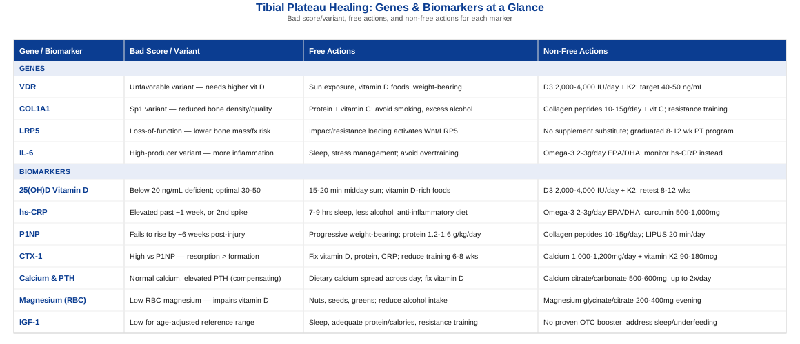
The Biomarkers Worth Tracking After a Tibial Plateau Bone Contusion
A tibial plateau bone contusion is, biologically, a form of trabecular micro-fracture — the MRI shows marrow edema because trabecular struts inside the bone have actually cracked, even though the outer cortical shell hasn't broken. Healing follows the same general biology as a stress fracture: an initial inflammatory phase, then a bone-formation phase where osteoblasts lay down new matrix, and a remodeling phase where the tissue is refined back toward normal density. Because that process depends directly on nutrient status, inflammatory control, and hormonal signaling, blood work can offer a genuinely useful window into how the injury is progressing — not a replacement for imaging or a surgeon's assessment, but a meaningful complement to both.
The following seven biomarkers are the ones with the clearest link to bone healing biology, a reasonable cost, and a plan of action when they come back abnormal. They split naturally into three groups: nutrient status (vitamin D, calcium/PTH, magnesium), the healing process itself (P1NP, CTX-1, IGF-1), and general inflammatory load (hs-CRP).
1. 25-Hydroxyvitamin D [25(OH)D]
Vitamin D status is the single most-studied nutrient marker in fracture and bone-injury literature, because it directly governs how much calcium your gut absorbs and how well osteoblasts mineralize new bone. A systematic review of human fracture-healing studies found that low 25(OH)D is common in fracture patients and is biologically linked to impaired bone formation, even though the clinical trial evidence on whether supplementation speeds healing is still mixed (role of vitamin D in human fracture healing). A more recent phase II randomized trial specifically tested vitamin D3 dosing strategies around acute fracture healing, reflecting how seriously this question is now being taken in orthopedic research (vitamin D3 supplementation and acute fracture healing trial).
How to Measure It
A serum 25(OH)D test is a standard blood draw, available through any primary care provider or directly through consumer lab services. Cost typically runs $30–$80 out of pocket if not covered by insurance, and turnaround is usually 1–3 days. Optimal ranges for bone healing generally sit between 30–50 ng/mL; below 20 ng/mL is considered deficient.If the Score Is Bad: The Plan Without Supplements
Fifteen to twenty minutes of midday sun exposure on bare arms and legs, several times a week, meaningfully raises vitamin D synthesis in most skin types, though this is slower and less predictable than supplementation and depends on latitude, season, and skin pigmentation. Pairing this with vitamin D-containing foods — fatty fish, egg yolks, fortified dairy — helps but rarely closes a real deficiency on its own.If the Score Is Bad: The Plan With Supplements or Equipment
A typical correction protocol uses 2,000–4,000 IU of vitamin D3 daily, or a weekly loading dose (such as 50,000 IU) for 6–8 weeks under a clinician's guidance for more significant deficiency, followed by a lower daily maintenance dose. Retest at 8–12 weeks. Because vitamin D3 is fat-soluble and stored, cycling isn't necessary at moderate doses, but sustained intake above 4,000 IU/day without monitoring risks hypercalcemia over months; pairing with vitamin K2 is commonly recommended to help direct calcium toward bone rather than soft tissue. Red-light or UV-lamp devices exist but are a poor substitute for supplementation in terms of cost-effectiveness and dosing precision.2. High-Sensitivity C-Reactive Protein (hs-CRP)
CRP is your body's general inflammation gauge, and it has a well-documented, predictable rhythm after bone trauma: it rises within hours, peaks around 48–72 hours, and returns to baseline within about a week in an uncomplicated case (CRP kinetics after long bone fractures). A bone contusion that keeps CRP elevated well past that window, or that produces a second unexpected spike, is worth flagging to a clinician — it may signal a complication rather than normal healing inflammation, and it also reflects how much systemic inflammatory "noise" your body is carrying into the repair process.
How to Measure It
A simple hs-CRP blood test, widely available and inexpensive at $15–$40, with results in 1–2 days. Repeat testing weekly during the first month after injury gives the most useful trend data.If the Score Is Bad: The Plan Without Supplements
Prioritizing 7–9 hours of consistent sleep, reducing alcohol, and avoiding unnecessary re-aggravation of the injury (overuse or premature loading reignites local inflammation) are the highest-leverage, no-cost levers. An anti-inflammatory eating pattern — more vegetables, fatty fish, olive oil, fewer ultra-processed foods — has consistent, if modest, effects on baseline CRP.If the Score Is Bad: The Plan With Supplements or Equipment
Omega-3 fatty acids (roughly 2–3 g/day of combined EPA/DHA) are the best-evidenced supplement for lowering CRP over 8–12 weeks; take with food to reduce GI side effects, and check with a physician if on blood thinners. Curcumin (500–1,000 mg with piperine) has more modest supporting evidence. Avoid cycling omega-3s; they work cumulatively and are safe for continuous use at these doses.3. P1NP (Procollagen Type I N-Terminal Propeptide)
P1NP is the reference marker for bone formation — it's released when osteoblasts are actively synthesizing new type I collagen, the main building block of bone. A study specifically tracking bone turnover markers after tibia and femur fracture fixation found that P1NP rises and peaks around six weeks post-injury, and that a healthy six-week elevation correlated with radiological healing by twelve weeks (bone turnover markers as surrogates of fracture healing). For a bone contusion, a P1NP that fails to rise appropriately in that window can be an early signal that the formation phase is sluggish.
How to Measure It
This requires a specialty lab (not a standard panel); cost runs $60–$150 and is best drawn in the morning, since P1NP has some diurnal variation. It's not routinely ordered by primary care, so you may need to request it specifically or work with a sports medicine or endocrinology provider.If the Score Is Bad: The Plan Without Supplements
Progressive, pain-free weight-bearing and resistance loading (once cleared) is the single strongest natural stimulus for osteoblast activity — mechanical load is a direct signal for bone formation. Adequate protein intake (roughly 1.2–1.6 g/kg bodyweight daily) supports the raw material supply for collagen synthesis.If the Score Is Bad: The Plan With Supplements or Equipment
Collagen peptides (10–15 g/day, ideally with a source of vitamin C to support collagen crosslinking) are commonly used, though the human evidence in bone-specific contexts is still early. Low-intensity pulsed ultrasound (LIPUS) bone-healing devices are FDA-cleared adjuncts for fracture healing and are sometimes used for slow-healing bone injuries under physician guidance; typical use is 20 minutes daily, and side effects are minimal, though cost (device rental or purchase) is a real consideration.4. CTX-1 (C-Terminal Telopeptide of Type I Collagen)
CTX-1 is the mirror-image marker to P1NP: it reflects bone resorption, the breakdown side of the remodeling cycle. Healthy healing needs both formation and controlled resorption in the right proportion — remodeling is how bone reshapes itself back toward normal architecture after the initial repair. The same tibia/femur fracture study that tracked P1NP found CTX also peaks around six weeks and that its trajectory tracks with radiographic healing, making the P1NP-to-CTX ratio a more informative picture than either marker alone (bone turnover markers as surrogates of fracture healing).
How to Measure It
Also a specialty lab test, usually ordered alongside P1NP, at a similar cost ($60–$150). Fasting, morning draws reduce variability, since CTX fluctuates more than P1NP throughout the day.If the Score Is Bad: The Plan Without Supplements
A persistently high CTX relative to P1NP (excess resorption without matching formation) often reflects poor nutrient status or excess inflammation rather than a standalone problem — addressing vitamin D, protein, and CRP (above) tends to bring the ratio back into balance more effectively than targeting CTX in isolation.If the Score Is Bad: The Plan With Supplements or Equipment
Adequate calcium (1,000–1,200 mg/day from diet plus supplementation as needed) and vitamin K2 (90–180 mcg/day) support the formation side of the ratio. Avoid excessive endurance training volume during the acute healing window, since very high training loads without adequate recovery can shift the balance toward resorption; this is a cycling consideration rather than a supplement one — scale training volume down for 6–8 weeks post-injury, then reintroduce gradually.5. Calcium and Parathyroid Hormone (PTH)
Calcium and PTH are best read together, because PTH is the hormone that keeps blood calcium stable by pulling from bone, gut absorption, and the kidneys as needed (regulation of bone remodeling by parathyroid hormone). A normal serum calcium with an elevated PTH often means the body is compensating for low vitamin D or low dietary calcium by drawing more aggressively on bone — precisely the wrong direction while a bone contusion is trying to rebuild.
How to Measure It
Both are standard blood tests, often bundled with a basic metabolic panel; combined cost is usually $30–$70. Since PTH is affected by vitamin D status, it's most informative when tested alongside 25(OH)D.If the Score Is Bad: The Plan Without Supplements
Dietary calcium from dairy, leafy greens, canned fish with bones, and fortified plant milks, spread across the day (absorption is capped at roughly 500 mg per sitting), is the first lever. Correcting vitamin D status (see above) is usually what actually normalizes an elevated PTH, since the two are mechanistically linked.If the Score Is Bad: The Plan With Supplements or Equipment
Calcium citrate or carbonate supplementation (500–600 mg per dose, up to twice daily) can close a dietary gap; take with meals, and be aware that excess supplemental calcium (above roughly 2,000 mg/day total) has been linked to kidney stone risk and possible cardiovascular concerns in some studies, so more isn't automatically better. No cycling is needed at recommended doses; retest calcium and PTH after 8–12 weeks of correction.6. Magnesium (RBC Magnesium)
Magnesium is a frequently overlooked cofactor in bone metabolism — it's required for vitamin D activation and influences PTH secretion. A long-term prospective cohort study found that low serum magnesium was independently associated with a substantially higher fracture risk, with more than a two-fold hazard ratio between the lowest and highest magnesium quartiles (low serum magnesium and fracture risk).
How to Measure It
Standard serum magnesium is cheap ($15–$30) but a poor reflection of total body stores, since most magnesium is intracellular; a red blood cell (RBC) magnesium test, at $40–$70, is more accurate and worth requesting specifically.If the Score Is Bad: The Plan Without Supplements
Nuts, seeds, legumes, leafy greens, and whole grains are reliably good magnesium sources; reducing alcohol intake also helps, since alcohol increases urinary magnesium loss.If the Score Is Bad: The Plan With Supplements or Equipment
Magnesium glycinate or citrate, 200–400 mg in the evening, is a common and well-tolerated correction; magnesium oxide is cheaper but less bioavailable and more likely to cause loose stools. This is a supplement that's reasonable to continue long-term without cycling, though doses above 350 mg from supplements (separate from food) can cause GI upset in sensitive individuals — start lower and titrate up.7. IGF-1 (Insulin-Like Growth Factor 1)
IGF-1 is a growth-hormone-dependent signaling molecule that drives osteoblast proliferation and has been directly studied as a therapeutic accelerant for fracture healing, with early research showing that systemic growth hormone combined with local IGF-1 application improved healing parameters (growth hormone and IGF-1 in fracture healing). It's worth being clear that most of the direct fracture-healing data on IGF-1 comes from preclinical and early-stage research rather than large human trials, so it belongs in this list as a marker of your general anabolic status rather than a proven lever to manipulate on its own.
How to Measure It
A standard blood test, typically $60–$100, available through most labs. Levels naturally decline with age, so results should be interpreted against age-adjusted reference ranges.If the Score Is Bad: The Plan Without Supplements
Adequate sleep (growth hormone and downstream IGF-1 pulse primarily during deep sleep), sufficient total protein and calorie intake (chronic undereating suppresses IGF-1), and resistance training are the most reliable natural levers.If the Score Is Bad: The Plan With Supplements or Equipment
There is no well-evidenced, safe over-the-counter supplement that reliably raises IGF-1 in humans — claims around certain amino acid blends or "growth hormone secretagogues" are not well supported. The more defensible approach is addressing the underlying driver (sleep debt, underfeeding, or insufficient resistance training) rather than supplementing directly; if IGF-1 is low alongside other symptoms of hormonal dysfunction, that's a conversation for an endocrinologist rather than a self-directed supplement plan.Tracking these seven markers together — rather than fixating on any single number — gives a genuinely useful picture of whether a tibial plateau bone contusion is healing on a normal biological timeline. But biomarkers only tell you what's happening right now; some of the variation in how efficiently different people build bone in the first place traces back to inherited differences in a small number of genes.
The Genetic Side: What Your DNA May Add to the Picture
Consumer genomics has made it easy to pull up raw genetic data and see variants in bone-related genes, and researchers like Ali Torkamani — whose work on polygenic risk scoring has pushed genetic risk assessment well beyond single-gene stories — along with health personalities like Gary Brecka, who popularized panel-based genetic testing for a broader audience, have both helped bring this kind of information out of research labs and into everyday health decisions. It's worth being precise about what that information can and can't tell you: these are genes with real, replicated associations with bone density and healing biology, but in almost every case the effect size is modest, the evidence is population-level rather than individually predictive, and lifestyle factors can meaningfully offset an unfavorable variant. Genetics here is a modifier, not a verdict.
VDR (Vitamin D Receptor)
The VDR gene encodes the receptor that vitamin D binds to in order to exert its effects on calcium absorption and osteoblast signaling. Common variants — FokI, BsmI, TaqI, and ApaI are the most studied — have been linked to differences in bone mineral density across many population studies, though a systematic review of this literature notes the associations are inconsistent across ethnic groups and study designs, and are strongly influenced by actual circulating vitamin D levels rather than acting independently of them (VDR gene polymorphisms and osteoporosis: a systematic review). In practical terms, an unfavorable VDR variant likely means your bone tissue needs a higher circulating vitamin D level to achieve the same signaling effect as someone with a more favorable variant.
If the Gene Variant Is Unfavorable: The Plan Without Supplements
Consistent sun exposure and a vitamin D-rich diet (fatty fish, egg yolks, mushrooms exposed to UV light) become more important, not less, since you may need to work harder to reach the same effective vitamin D signal. Regular weight-bearing activity also supports the downstream calcium-handling pathway independently of receptor sensitivity.If the Gene Variant Is Unfavorable: The Plan With Supplements or Equipment
Rather than guessing, this is a case where testing (see 25(OH)D above) and targeting the higher end of the optimal range (40–50 ng/mL rather than just clearing the deficiency cutoff) makes sense, using the same 2,000–4,000 IU/day D3 protocol described earlier, paired with vitamin K2. Retest every 8–12 weeks until stable, then check annually.COL1A1 (Collagen Type I Alpha 1)
COL1A1 encodes the primary protein chain of type I collagen, the structural scaffold of bone. A well-known variant affecting a binding site for the transcription factor Sp1 has been shown, across multiple studies including a landmark analysis, to reduce bone density and alter bone quality independent of density — meaning carriers can have measurably weaker bone even at a similar bone mineral density reading (COL1A1 Sp1 polymorphism and bone density/quality). For someone recovering from a bone contusion, this variant is a plausible reason why healing might run slower or the injury might have happened at lower impact than expected.
If the Gene Variant Is Unfavorable: The Plan Without Supplements
Since collagen quality — not just quantity — is at stake, the priority is providing collagen synthesis cofactors through diet (vitamin C-rich foods, adequate total protein) and avoiding known collagen-degrading habits: smoking and excess alcohol both impair collagen cross-linking and should be minimized during any healing window.If the Gene Variant Is Unfavorable: The Plan With Supplements or Equipment
Collagen peptides (10–15 g/day) taken with 100–200 mg of vitamin C are a reasonable, low-risk addition, though the direct human evidence for this combination in bone-specific outcomes is still early rather than definitive. Resistance training, once cleared by a clinician, remains the strongest mechanical stimulus for improving bone quality regardless of genotype, and should be viewed as the primary intervention with supplements as a secondary support.LRP5 (Low-Density Lipoprotein Receptor-Related Protein 5)
LRP5 is a co-receptor in the Wnt signaling pathway, which governs osteoblast proliferation and differentiation. Loss-of-function variants are associated with lower bone mass and higher fracture risk, while gain-of-function variants produce unusually high bone density; a study in elderly osteoporosis patients found LRP5 variants were associated with both bone mineral density and bone turnover marker levels (LRP5 gene, bone mineral density, and fractures). What makes LRP5 particularly actionable is that the Wnt pathway it feeds into is directly activated by mechanical loading — meaning exercise partially works around the genetic bottleneck rather than depending on it.
If the Gene Variant Is Unfavorable: The Plan Without Supplements
Impact and resistance loading — once medically cleared for a healing bone contusion — is the most direct non-supplement lever, since mechanical strain activates the same Wnt/LRP5 signaling axis. This is a case where consistency (regular loading sessions, not occasional intense ones) matters more than intensity alone.If the Gene Variant Is Unfavorable: The Plan With Supplements or Equipment
There's no supplement that meaningfully substitutes for mechanical loading in this pathway. Where equipment is useful, a graduated return-to-loading program — starting with body-weight and low-impact resistance work and progressing to higher-load training over 8–12 weeks — is the most evidence-aligned approach; a physical therapist can help calibrate the progression around the healing tibial plateau specifically.IL-6 (Interleukin 6)
IL-6 is a cytokine central to the inflammatory phase of bone healing — animal studies show that IL-6 knockout mice have delayed callus mineralization and maturation after fracture, confirming that some IL-6 signaling is actually necessary for normal healing, not just a byproduct of injury (IL-6 knockout mice and delayed fracture healing). Genetic variants in the IL-6 promoter region affect how much IL-6 an individual produces in response to injury; the practical concern isn't having IL-6 at all — it's producing too much for too long, since chronic elevation (as reflected by a persistently high hs-CRP) is associated with poorer healing environments.
If the Gene Variant Is Unfavorable: The Plan Without Supplements
Sleep quality, stress management, and avoiding chronic overtraining are the most direct levers on IL-6 regulation, since all three influence baseline inflammatory tone. Avoiding a second injury or re-aggravation during the healing window is particularly important for this pathway, since repeated insult keeps IL-6 elevated past its useful window.If the Gene Variant Is Unfavorable: The Plan With Supplements or Equipment
Omega-3 fatty acids (2–3 g/day EPA/DHA), the same protocol described under hs-CRP, are the best-supported option for moderating chronic inflammatory tone; there is no need to cycle this, and side effects are typically limited to mild GI upset or a fishy aftertaste, manageable by taking with food. Monitoring hs-CRP alongside this gene, rather than trying to test IL-6 directly (which is expensive and highly variable), is the more practical real-world approach.Genetics explains part of why healing speed varies between people, but it isn't the most actionable lever day to day — biomarkers and behavior are. With both of those pieces in place, it's worth looking at how a broader framework for bone health, popularized outside the orthopedic world, fits into this picture.
What Outlive Gets Right About Protecting Bone Through Injury and Age
Peter Attia's Outlive: The Science and Art of Longevity isn't written about bone contusions specifically, but its central argument — that bone and muscle are strategic assets you have to build well before you need them — is directly relevant to anyone recovering from one and thinking about future injury risk. Here are the ten ideas from that framework most worth carrying into a bone-injury recovery plan.
1. Bone Density Peaks Decades Before You Need It
Peak bone mass is typically reached by the late twenties to early thirties, and it's substantially harder to build back after that window than it is to lose. A bone contusion in your thirties, forties, or beyond is, in part, a test of how much reserve you built earlier — a reason to treat bone-building training as urgent regardless of your current age.2. Muscle Is Medicine — and Armor for Bone
Attia frames muscle mass not as an aesthetic goal but as a protective organ system: stronger surrounding musculature reduces the mechanical load transmitted directly into bone during falls and impacts, and supports faster, more stable rehabilitation once weight-bearing resumes.3. Strength Training Beats Cardio for Skeletal Loading
Cardiovascular exercise is essential for longevity, but it doesn't load bone the way resistance training does. Heavy, progressive resistance work stimulates osteoblast activity through direct mechanical strain in a way that low-impact cardio simply doesn't replicate.4. Train for the "Centenarian Decathlon," Not Just Today
Attia's exercise philosophy asks: what physical tasks do you want to be able to do in your final decade of life, and are you training for them now? Applied to a tibial plateau injury, that means rehab shouldn't stop at "pain-free," it should extend to the loaded, dynamic movement patterns you'll need decades from now.5. Fall Prevention Starts Long Before You're Old
Balance, proprioception, and stability training are framed as an early-life investment rather than a late-life necessity. After a tibial plateau contusion — an injury often caused by exactly the kind of unstable loading or fall that balance training helps prevent — this becomes a directly relevant part of rehab, not an afterthought.6. Track Objective Markers, Not Just How You Feel
A recurring theme is that subjective feeling is a poor guide to underlying bone and metabolic health — Attia advocates annual DEXA scans to track bone mineral density objectively. The same logic supports the biomarker-tracking approach in this article: numbers catch what "feeling fine" can miss.7. Protein Intake Is a Lever Most People Underuse
Attia repeatedly emphasizes that most adults, especially as they age, under-consume protein relative to what's needed to support muscle and connective tissue repair — directly relevant to the collagen-synthesis needs of a healing bone contusion.8. Stability Work Reduces Reinjury Risk
Beyond strength, Attia highlights stability and mobility training as protective against the kind of awkward, uncontrolled movement that causes bone contusions and ligament injuries in the first place — a case for building this into long-term training, not just acute rehab.9. Medicine 3.0: Be Proactive, Not Reactive
The book's broader philosophy — acting before a problem becomes acute rather than waiting for a diagnosis — maps directly onto the idea of tracking biomarkers and genetic risk before and after an injury, instead of only reacting to pain.10. Small, Consistent Inputs Compound More Than Dramatic Interventions
Attia is skeptical of quick fixes and consistently favors compounding, unglamorous habits — sleep, strength training, protein, consistent movement — over dramatic interventions. That's a useful corrective for anyone tempted to chase an expensive gadget instead of the basics covered in the biomarker sections above.These lessons sit comfortably alongside conventional orthopedic advice — they don't contradict it, they extend it into a longer time horizon. The same is true of a handful of complementary approaches that have real, if often modest, human evidence behind them for bone and joint recovery.
Complementary Approaches Worth Considering
None of the following replace medical clearance, weight-bearing guidance from an orthopedic provider, or physical therapy — they sit alongside that care, and the evidence for each is honestly mixed rather than definitive.
Photobiomodulation (Low-Level Laser Therapy)
Photobiomodulation uses specific wavelengths of red or near-infrared light, believed to stimulate osteoblast activity, collagen synthesis, and local blood flow — mechanisms that are directly relevant to a healing bone contusion, since they target the same cellular processes involved in trabecular bone repair.
A human trial using 830 nm low-level laser therapy applied five times weekly for two weeks to closed wrist and hand fractures found improvements in pain, functional disability, grip strength, and radiographic healing parameters compared to placebo (LLLT for closed bone fracture in the wrist and hand); a broader review of the fracture-healing literature notes the mechanism is plausible and consistent across studies, even as clinical effect sizes vary (effect of low-level laser therapy on fracture healing).
Realistically, this means discussing a licensed photobiomodulation device with a physical therapist or sports medicine provider rather than self-treating with an uncalibrated consumer device; sessions are typically short (a few minutes per area, several times weekly) and side effects are minimal, but the evidence supports it as an adjunct to standard care, not a substitute for weight-bearing progression or time.
Tai Chi
Tai chi combines slow, weight-shifting movement with balance and proprioceptive training — relevant both to later-stage rehabilitation after a tibial plateau injury and to reducing the fall risk that often causes these injuries in the first place.
A systematic review and meta-analysis found tai chi produced measurable improvements in bone mineral density at the lumbar spine and femoral neck in postmenopausal women across 23 randomized trials (tai chi and bone mineral density in postmenopausal women), while a separate meta-analysis confirmed consistent, safe benefits for balance and fall prevention in older adults, with no adverse events reported across trials (tai chi for fall prevention and balance).
For someone past the acute phase of a bone contusion, two to three tai chi sessions weekly, once weight-bearing is cleared, is a low-impact way to rebuild balance and confidence in the joint without the loading risk of higher-impact activity; it complements rather than replaces progressive resistance training.
Guided Imagery
Guided imagery — structured mental rehearsal of movement, healing, or relaxation — has been studied specifically in orthopedic surgical and rehabilitation populations as a low-cost way to reduce pain perception and anxiety during recovery, both of which can indirectly slow rehab engagement.
A randomized controlled trial in geriatric orthopedic patients found guided imagery significantly reduced postoperative pain and improved comfort compared to standard care (guided imagery and postoperative pain in geriatric orthopedic patients), and similar mental-simulation approaches have shown favorable effects on functional outcomes after knee procedures.
Applied to a bone contusion, a 10–15 minute guided imagery session before physical therapy or at bedtime — free audio programs or a therapist-led script both work — is a reasonable, risk-free addition, particularly if pain or movement anxiety is slowing rehab progress; it's a supplement to pain management, not a replacement for appropriate loading or medication when needed.
Massage Therapy
Massage therapy is most directly relevant to the soft-tissue swelling, guarding, and secondary muscle tension that commonly accompany a tibial plateau bone contusion, rather than to the bone injury itself.
A randomized controlled trial in athletes found that sports massage combined with cold therapy accelerated recovery markers after intense exertion, and a broader body of research on massage for exercise-related soreness shows consistent, moderate reductions in pain and improvements in range of motion (sports massage and cold therapy for muscle recovery).
For a bone contusion, gentle massage focused on the surrounding musculature (quadriceps, hamstrings, calf) — never directly over the injured bone in the acute phase — can help manage compensatory tension and swelling; one to two sessions weekly during active rehab, performed by a licensed therapist familiar with the injury, is a reasonable cadence.
Conclusion
A tibial plateau bone contusion heals through a biological process that responds to specific, measurable inputs — vitamin D status, inflammatory load, bone turnover balance, and mechanical loading chief among them. Genetics shapes the baseline efficiency of that process, but for almost every gene discussed here, the actionable lever is the same behavior or nutrient status that would help regardless of genotype. The biomarkers matter because they turn "how is it healing?" from a guess into a trackable answer, and the complementary approaches matter because they address the pain, anxiety, and deconditioning that often slow recovery as much as the bone injury itself.
The next useful step isn't dramatic: order the 25(OH)D and hs-CRP panels if you haven't already, ask your orthopedic provider whether P1NP/CTX-1 tracking makes sense for your specific case, and bring this information into your next appointment rather than treating it as a replacement for one. Better data, reviewed with a qualified professional, is what actually turns "wait and see" into a plan.
Musculoskeletal: Bone Conditions Joint Conditions